Biopsychology
1/158
There's no tags or description
Looks like no tags are added yet.
Name | Mastery | Learn | Test | Matching | Spaced |
|---|
No study sessions yet.
159 Terms
Nervous System
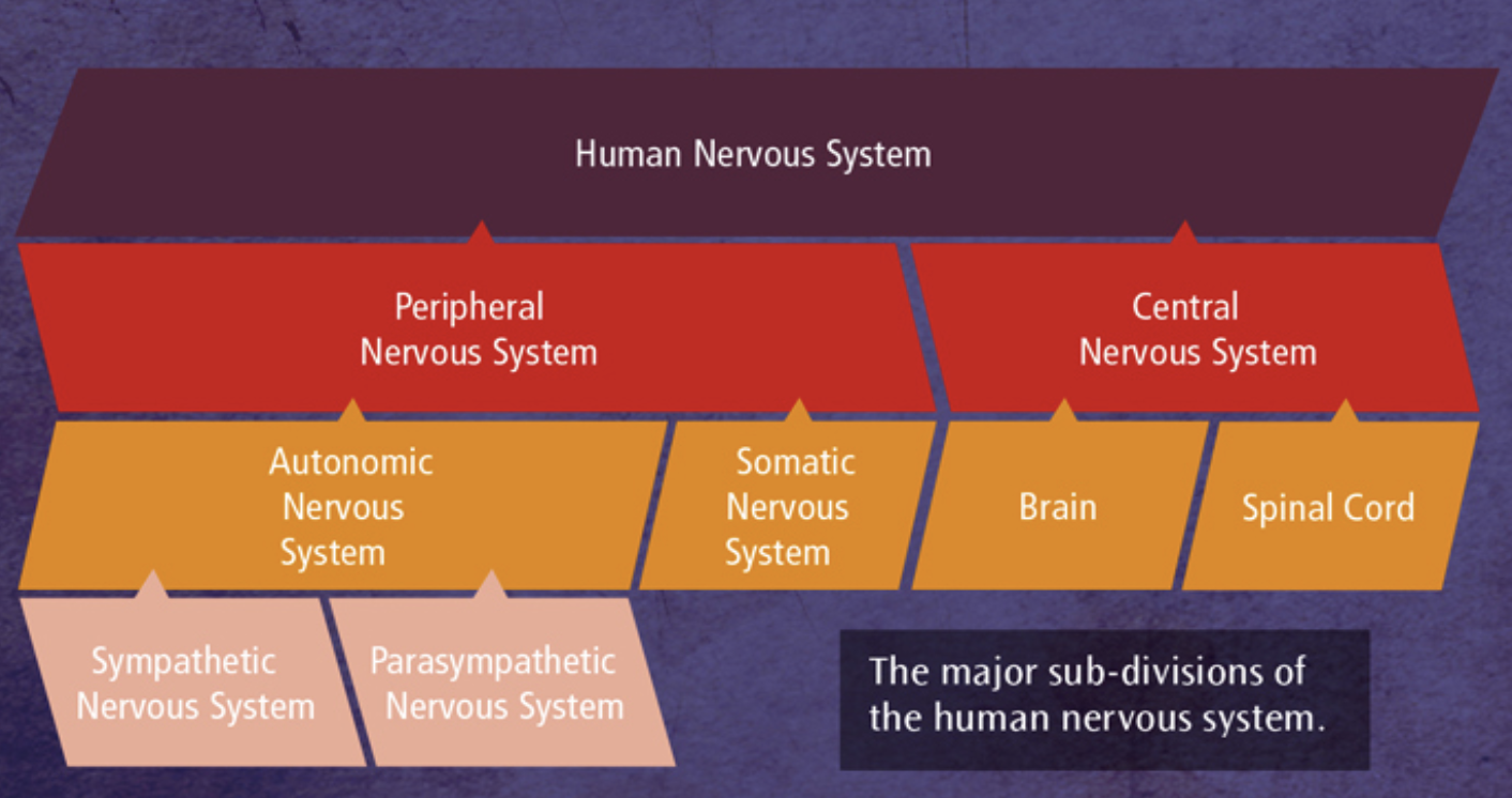
Consists of the central nervous system and peripheral nervous system
Central Nervous System (CNS)
Consists of the brain and the spinal cord and is the origin of all complex commands and decisions
Peripheral Nervous System (PNS)
Sends information to the central nervous system (CNS) from the outside world, and transmits messages from the CNS to muscles and glands in the body
Somatic Nervous System
Transmits information from receptor cells in the sense organs to the CNS. It also receives information from the CNS that directs muscles to act
Autonomic Nervous System
Transmits information to and from internal bodily organs. It’s autonomic as the system operates involuntarily. It has 2 main divisions: the sympathetic and parasympathetic nervous systems
The functions of the nervous system
The nervous system is a specialised network of cells in the human body and is our primary internal communication system. It has 2 main functions:
To collect, process and respond to information in the environment
To coordinate the working of different organs and cells in the body
What is the nervous system divided into?
Central Nervous System
Brain
Spinal Cord
Peripheral Nervous System
Somatic Nervous System
Autonomic Nervous System
Sympathetic Nervous System
Parasympathetic Nervous System
The Brain
The centre of all conscious awareness. The brain’s outer later, the cerebral cortex, is highly developed in humans and is what distinguishes our higher mental functions from those of animals. The brain is divided into 2 hemispheres
The Spinal Cord
An extension of the brain and is responsible for reflex actions such as pulling your hand away from a hot plate
The function of the peripheral nervous system
It transmits messages, via millions of neurons (nerve cells), to and from the CNS. The PNS is divided further into:
Autonomic Nervous System (ANS)
Somatic Nervous System (SNS)
The function of the autonomic nervous system
Governs the vital functions in the body such as breathing, heat rate and digestion
The function of the somatic nervous system
controls muscle movement and receives information from sensory receptors
3 layers of the brain
Central Core- regulates involuntary actions
Cerebrum- regulates our higher intellectual processes
Limbic System- regulates our emotions
4 lobes of the brain
Frontal lobe- planning, decision-making and problem solving
Temporal lobe- processing auditory information
Occipital lobe- visual processing area
Parietal lobe- sensory perception and integration, including the management of the senses
The Endocrine System
Instructs glands to release hormones directly into the bloodstream. The endocrine system acts more slowly but has very widespread and powerful effects
Thyroid Gland
Produces thyroxine which affects the heart rate by increasing it. This increases the metabolic rates and in turn affects growth rates
Pituitary Gland
The master gland because it controls the release of hormones from all the other endocrine gland. It’s located in the brain
Adrenal Gland
Produces adrenaline responsible for the fight-or-flight response
Ovaries
Produces oestrogen and progesterone
Testes
Produces testosterone
Process of fight-or-flight
The hypothalamus triggers activity in the sympathetic branch of the autonomic system
The ANS changes from its resting state (parasympathetic state) to the physiologically aroused state (sympathetic state)
Adrenaline is released from the adrenal medulla into the bloodstream
Once the threat has passed, the parasympathetic nervous system returns the body to its resting state
The parasympathetic nervous system acts as a ‘brake’ and reduces the activities of the body that were increased by the actions of the sympathetic branch. This is referred to as the rest and digest response
Psychological effects of adrenaline
Increased anxiety, attention and alertness
Physical effects of adrenaline
Increases heart rate and breathing rate, pupils dilating. Digestion and immune system slow down and decreased blood flow to the skin
Neuron
The basic building blocks of the nervous system, neurons are nerve cells that process and transmit messages through electrical and chemical signals. They are specialise to carry neural information. 80% of neurons are located in the brain
3 types of neurons:
Sensory neuron
Relay neuron
Motor neuron
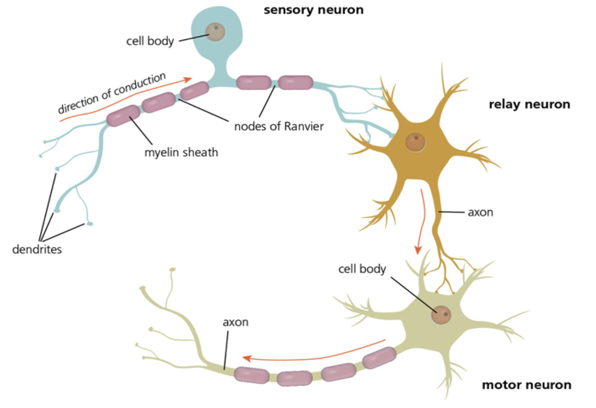
Sensory neuron
Carries messages from the PNS (sensory receptors) to the CNS
Long dendrites and short axons
Relay Neuron
They connect the sensory neuron to the motor neurons or other relay neurons.
They have short axons and short dendrites
Motor Neurons
Connects the CNS to effectors such as muscles and glands
Short dendrites and long axons
Cell body
Includes a nucleus and contains genetic material of the cell. D
Dendrites
They carry nerve impulses from neighbouring neurons towards the cell body
Axon
Carries the impulses away from the cell body down the length of the neuron. It’s covered by a fatty layer of myelin sheath
Myelin Sheath
Covers the axon to protect it and speeds electrical transmission of the impulse. It’s segmented by gaps called the nodes of ranvier
The Nodes of Ranvier
Speeds up transmission of the impulse by forcing it to ‘jump’ across the gaps along the axon
Terminal Buttons
Communicate with the next neuron in the chain across a gap known as synapses
Electric Transmission- the firing of a neuron
When a neuron is in their resting state, the inside of the cell is negatively charged compared to the outside. When a neuron is activated by a stimulus, the inside of the cell becomes positively charged for a second causing an action potential to occur
Synaptic Transmission
Synaptic transmission is how neurons communicate with each other. Information is passed down the axon of the presynaptic neuron as an electrical impulse known as an action potential. Once the action potential reaches the end of the axon, where the synaptic vesicles, it releases chemical messengers known as neurotransmitters. The neurotransmitters carry the signal across the synaptic gap and binds to the receptor sites on the post-synaptic neuron. Once the receptors have been activated, summation happens to determine whether the post-synaptic neuron produces an inhibitory or excitatory effect making them more or less likely to fire.
Excitation
A neurotransmitter, like adrenaline, increases the positive charge of the post-synaptic neuron. This increases the likelihood that the neuron will fire and pass on the electrical impulse
Inhibitor
A neurotransmitter, like serotonin, makes the charge on the post-synaptic neuron more negative. This decreases the likelihood that the neuron will fire and pass the electrical impulse
Evaluation of the fight-or-flight response
The ‘tend and befriend’ response
Negative consequences
‘Fight or flight’ doesn’t tell the whole story
Positive rather than ‘fight or flight’ behaviours
A genetic basis to sex differences in the fight-or-flight response
The ‘tend and befriend’ response
Taylor et al (2000) suggests that female have a different reposes than ‘fight or flight’ called ‘tend and befriend’. This involves protecting themselves and their young through nurturing behaviours and forming protective alliances with other women. Fleeing at any sign of danger would put the female’s offspring at risk
Negative consequences of fight or flight response
It’s maladaptive in modern world situations. Frequently triggered by stimuli that you can’t run away from or fight like exams. Causing acute stress in the short term and chronic stress in the long term
Fight or flight doesn’t tell the whole story
Gray (1988) argues that the first phase of reaction to a threat is not fight or flee but to avoid confrontation. He suggests prior to attacking or running away, we display the ‘freeze response’ which focuses attention and makes them look for new information in order to make the best response for that particular threat
Positive rather than fight or flight behaviours
Von Dawans et al (2012) challenged the view that under stress, men respond only with ‘fight or flight’ while women are prone to ‘tend and befriend’. Von Dawans et al found that acute stress can lead to greater cooperative and friendly behaviour in both men and women.
A genetic basis to sex differences in the fight or flight response
Lee and Harley (2012) found the SRY gene, found exclusively on the male Y chromosome directs male development promoting aggression and resulting in the fight or flight response to stress. The SRY gene may prime males to respond to stress in this way but the absence of the SRY gene in females prevents this response to stress leading to the ‘tend and befriend’ behaviour
Localisation of Function
Specific areas of the brain are associated with particular physical and psychological function
Contralateral
Each hemisphere of the brain control the opposite side of the body
Motor Cortex
Voluntary muscle motor movements across the body. Its contralateral. It’s in the frontal lobe
Damage: loss of muscle function or if severe then paralysis
Somatosensory Cortex
An area of the parietal lobe that receives sense impressions from around the body. Its contralateral
Damage: loss of sensation in opposite side to damage
Broca’s Area
An area of the frontal lobe in the left hemisphere that is responsible for speech production.
Damage: expressive aphasia/ difficulty producing fluent speech
Wernicke’s Area
An area of the temporal lobe in the left hemisphere that is responsible for speech comprehension
Damage: receptive aphasia/ difficulty understanding speech
Auditory Cortex
Located in the temporal lobe and receives and processes sound information from ears
Visual Cortex
Visual processing each hemisphere receives information from opposite visual field. Located in the occipital lobe
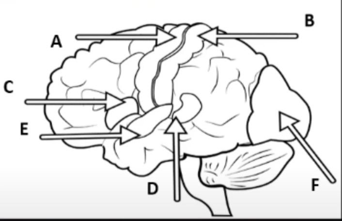
Label areas of the brain
A- Motor Cortex
B- Somatosensory Cortex
C- Broca’a Area
D- Wernicke’s Area
E- Auditory Cortex
F- Visual Cortex
Holistic Theory
Before the discoveries made by Broca and Wernicke, scientists believed that all parts of the brain were involved in the processing of thoughts and actions
The Cerebral Cortex
This separates us from animals as human cortex is much more developed. The cortex appears grey due to the location of the cell bodies, hence the name grey matter
Evaluation of Localisation
Brain scans evidence
Neurosurgical evidence
Case Study: Phineas Gage
Equipotentiality Theory
Aphasia studies
Individual Differences
Language production may not be confined to Broca’s area
Brain scans evidence
Peterson et al (1988) used brain scans to show how Wernicke’s area was active during a listening task and how Broca’s area was active during a reading task. Tulving et al (1994) revealed semantic and episodic memories reside in different parts of the prefrontal cortex
Neurosurgical evidence
Post-mortem confirm the function of Broca’s area. Walter Freeman tried severing connections in the frontal lobe in an attempt to control aggression. Dougherty et al (2002) reported 44 people with OCD had lesioned their cingulate gyrus. After 32 weeks, a third had a successful response and 14 had a partial successful response
Case Study evidence
Clinical case study research demonstrate the loss of a function if damage is caused to particular areas e.g Clive Wearing and Expressive and Receptive Aphasia Studies
Phineas Gage turned from a calm, reserved person to a quick-tempered and rude person after most of his frontal lobe was removed by the iron pole through his brain
Equipotentiality Theory
Lashley suggested that basic motor and sensory functions are localised but higher mental functions aren’t. He claimed that intact areas of the cortex could take over responsibility for specific cognitive functions following injury to the are normally responsible for that function
Aphasia Studies
Expressive Aphasia is an impaired ability to produce language. This is caused by damage to Broca’s area
Receptive Aphasia is an impaired ability to understand language; an inability to extract meaning from spoken or written words. This is caused by damage to Wernicke’s area
Individual Differences in Broca and Wernicke’s area
Bavelier et al (1997) found considerable variability of activation across different individuals when reading. They observed activity in the right temporal, left frontal, temporal and occipital lobes.
Harasty et al (1997) found that women have larger Broca and Wernicke’s areas
Language production may not be confined to Broca’s area alone
Dronkers et al (2007) re-examined the preserved brain of 2 of Broca’s patients including Tan. They used modern high-resolution brain MRI imaging. The MRIs found that other areas besides Broca’s area could have contributed to the reduced speech abilities. Damage to Broca’s area causes temporary speech disruption not severe disruption of spoken language
Hemispheric Lateralisation
Each hemisphere of the brain is specialised to perform different functions
Split-brain research
Research that studies individuals who have been subjected to the surgical separation of the 2 hemispheres of the brain as a result of severing the corpus callosum
Split-brain study
Sperry (1968) studied a unique group of individuals, all of whom have had their corpus callosums severed to separate the 2 hemispheres and control frequent and severe epileptic seizures.
Procedure of Sperry (1968)
An image or word will be projected to an individual’s right visual field and the same or different image could be projected to the individual’s left visual field. In the normal brain, the information would be conveyed to the other hemisphere through the corpus callosum, but for split brain patients the information can’t be transferred between the 2 hemispheres
Findings of Sperry (1968)
When a picture of an object was shown to the right visual field, they could easily describe what they saw. If it was shown to the left visual field, they couldn’t describe it but could draw it and recognise it.
Evaluation of lateralisation
Related to neural capacity
Lateralisation and immune system functioning
Lateralisation changes with age
Related to increased neural capacity
By using only 1 hemisphere to engage in a particular task, this would leave the other hemisphere free to engage in another function. Rogers et al (2004) found that in chickens brain lateralisation has an enhanced ability to perform 2 tasks simultaneously
Lateralisation and immune system functioning
Architects and the mathematically gifted tend to have superior right-hemispheric skills but are also much more likely to be left-handed and to suffer higher rates of allergies and immune system problems.
Tonnessen et al (1993) found a small but significant relationship between handedness and immune system disorder
Lateralisation changes with age
Lateralised patterns found in younger individuals tend to switch to bilateral patterns in healthy older adults. Szaflarski et al (2006) found that language became more lateralised to the left hemisphere with increasing age but after the age of 25, lateralisation decreases with each decade
Evaluation of split-brain research
Demonstrated lateralised brain functions
Strengths of the methodology
Issues with generalisability
Language may not be restricted to the left hemisphere
Demonstrated lateralised brain functions
Sperry and Gazzaniga’s work produced a lot of evidence that the left hemisphere is geared towards analytic and verbal tasks while the right hemisphere performs spatial and music tasks. The right hemisphere can only produce rudimentary words and phrases but contributes emotional and holistic content to language
The left hemisphere is the analyser and the right hemisphere is the synthesiser
Strengths of the methodology
They used high specialised and standardised procedures. This increased the replicability of the study. Sperry varied aspects of the basic procedure and ensured only hemisphere was receiving the information. Thus he developed a very useful and well-controlled procedure
Issues with generalisability
Split-brain research is rarely carried out nowadays. Andrewes (2001) pointed out that many studies are presented with as few as 3 participants or even just 1 participant. Some conclusions have been drawn from individuals who either have a confounding physical disorder that made split-brain procedure necessary. Only 11 took part in all the variations of the studies
Language may not be restricted to the left hemisphere
Turk et al (2002) found that a patient J.W. developed the capacity to speak out of the right hemisphere with the result that he can now speak about information presented to the left or right brain
Plasticity
The brain adapts its function and structure as a result of a change in the environment
4 reasons for plasticity
Learning new skills
A result of developmental changes
Response to direct trauma to the area of the brain
Response to indirect effects of damage like brain swelling
Synaptic Pruning
Synapses that are used frequently become stronger over time and unused synaptic connections are lost
Functional Recovery
The functions that were performed by areas of the brain that are lost or damaged are performed by undamaged areas of the brain
Axonal sprouting
Existing neurons grow new axons to connect to other neurons
Research into plasticity
Maguire et al (2000) studied the brains of London taxi drivers and found significantly more grey matter in the posterior hippocampus. This part of the brain is associated with the development of spatial and navigational skills in humans and animals.
Draganski et al (2006) imaged the brains of medical students 3 months before and after exams. Learning-induced changes were seen to have occurred in the posterior hippocampus and the parietal cortex
Factors affecting functional recovery
Age- children are more likely to recover
Gender- women are more likely to recover
Access to rehabilitative therapy
What happens in the brain during recovery?
The brain is able to rewire and reorganise itself by forming new synaptic connections close to the area of damage. Secondary neural pathways that wouldn’t typically be used to carry out certain functions are activated or ‘unmasked’ to enable functioning to continue often in the same way as before. There are a number of structural changes in the brain:
Axonal Sprouting: the growth of new nerve endings which connect with other undamaged nerve cells to form new neuronal pathways
Reformation of blood vessels
Recruitment of homologous (similar) areas on the opposite side of the brain to perform specific tasks. E.g. if Broca’s area was damaged, a right-side equivalent will carry out its functions and then after a while functionality might shift back to the left
Evaluation of Plasticity
Practical Application
Support from animal studies
Negative plasticity
Age and plasticity
Practical Application
Following illness or injury to the brain, spontaneous recovery tends to slow down after a number of weeks so forms of physical therapy may be required. Techniques may include movement therapy and electrical stimulation of the brain. This shows that although the brain has the capacity to fix itself to a point, the process requires further intervention to be completely successful
Negative Plasticity
The brain’s ability to rewire itself may be maladaptive. Prolonged drug use has been shown to result in poorer cognitive functioning as well as an increased risk of dementia later in life.
60-80% of amputees develop phantom limb syndrome
Age and Plasticity
Functional plasticity reduces with age. The brain has a greater propensity for reorganisation in childhood as it’s constantly adapting to new experiences and learning. Ladine Bezzola et al (2012) demonstrated how 40 hours of golf training produced changes in the neural representation of movement in participants aged 40-60. This showed that neural plasticity does continue through the lifespan
Support from animal studies
Kempermann et al (1998) suggested that an enriched environment could alter the number of neurons in the brain. They found evidence of an increased number of new neurons particularly in the hippocampus in rats housed in complex environments compared to rats in laboratory cages
Evaluation of functional recovery
Support from animal studies
Age differences in functional recovery
Educational attainment and functional recovery
Support from animal studies
Tajiri et al (2013) randomly assigned rats with traumatic brain injury to 1 of 2 groups. 1 group received transplanted stem cells and the other received no stem cells. 3 months later, the brains of stem celled rats showed clear development of neurone-like cells in the area of injury
Age differences in function recovery
Functional plasticity reduces with age. According to this view, the only option following an injury beyond childhood is to develop compensatory behavioural strategies to work around the deficit (such as seeking social support). However, studies have suggested that even abilities commonly thought to be fixed in childhood can still be modified in adults with intense retraining
Educational Attainment and Functional Recovery
Schneider et al (2014) found that patients with the equivalent of a college education are 7x more likely than those who didn't finish high school to be disability-free one year after a moderate to severe traumatic brain injury
4 ways of investigating the brain
fMRIs
EEGs- Electroencephalogram
ERPs- Event-related potentials
Post-mortems
fMRIs
Works by detecting the changes in the blood oxygen levels and flow that occur as a result of neural (brain) activity in specific parts of the brain. When a brain area is more active it consumes more oxygen and to meet this increased demand, blood flow is directed to the active area.
fMRI produces 3-dimensional images (activation maps) showing which parts of the brain are involved in a particular mental processes and this has important implications for our understanding of localisation of functions
Strengths of fMRIs
It doesn’t use radiation- it’s risk-free, non-invasive and straightforward to use
Produces images with very high spatial resolution- depicting detail by mm and provides a clear picture of how brain activity is localised
Offers a more objective and reliable measure of psychological processes than is possible with verbal reports
Precisely identifies brain regions and patterns of activation over time
Weaknesses of fMRIs
It’s expensive and can only capture a clear image if the person stays still
Poor temporal resolution- there’s a 5 second time lag behind the image on screen and the initial firing of neuronal activity
Only measures blood flow in the brain and doesn’t hone in on the activity of individual neurons, so it’s difficult to tell exactly what kind of brain activity is being represented
EEGs (Electroencephalogram)
Measure electrical activity in the brain via electrodes that are fixed to an individual’s scalp using skull caps. The scan recording represents the brainwave patterns that are generated from the action of millions of neurons, providing an overall account of brain activity. EEG is often used by clinicians as a diagnostic tool as unusual rhythmic patterns of activity may indicate neurological abnormalities like epilepsy, tumours or disorders of sleep
Strengths of EEGs
Invaluable in the diagnosis of conditions like epilepsy
It has contributed to our understanding of the stages of sleep
High temporal resolution
Can accurately detect brain activity at a resolution of a single millisecond
Cheaper than fMRI
Can be used while the participant completes a task
Provides a recording of the brain’s activity in real time rather than a still image of the passive brain