MHSB SHORT QUIZ #1
 Call Kai
 Learn
Learn Practice Test
 Spaced Repetition
 Match
Match Flashcards
 Knowt Play
1/15

There's no tags or description
Looks like no tags are added yet.
Name | Mastery | Learn | Test | Matching | Spaced | Call with Kai | Chat |
|---|
No analytics yet
Send a link to your students to track their progress
16 Terms

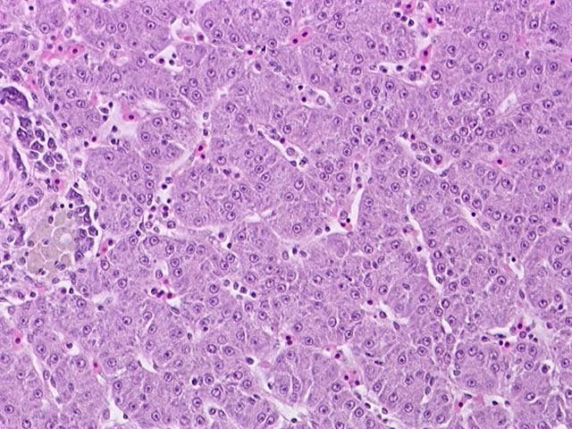
a.) What is this organ?
b.) What is the pointed structure?
c.) What is the parenchyma of the cell?
d.) How many nucleus?
e.) What is the stroma?
f.) What is the arrangement of the cell?
g.) location of the nucleus?
h.) shape?
Liver, Central vein, Hepatocytes, mononucleated or binucleated, Reticular Fibers, loosely arranged, central, polygonal
a.) What is this organ?
b.) What are the pointed structures?
liver, central vein, hepatocytes , sinusoids

a.) What is this organ?
b.) What is the pointed structure?
c.) What is the parenchyma of the cell?
d.) How many nucleus?
e.) What is the stroma?
f.) What is the arrangement of the cell?
g.) location of the nucleus?
h.) Shape?
Skeletal Muscle, Epimysium, Perimysium,Skeletal myoctes, multinucleated, endomysium, epimysium, perimysium, bundles/fasicles, peripheral, Cylindrical/Tubular
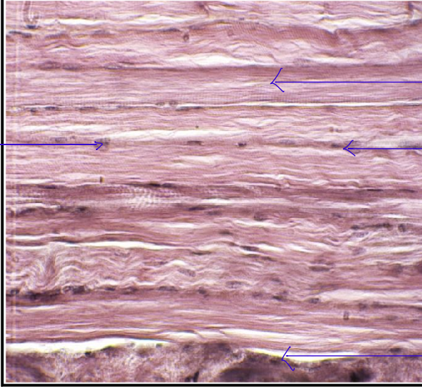
What is/are the structure/s visible in HPO which is/are barely recognizable in LPO?
muscle fibers, nucleus (skeletal myoctes), endomysium

a.) What is this organ?
b.) What is the pointed structure?
c.) What is the parenchyma of the cell?
d.) How many nucleus?
e.) What is the stroma?
f.) What is the arrangement of the cell?
g.) location of the nucleus?
h.) Shape?
trachea, epithelium, tracheal gland, Columnar epithelial Cells with cilia, mononucleated, basement membrane, layers/sheets, central, columnar

What is the cell surface specialization present in these pictures?
Cilia
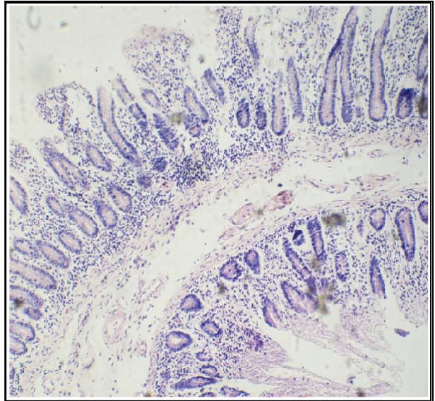
What is the cell surface specialization present in this organ?
Microvili
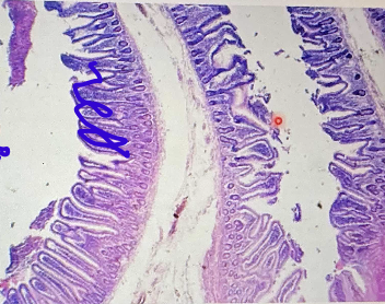
a.) What is this organ?
b.) What is the parenchyma of the cell?
c.) How many nucleus?
d.) What is the stroma?
e.) What is the arrangement of the cell?
f.) location of the nucleus?
g.) Shape?
Small Intestine-Jejunum, Columnar Epithelial Cells with Microvili/Enterocytes, mononucleated, basement membrane, Layers/Sheets, Central, columnar
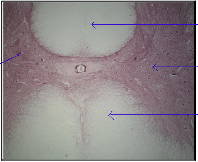
a.) What is this organ?
b.) What is the parenchyma of the cell?
c.) How many nucleus?
d.) What is the stroma?
e.) What is the arrangement of the cell?
f.) location of the nucleus?
g.) Shape?
Spinal Cord, Neurons, mononucleated, neuroglial cell, random/haphazard, central, stellate
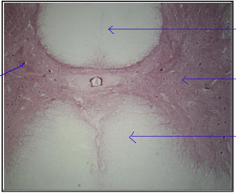
What are the structures seen in this organ? top to bottom
white matter, neurons, gray matter
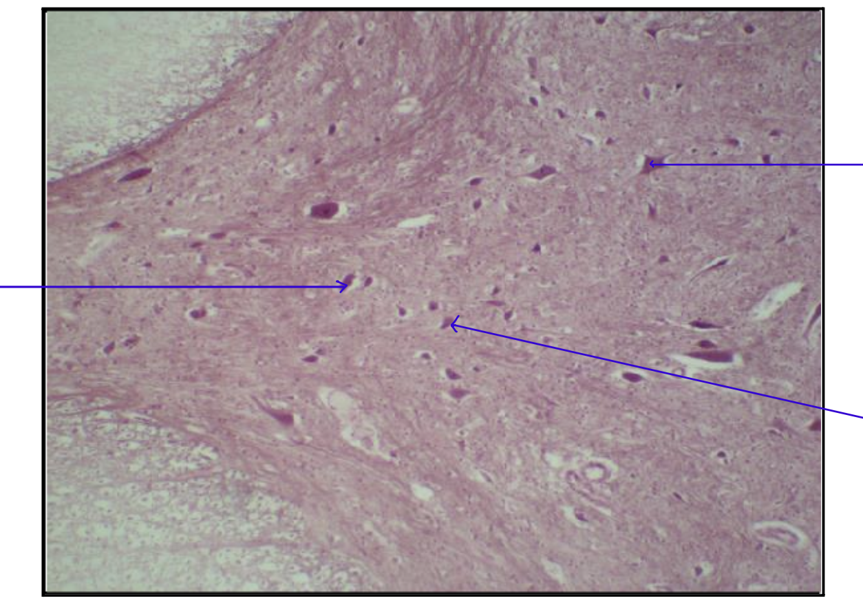
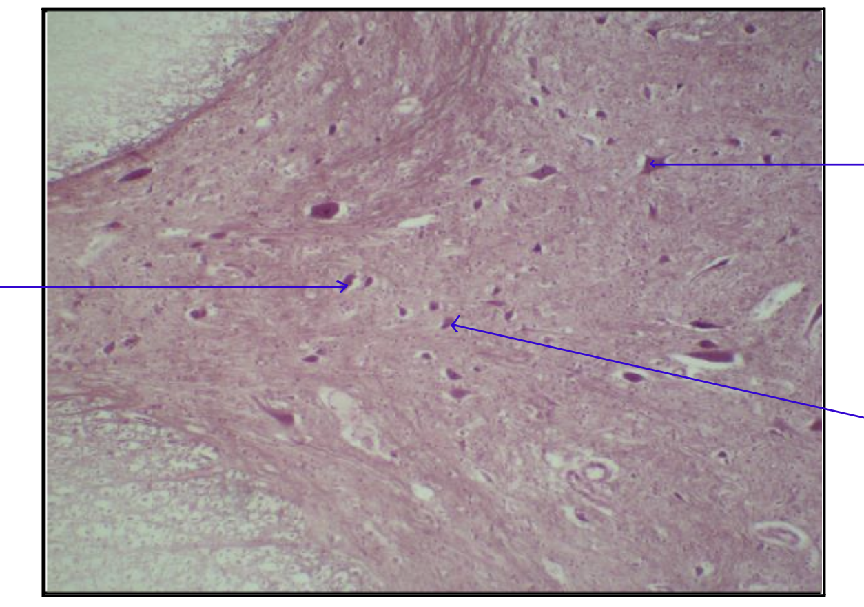
a.) What is the organ?
b.) What structures are seen?
spinal cord, astrocytes, oligodendrocytes, microglia

a.) What is this organ?
b.) What is the pointed structure?
c.) What is the parenchyma of the cell?
d.) How many nucleus?
e.) What is the stroma?
f.) What is the arrangement of the cell?
g.) location of the nucleus?
h.) shape?
Skin, epidermis, basement membrane, keratinocytes, mononucleated, basement membrane, layers/sheets, central, flat/squamous

a.) What organ is this?
b.) What are the structures observed?
skin, epthelial, keratin, epidermis, dermis

a.) What is this organ?
b.) What is the pointed structure?
c.) What is the parenchyma of the cell?
d.) How many nucleus?
e.) What is the stroma?
f.) What is the arrangement of the cell?
g.) location of the nucleus?
h.) shape?
compact bone, haversian canal, astrocytes, osteocytes, mononucleated, concentric lamellae, concentric, central, ovoid almond, irregular

a.) what is this organ?
b.) What is the parenchyma?
c.) What is the stroma?
tendon, fibroblasts, collagen fibers
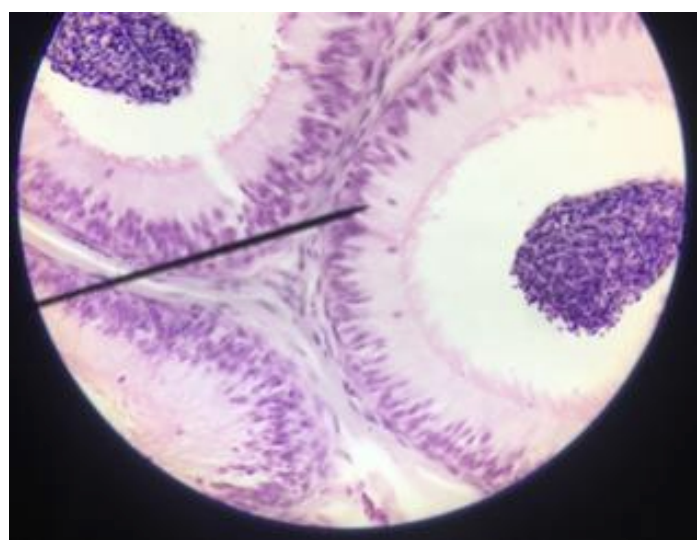
a.) What is this organ?
b.) What is the parenchyma of the cell?
c.) How many nucleus?
d.) What is the stroma?
e.) What is the arrangement of the cell?
f.) location of the nucleus?
g.) Shape?
Epididymis, Columnar Epthelial Cells with stereocilia, mononucleated, basement membrane, layers/sheets, central, columnar