Spinal Cord and Spinal Nerves
1/43
There's no tags or description
Looks like no tags are added yet.
Name | Mastery | Learn | Test | Matching | Spaced |
|---|
No study sessions yet.
44 Terms
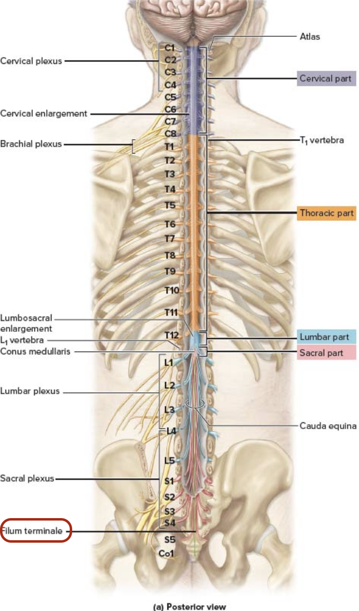
thin strand of pia mater at the end
Filum terminale
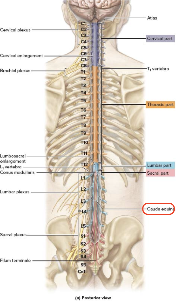
nerve roots that project inferiorly from the spinal cord
Cauda equina
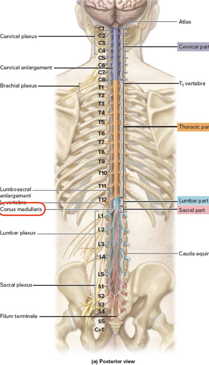
terminal spinal cord at L1, stops at age 4
Conus medullaris
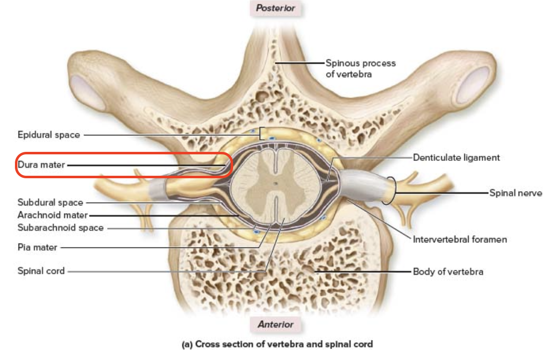
above the dura
Spinal Cord Meninges - Epidural space
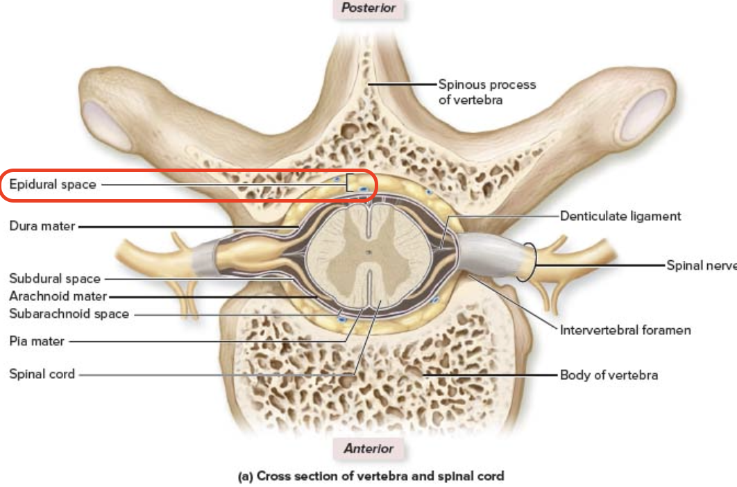

below epidural space
Spinal Cord Meninges - dura matter
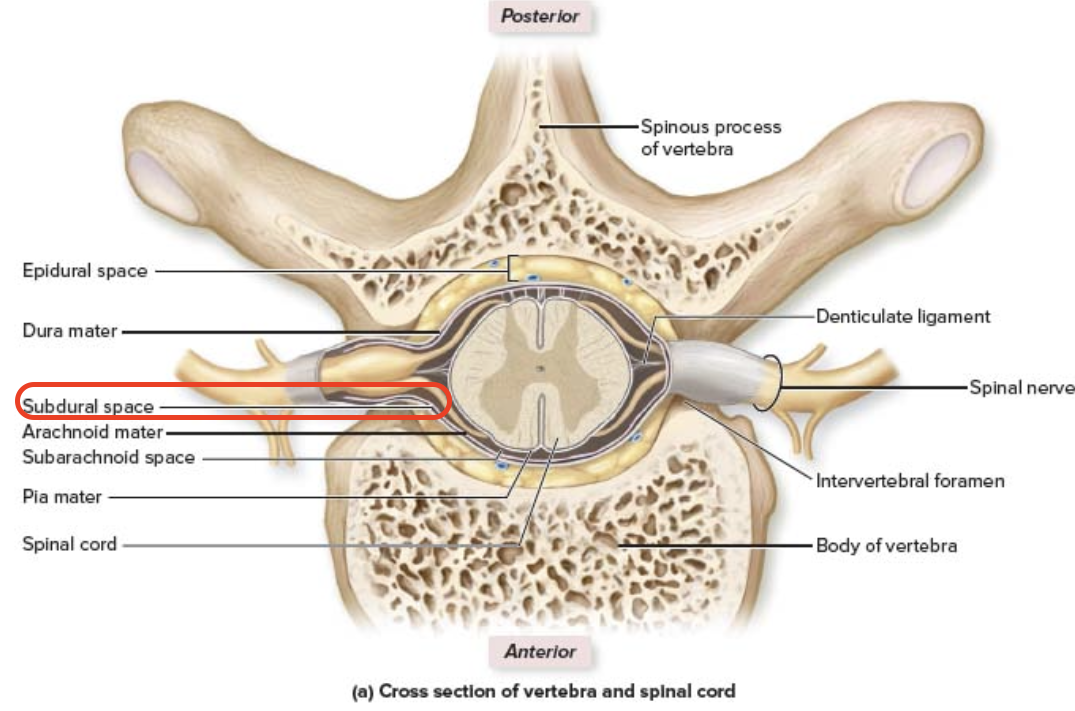
potential space between dura mater and arachnoid mater
Spinal Cord Meninges - Subdural space:
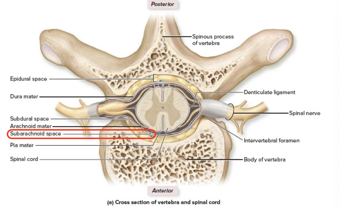
middle membrane, delicate/web-like extension
Spinal Cord Meninges - Arachnoid mater

(contains cerebral fluid CSF)
Subarachnoid space
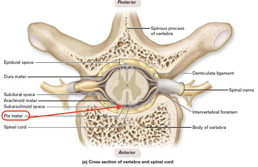
innermost layer, inseparable from spinal cord/brain
Pia mater
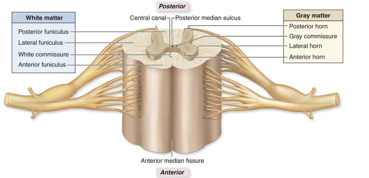
centrally located, and its shape resembles a letter H or a butterfly
gray matter
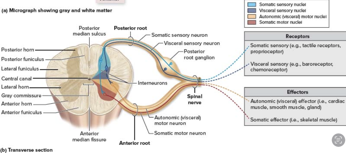
contains axons of sensory neurons
cell bodies (nuclei) of interneuron
gray matter - posterior horns

posterior horns - Aferent or Eferent?
aferent (sensory)
posterior horns - Structural classification of neuron
somatic sensory
pain & pressure
visceral sensory
smooth muscle & ligands
posterior root ganglion
collection of cell bodies of sensory neurons
posterior horns inntervates
pain
pressure
other sensory inputs

gray matter - anterior horns
somatic motor neurons that innervate skeletal muscles
Eferent
Anterior Horn - Aferent or Eferent?
Motor
Anterior Horn - Sensory or Motor?
Anterior Horn - Contains cell bodies, axons, both or neither?
cell bodies of somatic motor neurons -
innervate skeletal muscle
Somatic motor neurons
Anterior Horn - Structural classifcaton of neuron
innervates skeletal muscle
Anterior Horn - Innervates
contains cell bodies of autonomic (visceral) motor neurons
innervate smooth muscle, cardiac muscle, glands (unaware)
gray matter - lateral horns
Eferent
lateral horns - Aferent or Eferent?
Motor
lateral horns - Sensory or Motor?
lateral horns - Contains cell bodies, axons, both or neither?
cell bodies of autonomic (visceral) motor neurons
smooth muscle
cardiac muscle
glands
lateral horns - Innervates
autonomic visceral motor neurons
lateral horn - Structural classifcaton of neuron
collection of cell bodies of sensory neurons
Posterior Horn –Posterior root ganglion

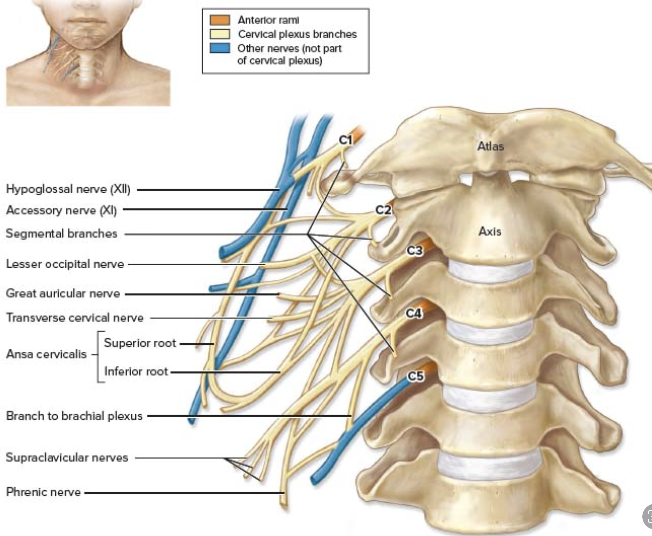
Innervate the muscles of the neck - C1–C4t
Phrenic nerve: extends down, diaphragm
Cervical Plexus
Supply upper limbs
Brachial Plexus
1. axillary nerve
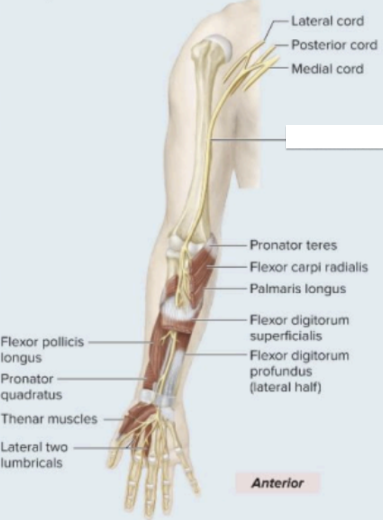
2. median nerve
3. musculocutaneous nerve
4. radial nerve
5. ulnar nerve
Brachial Plexus - fivevmajor terminal branches
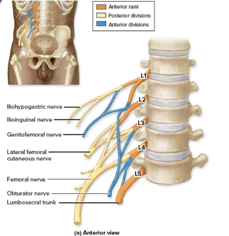
Innervate the abdomen, some external genitalia, anterior & medial thigh
Lumbar Plexus
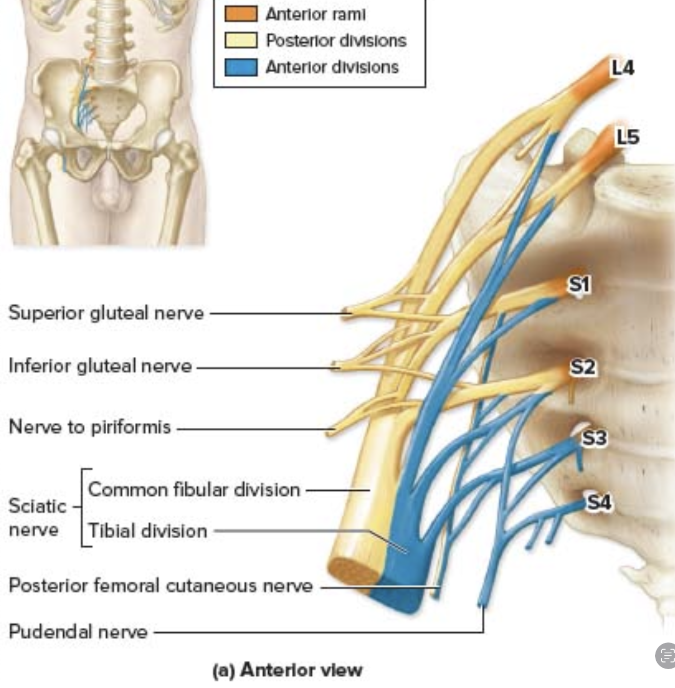
Sacral Plexus
Innervate
Pelvis
Posterior thigh
Leg
Foot
main nerve: sciatic nerve
largest & longest nerve in the body
No thoracic pletus
Intercostal nerve
Reflexes - Characteristcs:
stimulus
Rapid
automatic
involuntary
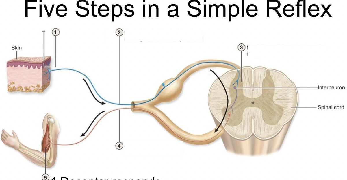
Receptor responds
Sensory neuron to spinal cord Integration –sometimes by
integration - Interneuron (monosynaptic & polysynaptic)
Motor neuron to effector
Effector responds
Five Steps in a Simple Reflex
receptor and effector on same side
Ipsilateral–
receptor & efector on opposite sides
Contralateral
does not involve interneuron
Ex. Knee-jerk (stretch reflex)
monosynaptic
involve interneurons Ex. With drawl reflex
polysynaptic –
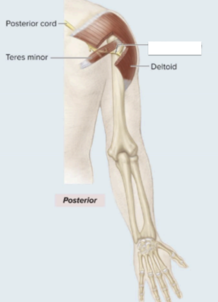
Motor innervation: Deltoid & teres minor
Action: Arm Abduction
sensory innervation: superolateral arm (shoulder)
Axillary Nerve

Motor innervation: anterior forearm muscle, thumb, lumbricals
Action: wrist, digit flexors
sensory innervation: palmer aspect, lateral 3 ½ digits (thumb, index, middle, & ½ ring finger)
median nerve
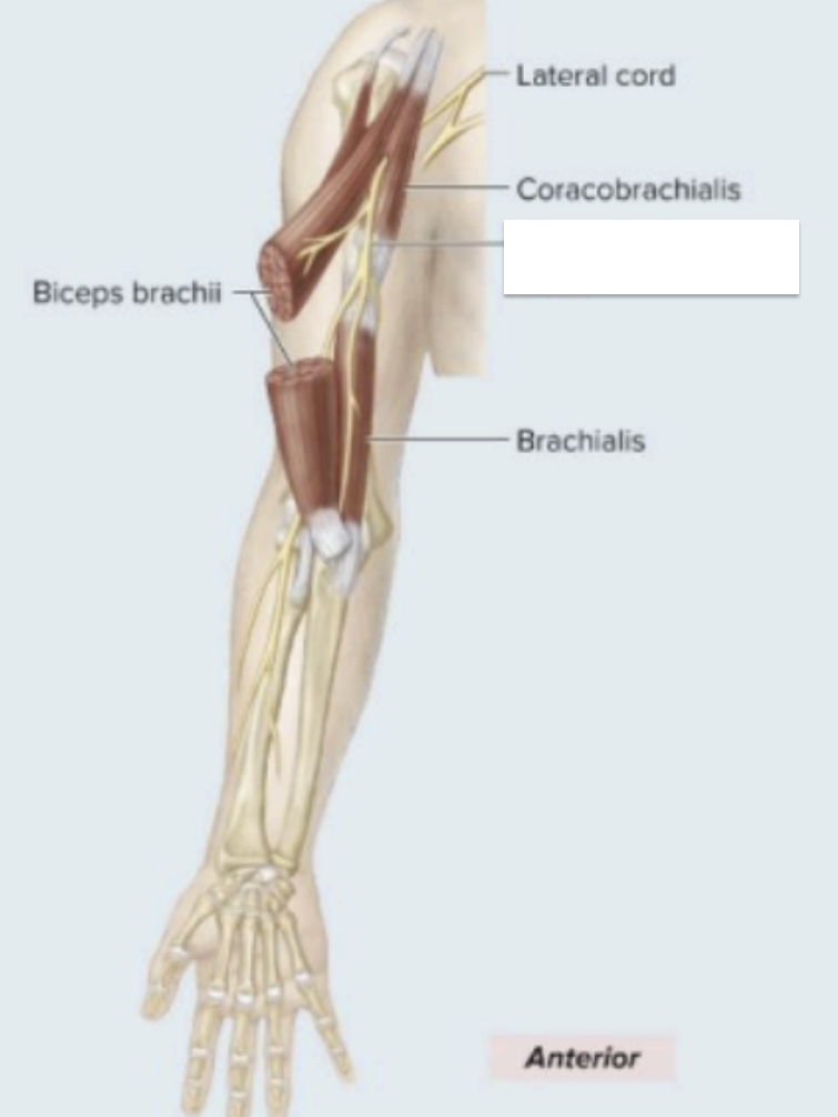
Motor innervation: anterior arm muscles - Iflex humerus, flex elbow joint, supinate forearm)
coracobrachialis
bicep brachii
brachialis
Action: forearm flexors
sensory innervation: later region of forearm
Musculocutaneous Nerve
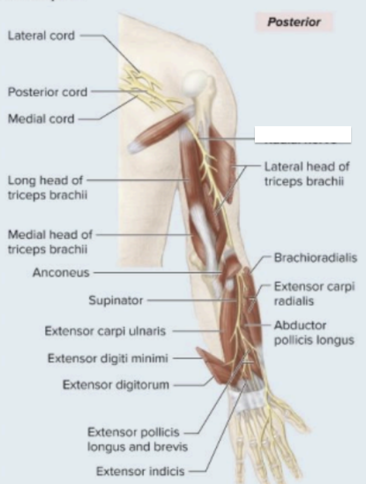
Motor innervation:
posterior arm muscle
posterior forearm muscle
brachioradialis
Action: forearm, wrist, digit, extensor
sensory innervation: arm, forearm, dorsal aspect of lateral 3 digits (except distal tips)
Radial Nerve
Motor innervation:
anterior forarm muscle
intrinsic hand muscle
Action: wrist, digit flexors
sensory innervation: dorsal and palmar aspects of medial 1 ½ digits (little finger, medial aspect of ring finger)
ulnar nerve