Pharynx & Pharyngeal Muscles - Module 7
1/24
There's no tags or description
Looks like no tags are added yet.
Name | Mastery | Learn | Test | Matching | Spaced |
|---|
No study sessions yet.
25 Terms
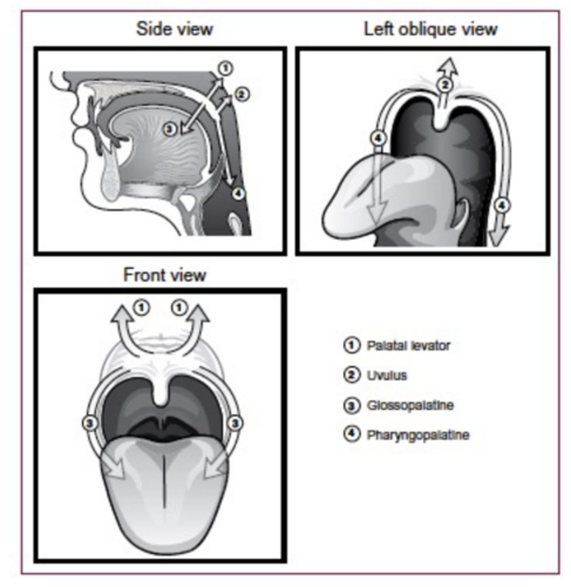
Velum Muscles
Velum Muscle Actions
Pharynx
An oval tube, larger side to side then front to back
Connective tissue
predominates at the top;
muscle predominates at
the bottom
Continuous with
esophagus at lower end

Pharynx
Three cavities
Lower boundaries are level of hard palate
(nasopharynx), hyoid bone
(oropharynx), and base of
the cricoid cartilage
(laryngopharynx)
Nasopharynx contains the
auditory tubes and
nasopharyngeal tonsils
(adenoids)
Pharynx
Opening of oropharynx is
through the faucial isthmus
(bounded by anterior
faucial pillars)
Oropharynx contains the
palatine tonsils and lingual
tonsil

Pharynx
Pharyngeal tube has 3 layers
- Fibrous layer (aponeurosis)—predominately in upper part of pharynx
- Muscular layer
- Mucous layer
Aponeurosis attached to base of skull anterior to foramen magnum
- Pharyngeal muscles attached to this aponeurosis
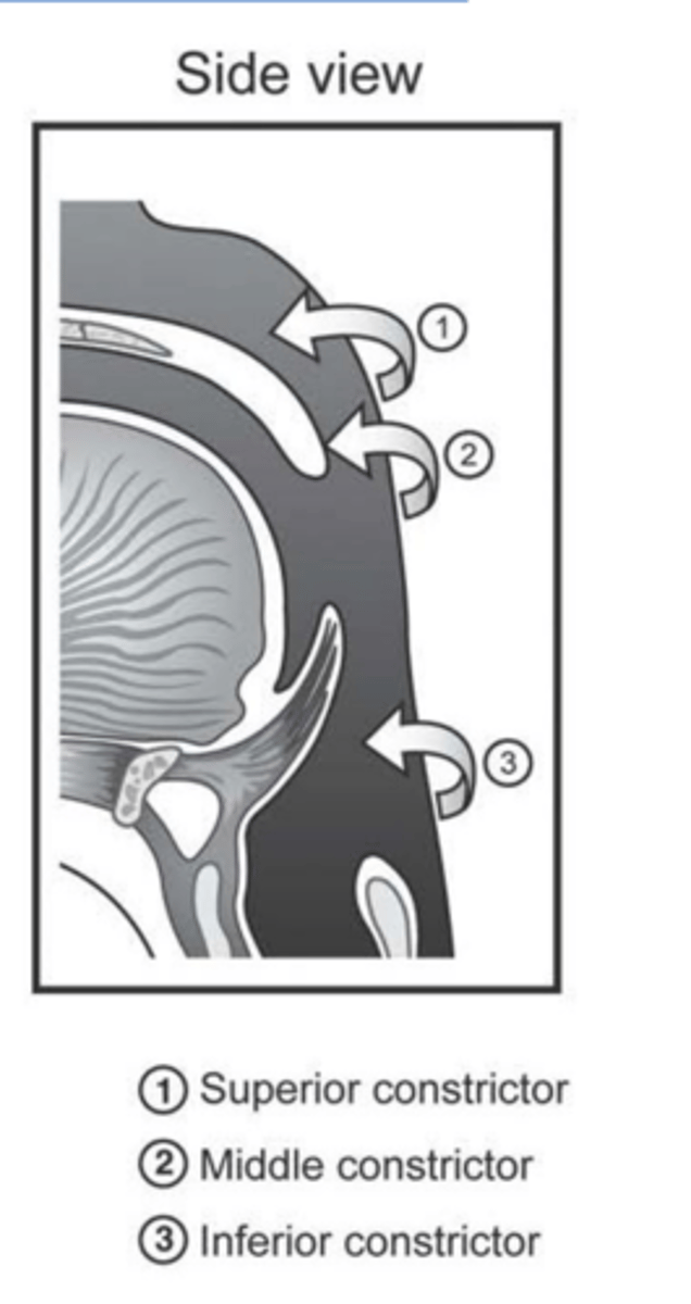
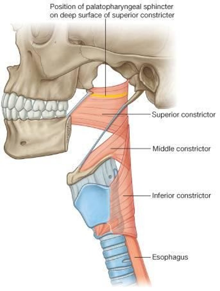
3 pairs of muscles, referred to as constrictors
Action of each is to reduce the diameter of a portion of the pharyngeal cavity

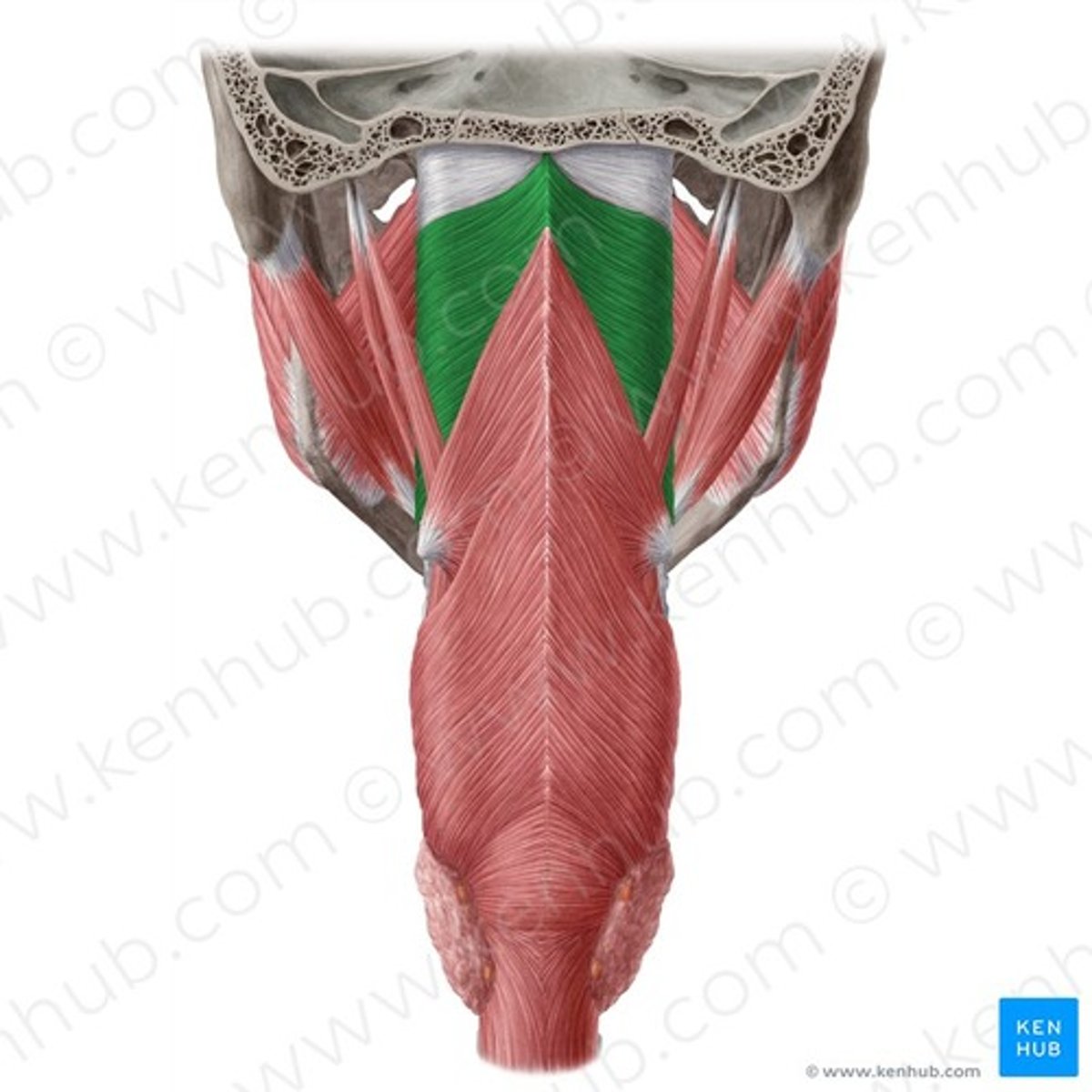
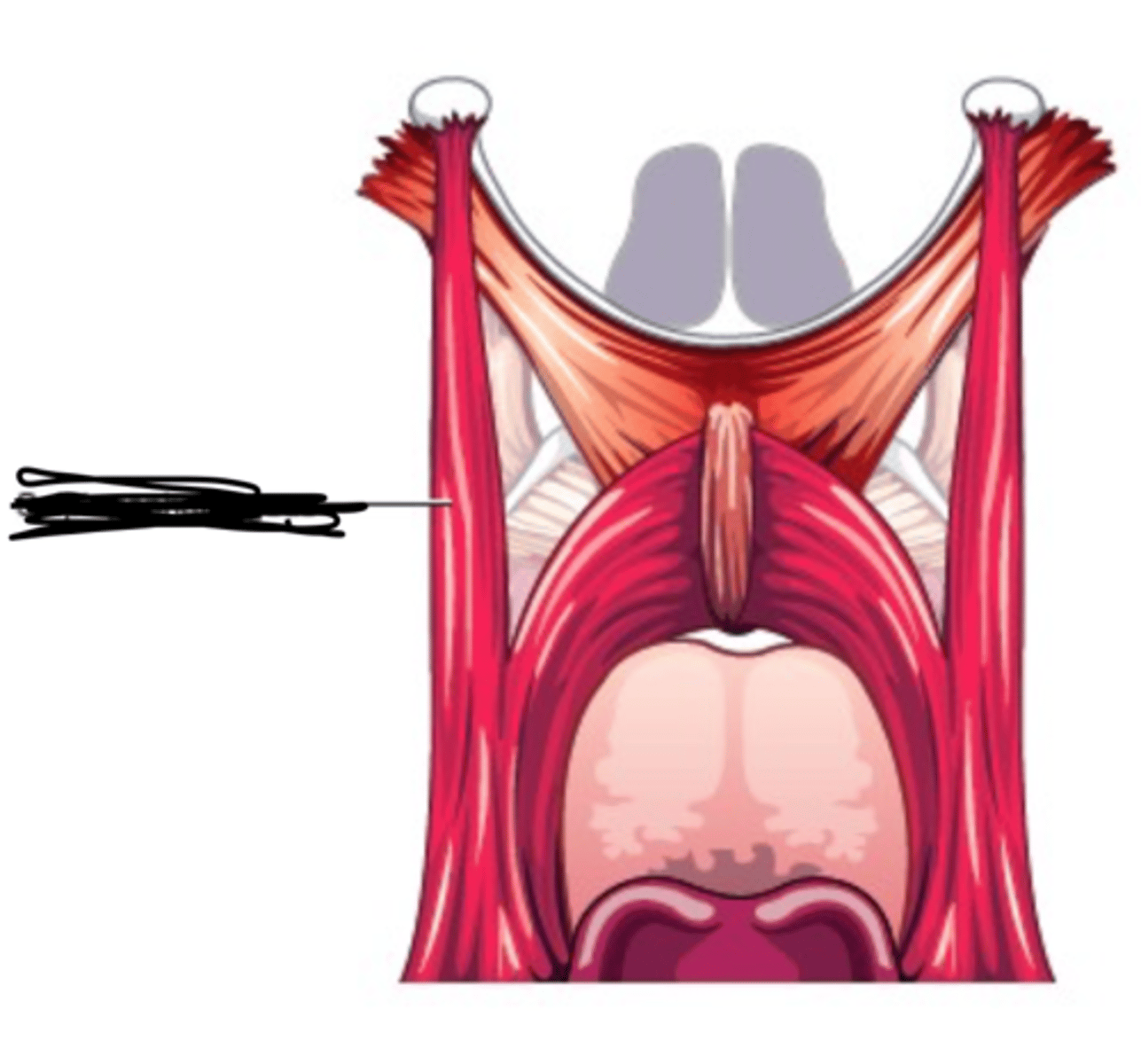
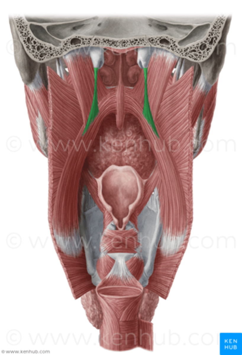
Superior Constrictor Description
Weakest, but most complex of the three
Forms nasopharyngeal and upper
oropharyngeal walls
Origin - sphenoid, mandible, pterygomandibular ligament
Course - posterior, then medial
Insertion - midline raphe
Action - may contribute to velopharyngeal closure by moving posterior wall of pharynx
anteriorly
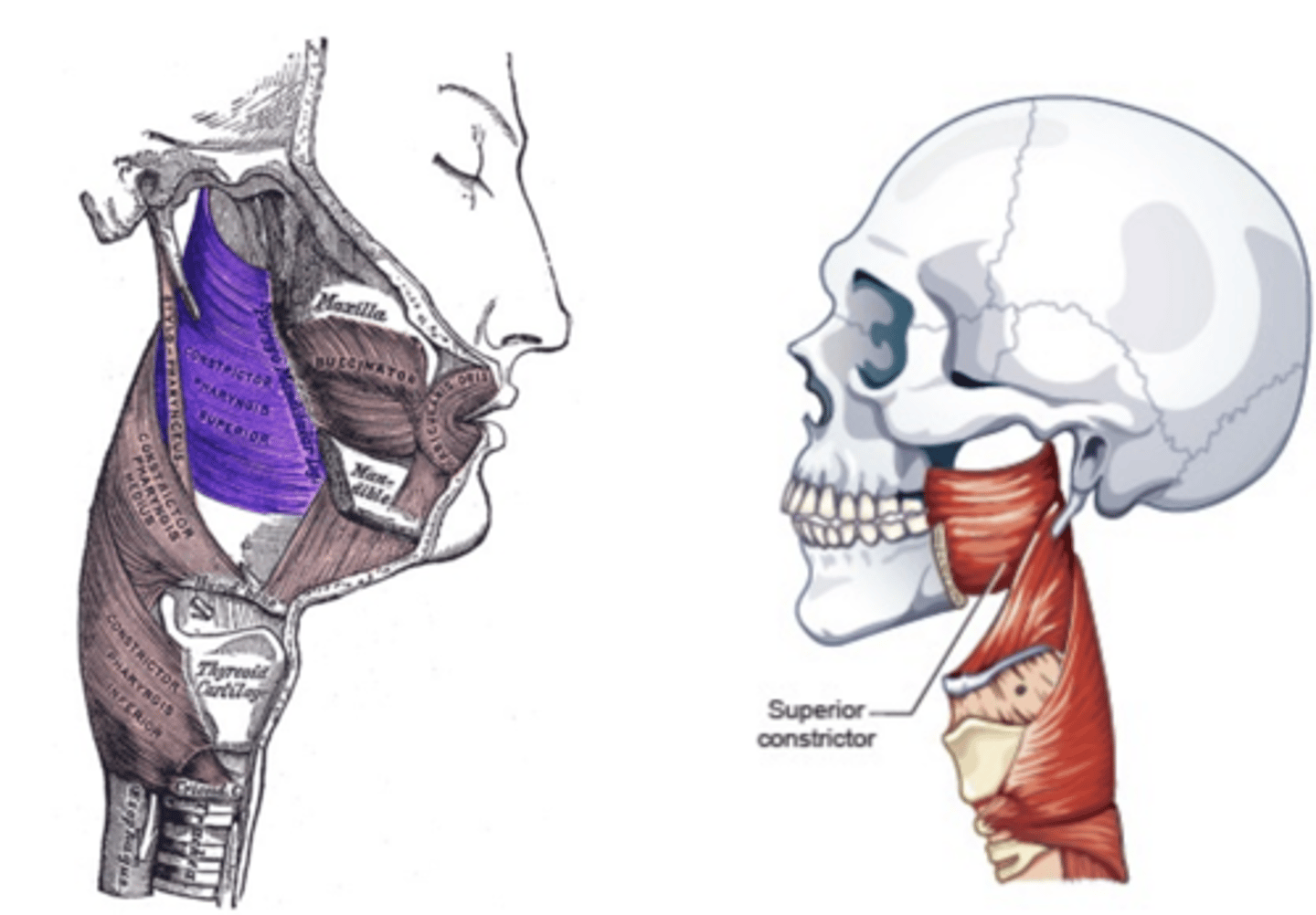
Superior Constrictor Figure
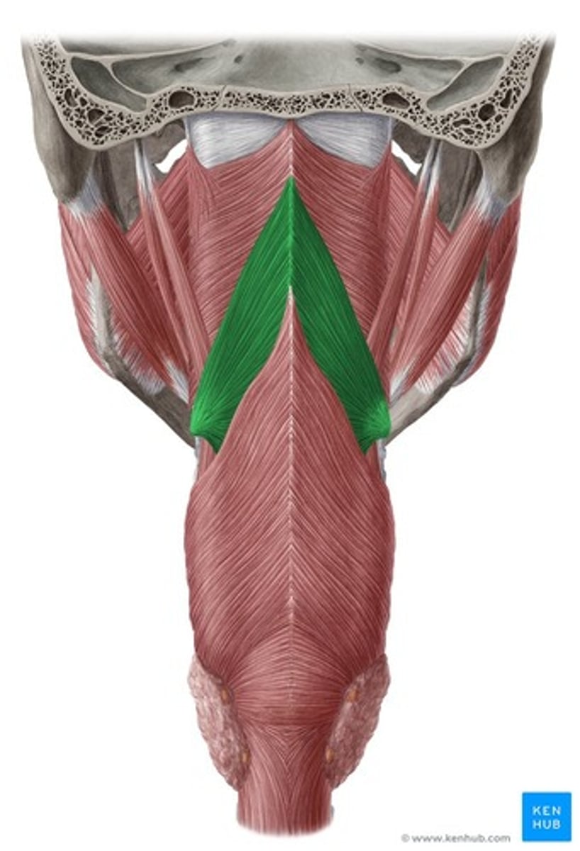
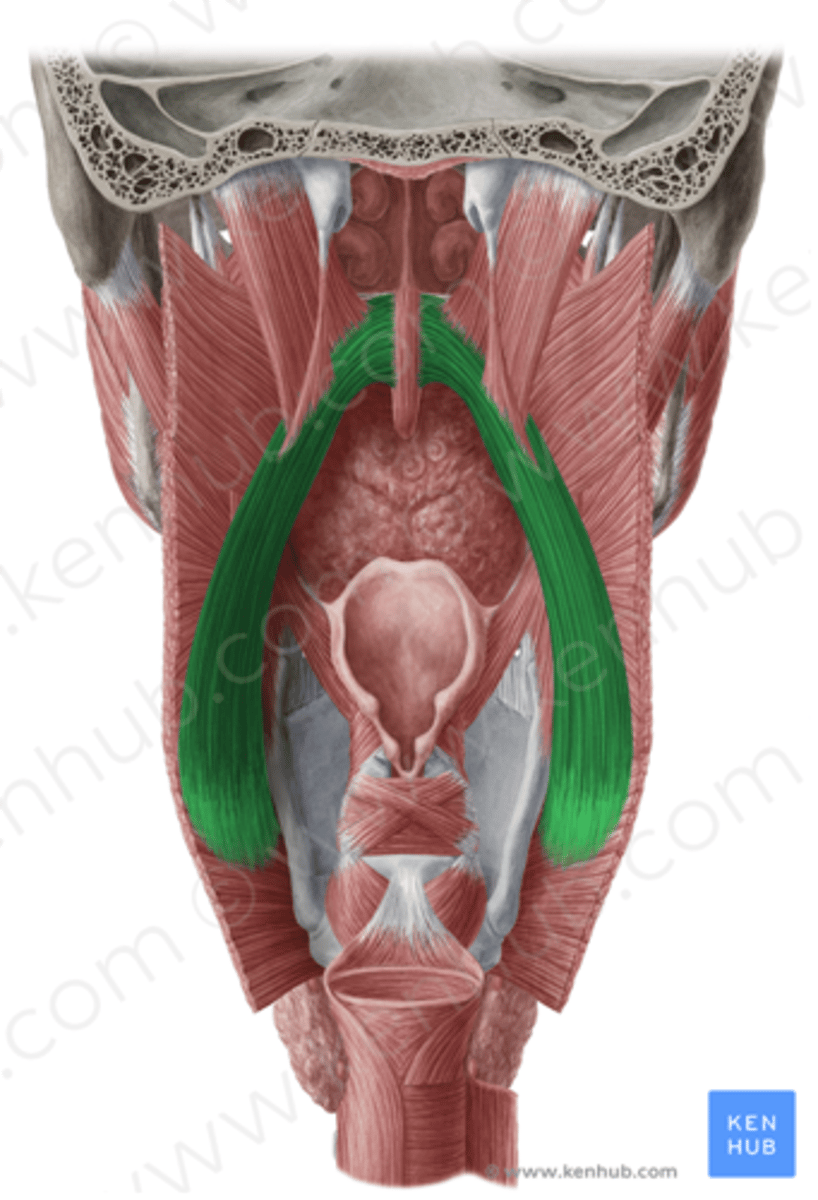
Middle Constrictor Description
Somewhat fan shaped
Origin - hyoid bone
Course - fan out posteriorly and medially
Insertion - midline raphe
Action - reduce diameter of pharynx
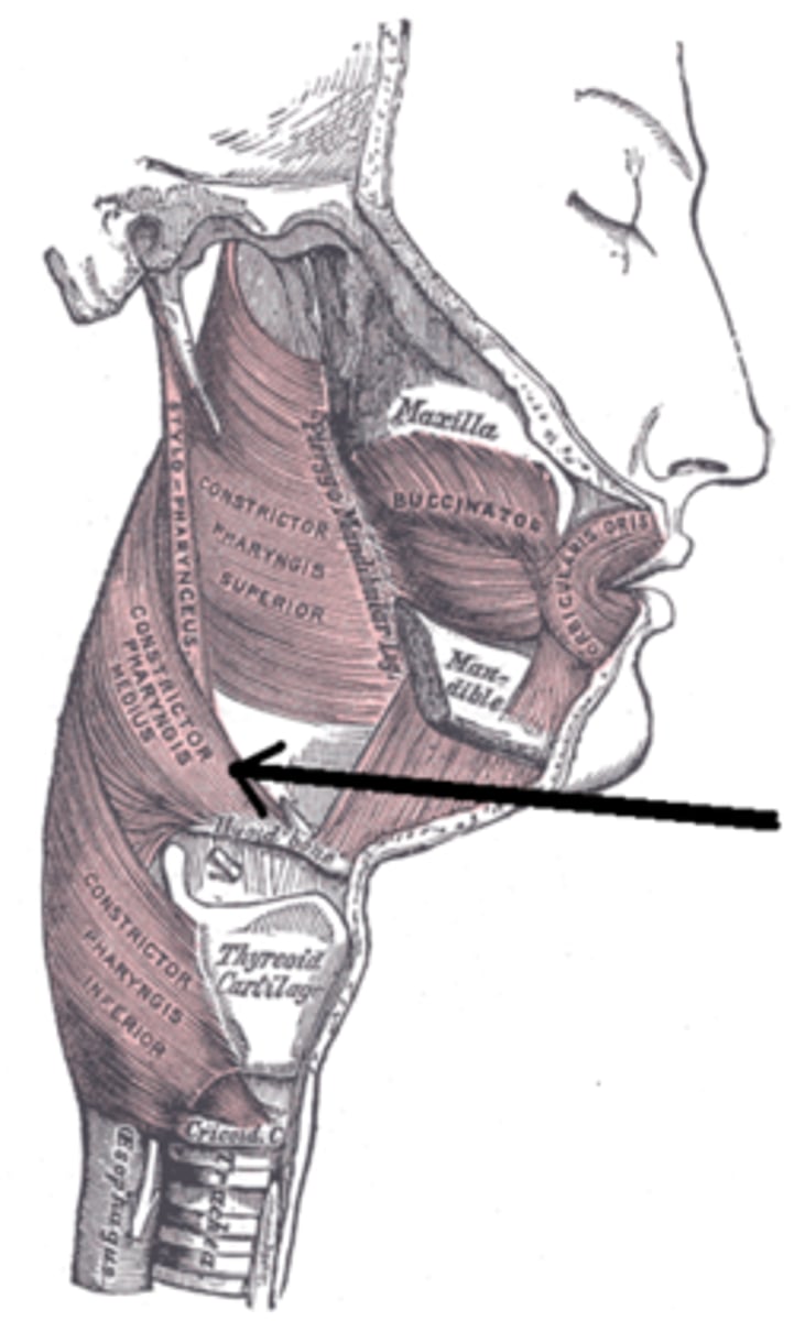
Middle Constrictor Figure
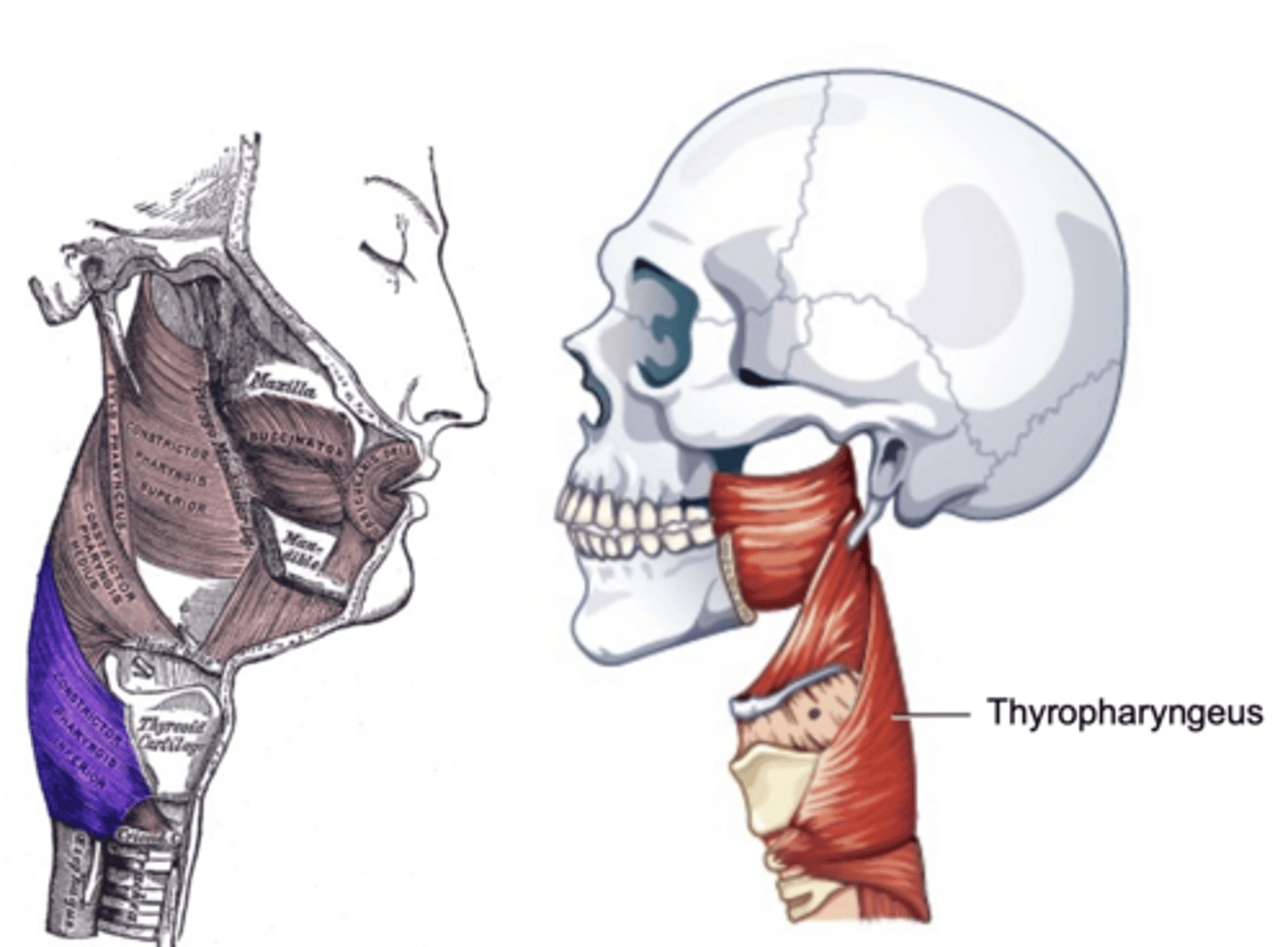
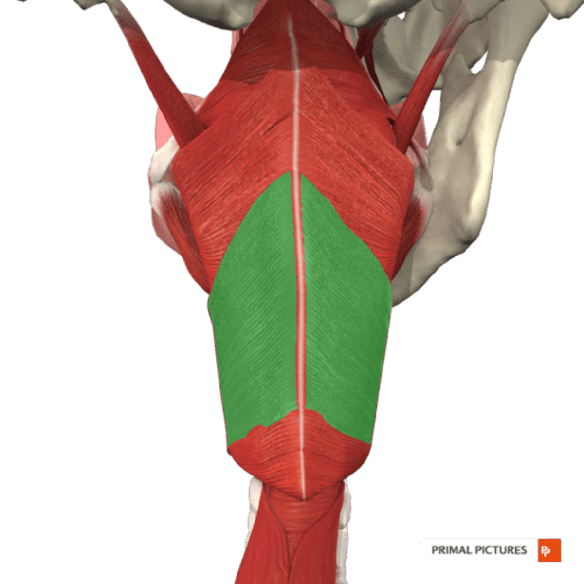
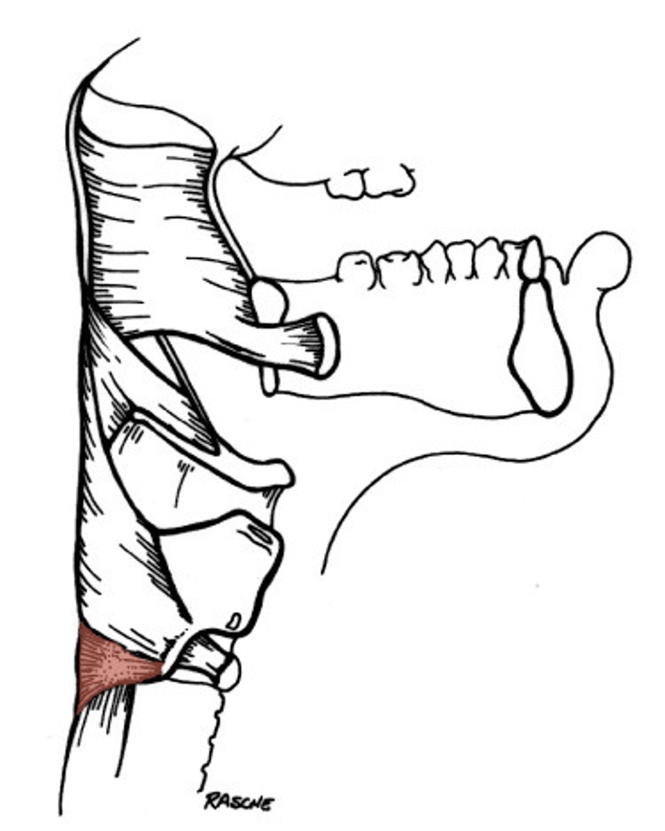
Inferior Constrictor -
Thyropharyngeus Description
Inferior Constrictor consists of the
Thyropharyngeus and the Cricopharyngeus
Inferior Constrictor is thickest and strongest of constrictor muscles
Thyropharyngeus is the majority of the Inferior Constrictor
Origin -thyroid cartilage
Course - fans out posteriorly and medially
Insertion - midline raphe
Action - reduce diameter pharynx
Inferior Constrictor -
Thyropharyngeus Figure
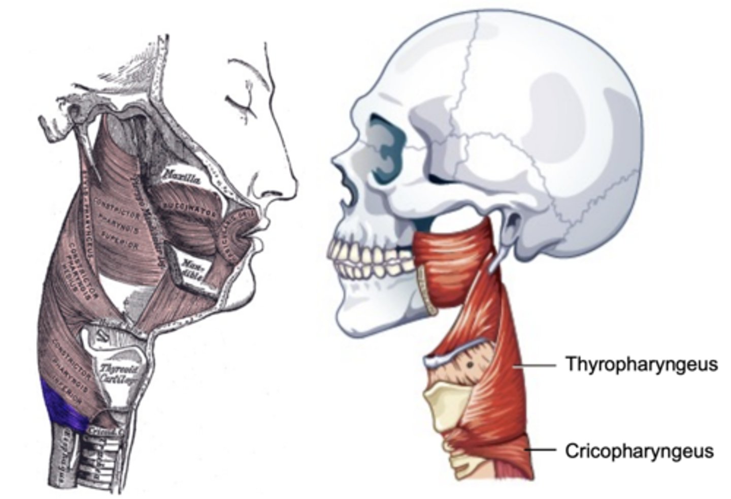
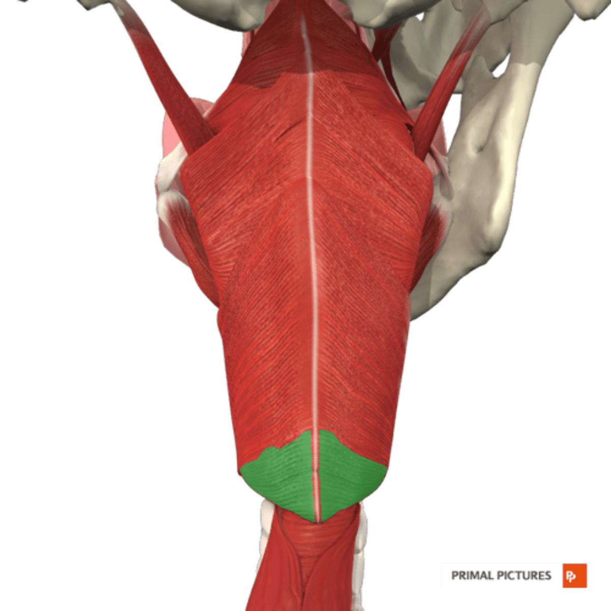
Inferior Constrictor -
Cricopharyngeus Description
Part of Inferior constrictor
Muscular component of upper esophageal
sphincter (UES)
Origin - cricoid cartilage
Course - fans out posteriorly and medially
Insertion - midline raphe
Action - open and close upper esophageal
sphincter
Inferior Constrictor -
Cricopharyngeus Figure
Constrictor Muscle Actions
Constrictor muscles all pull pharyngeal
walls inward and forward to constrict
the pharyngeal tube
Cricopharyngeus also assists in closing
the upper esophageal sphincter
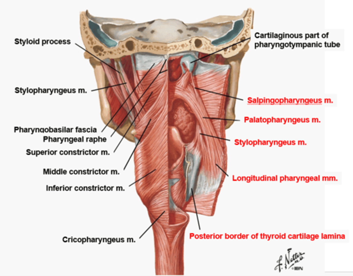
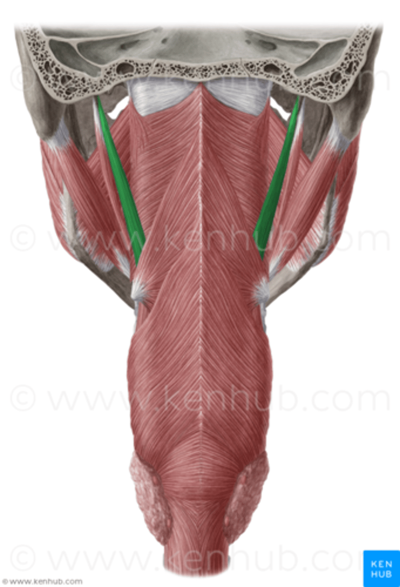
Stylopharyngeus Description
Long thin muscle
Origin - styloid process of temporal bone
Course - inferior, entering between
superior and middle constrictor
Insertion - blend with constrictors, with
some fibers inserting on thyroid cartilage
Action - elevate and dilate pharynx
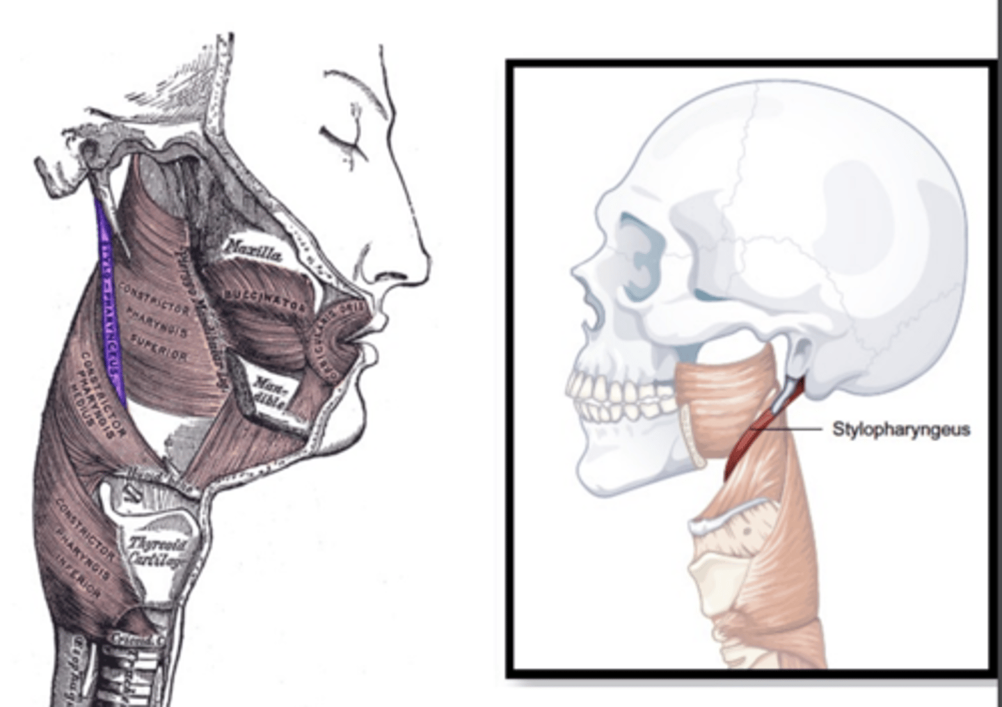
Stylopharyngeus Figure
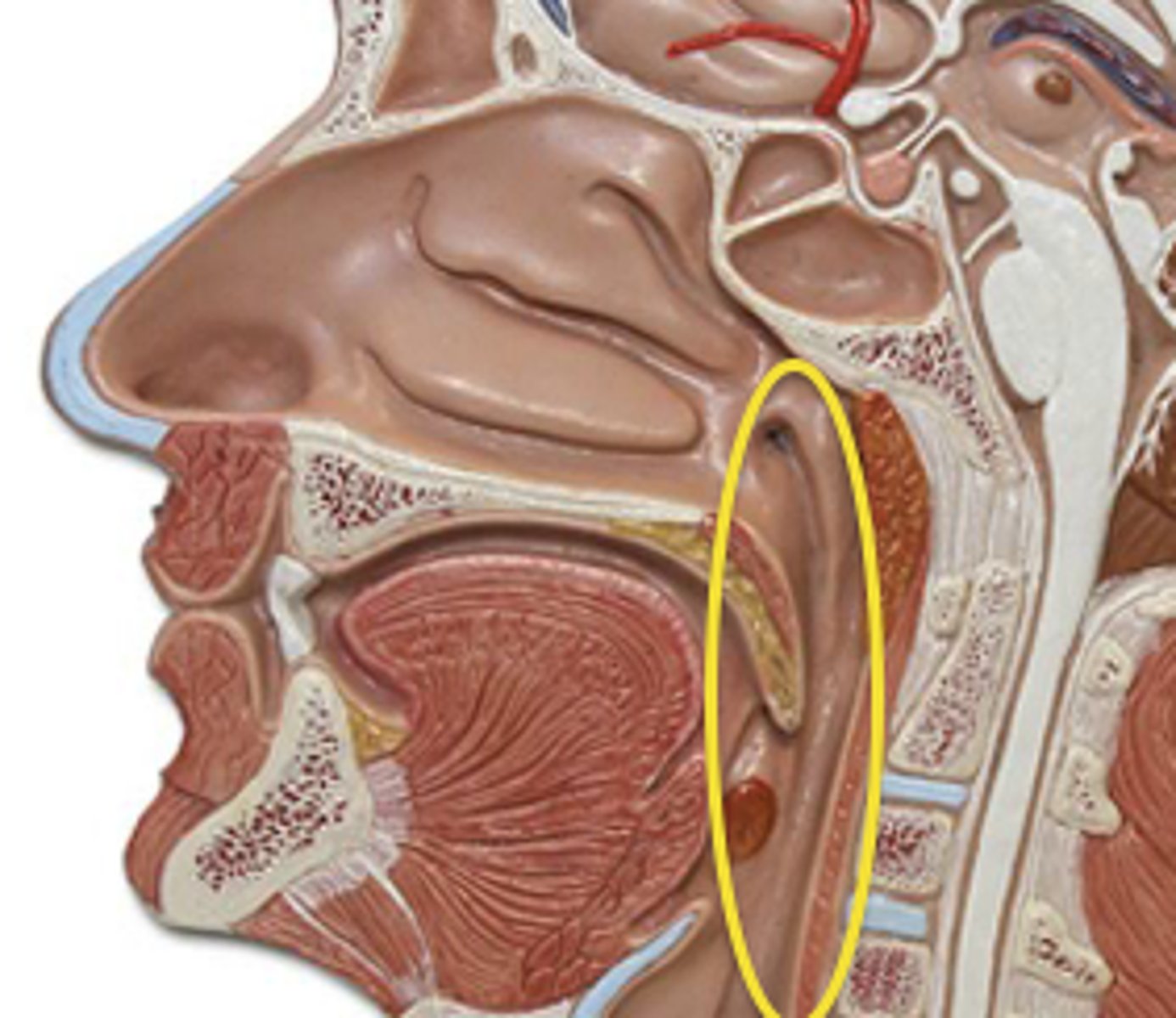
Salpingopharyngeus Description
Short thin muscle
Origin - lower border of the pharyngeal
orifice of the Eustachian tube
Course - inferior, deep to superior
constrictor
Insertion - blends with fibers of palatopharyngeus muscle
Action - elevate and dilate pharynx
Salpingopharyngeus Figure
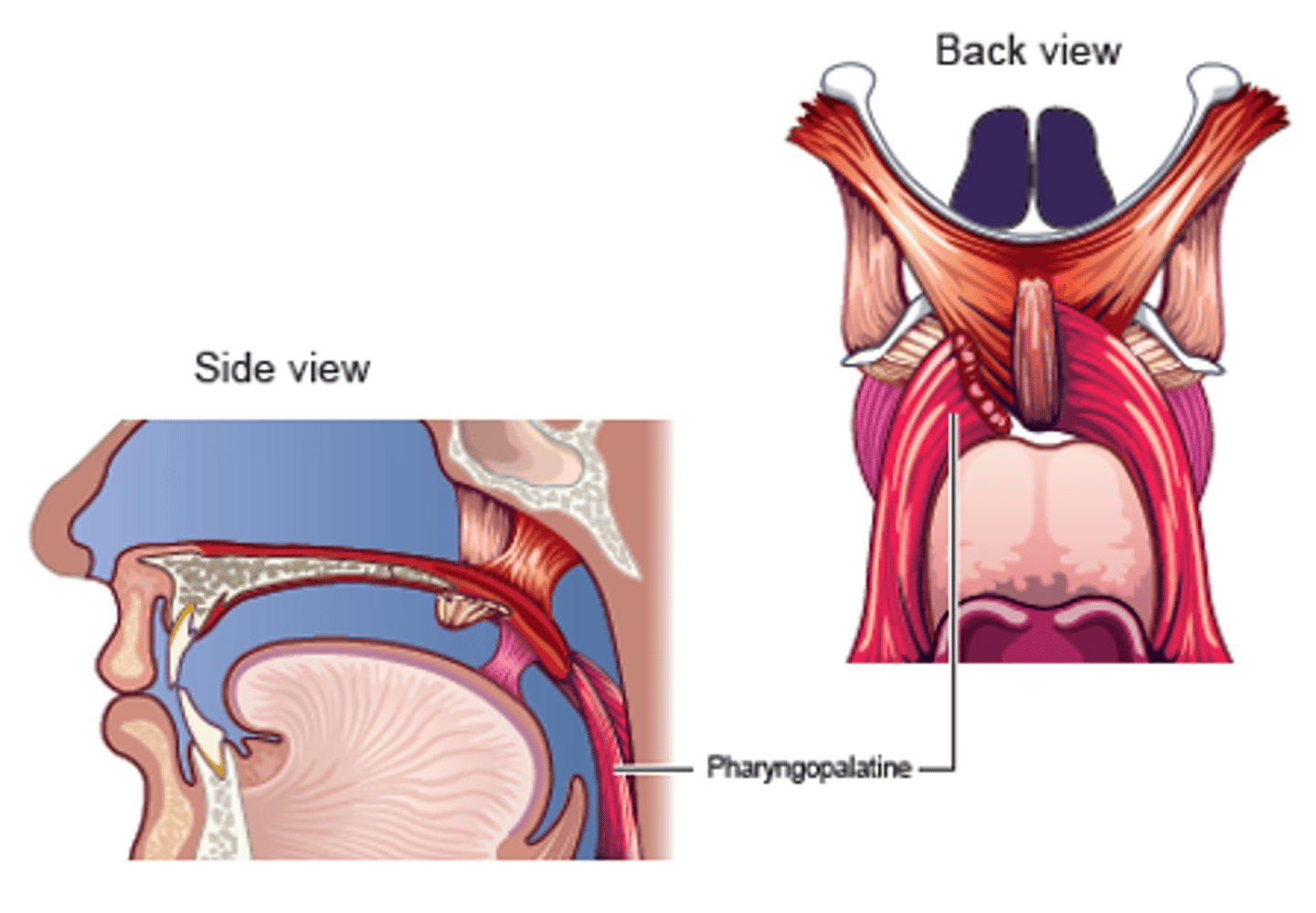
Palatopharyngeus Description
Also known as Pharyngopalatine
Posterior faucial pillar
- Longer muscle
Origin
- Soft palate
Course
- Superior through posterior faucial pillar
Insertion
- Lateral walls of pharynx, thyroid cartilage
Action
- Principally to guide material through pharynx
- May contribute to palatal lowering
- May contribute to VP seal

Palatopharyngeus Figure
What is the purpose of the pharyngeal muscles?
Constrict (reduce the diameter) of the pharynx

What are the three major muscles?
Superior
Middle
Inferior - divided into Thyropharyngeus and Cricopharyngeus
What is the purpose of the Cricopharyngeus?
Helps to close the Upper Esophageal Sphincter
What is the purpose of the Stylopharyngeus and Salpingopharyngeus?
Elevate and dilate (increase the diameter) of the pharynx