pathology quiz NMU
1/92
There's no tags or description
Looks like no tags are added yet.
Name | Mastery | Learn | Test | Matching | Spaced | Call with Kai | Chat |
|---|
No analytics yet
Send a link to your students to track their progress
93 Terms
compare between risk factor and exciting factor
risk factor make an individual more susceptible to a disease than others (فرصه ان يجيله مرض اعلي ) like obesity, poor ventilation, etc.
while exciting factor is directly responsible for a disease (bacterium, virus, trauma, etc)
what are the 3 types of a biopsy just the name
1-excision biopsy
2-incision biopsy
3-true cut biopsy
what is an excision biopsy
Therapeutic surgical resection of the entire lesion.
what is an incision biopsy
Surgical resection of part of the lesion for diagnosis.
what is a true cut biopsy
Core of tissue obtained by large bore needles, sometimes radiologically guided
what is the widely used fixative for specimen
Immediate specimen fixation is mandatory.
The widely used fixative is 10%
formaldehyde (Formalin) buffered to a neutral pH.
why is immediate specimen fixation important 4 points ?
• Fixation will preserve the morphology.
• Prevent decomposition and autolysis.
• Minimize microbial/fungal growth.
• Minimize the loss of molecular components.
fresh specimen without fixation is mandatory for ? 4
▪Frozen section
▪Immunofluorescence
▪Electron microscopy
▪Chromosome studies
what are the 7 causes for cell injury ?
just the name
هقول انا مثال علي الحاجات التافهه ولو قولته انت هيبقي احسن
1-hypoxia
2-infectious agents (virus, bacteria, fungi)
3-immunologic reaction (autoimmune diseases)
4-phyical agents (trauma, heat, cold)
5-chemical agents (conc. acids, alkalies, poisons)
6-genetic and chromosomal defects
7-nutritional disturbances
what are the 3 causes of hypoxia ?
a) Ischemia : arterial occlusion
b) Inadequate oxygenation: heart or lung disease
c) Decreased oxygen carrying capacity: anemia
the effects of cell injury could be in the form of ? 4
1- adaptation
2- reversible cell injury
3- irreversible cell injury
4- Intracellular and extracellular deposits.
adaptation is ?
irreversible ?
too much stress leads to ?
adaptation def: modification of cell structure and function in response to excess physiologic or pathologic stress to achieve a new steady state that preserve the vitality of cells
.
its reversible
.
too much stress exceeds the cell's adaptive capacity leads to injury
enumerate the 4 types of cell adaptation
1-atrophy
2-hypertrophy
3-hyperplasia
4-metaplasia
atrophy means ?
why it happens (mechanism يعني )
decrease in size of mature organ due to decrease in cell size&/ or number
pathogenesis:
cells become smaller DUE TO
1-decrease protein synthesis
2-increase its degradation
mention the 3 physiological atrophy examples and the 3 pathological atrophy examples
physiological:
1-aging
2-uterus atrophy: after labor
3-thymus atrophy: after puberty
pathological:
1-disuse atrophy (after bone fracture)
2-neurogenic atrophy: (denervation)
3-thermal atrophy: undescended testis
hypertrophy is ?
what happens inside
اسمها هنا وفي اللي فاتت pathogenesis
القصد يعني من الجمله اللي بعدها اي اللي بيحصل
def: increase in size of mature organ due to increase in cell size
pathogenesis: increased RNA and structural proteins
mention 2 physiological examples and one pathological example of hypertrophy
physiological:
- Uterine hypertrophy
- Muscle hypertrophy in athletes.
pathological (compensatory)
- Left ventricular hypertrophy in systemic hypertension
hyperplasia means ?
pathogenesis ? (اي اللي بيحصل يعني)
def: increase in size of mature organs due to increase in cell number
pathogenesis :
cell proliferation with increased DNA synthesis
mention one physio. and one patho. example for hyperplasia
physio.
- Hyperplasia of female breast
in puberty, pregnancy and lactation
patho.
- Prostate and endometrium ( hormones)
metaplasia is ?
pathogenesis ?
example ?
def:change of one type of tissue to another type of
same category, epithelium to epithelium or C.T to C.T,
ALWAYS PATHOLOGICAL
pathogenesis: reprogramming of stem cells differentiate along a new pathway to tolerate stress
for an example squamous metaplasia of respiratory epithelium in smokers
enumerate the 2 types of reversible cell injury
what does it affect ?
1-cloudy swelling & hydropic degeneration هم اه دول اتنين بس بيتحطو سوا طول الوقت
2-fatty change
.
Affects cells with higher rate of
metabolism more than supporting stromal cells.
cloudy swelling & hydropic degeneration is ?
caused by ?
its a reversible cell injury characterized by
-mild (in cloudy) and لو زياده المياه بسيطه
-excess (in hydropic) لو زياده المياه كتير
intracellular water accumulation
caused by mild injury or injury of short duration
cloudy swelling & hydropic degeneration
pathogenesis ?
Mitochondrial oxidative phosphorylation is disrupted
first → Decreased ATP leading to;
1- Decreased Na/K pump → gain of intracellular Na.
2- Anaerobic respiration → Lactic acid accumulation.
3- Release of mitochondrial protein.
• The increase in the cytoplasmic osmotic pressure helps intra-cellular water accumulation → cell swelling
in cloudy swelling & hydropic degeneration
how does the affected organ appear (gross picture) ?
size ?
shape ?
surface ?
capsule ?
color ?
consistency ?
cut surface ?
دي النقط بتاعت السؤال
size: swollen
shape: perserved
surface: smooth & has rounded borders
capsule: tense stretched
color: pale due to compression of the capillaries by the swollen cells
consistency: soft
cut surface: appears cloudy (less glistening لمعان اقل يعني) & bulges outwards
in cloudy swelling & hydropic degeneration
how does the affected organ appear (under microscope) ?
cell size ?
cytoplasm ?
nucleus ?
فيه اختلاف بسيط بين الاتنين قوله
Cloudy swelling
• The cells are swollen with compressed capillaries in between.
• The cytoplasm is granular
• The nucleus is normal
Hydropic degeneration
• similar lo cloudy swelling but the cytoplasm shows multiple vacuoles
fatty change is ?
a type of reversible cell injury characterized by accummulation of neutral fat (triglycerides!!!!!!!!!!!!) in parenchymatous cells
fatty change
effect on cell or tissue morphology
size ?
shape ?
surface ?
capsule ?
color ?
consistency ?
cut surface ?
size: swollen
shape: perserved
surface: smooth & has rounded borders
capsule: tense stretched
color: pale yellow
consistency: soft
cut surface: appears yellow, greasy to touch & bulges outwards
fatty change
effect on cell or tissue morphology UNDER MICROSCOPE
• The cells appear swollen and compress the intercellular
capillaries.
• The cytoplasm show fat globules fuse together
forming a big globule that pushes and flattens the
nucleus against the cell membrane giving the cell a
signet ring appearance.
• Fat special stains done on frozen section
what are the 2 types of irreversible cell injury
واشرح كل واحد
• Necrosis: is death of group of cells within the living body
• Apoptosis: is programmed single cell death
necrosis cause ?
pathogenesis )3( (just the name) اسم الحاجه اللي بتحصل جوا عشان تسبب الموضوع دا
Severe injury or injury of long duration causing damage of the nucleus and cell death
.
1-mitochondria damage
2-increased calcium ions
3-increase in reactive oxygen metabolites
in necrosis mitochondria damage leads to ?
اي اللي بيحصل جوا بالتفصيل بقا
• Decrease ATP with decrease in energy dependent
functions:
• decrease protein synthesis
• decrease in Na pump lead to cell swelling
• Anaerobic glycolysis with decrease in PH
in necrosis increased calcium ions leads to
leading to activation of all enzymes; phospholipases, proteases, endonucleases, ATPases
Finally damage of protien, membranes & DNA occur
in necrosis increase in reactive oxygen metabolites leads to ?
Increase in reactive oxygen
metabolites
(ROS) that damage the membranes
and various cell components.
pathology of necrosis ? يعني هي بتعمل اي
•Necrotic cells liberate chemicals that irritate the adjacent living tissue leading to an inflammatory reaction.
post necrotic changes (under microscope)
cell membrane ?
cytoplasm ?
nucleus ? 3( just the name )
-cell membrane disappears
-cytoplasm: Swollen, coagulated (denaturation of protein), homogenous (due to loss of glycogen particles) and deeply eosinophilic (loss of normal basophilia of RNA in the cytoplasm)
nucleus
1-pyknosis
2-karyorrhexis
3-karyolysis
pyknosis of nucleus is ?
nucleus is Shrunken, dense, deeply basophilic.
karyorrhexis of nucleus is ?
fragmented nucleus
karyolysis of nucleus means ?
the nuclear fragments fades and disappears due to chromatin hydrolysis
enumerate the 5 types of necrosis
✓ Coagulative necrosis
✓ Liquefactive necrosis
✓ Caseation necrosis
✓ Fat necrosis
✓ Fibrinoid necrosis
necrosis pathology tissue changes are either (2)
-denaturation of proteins
-enzymatic digestion of the cell
necrosis pathology (denaturation of proteins) اشرحها
The cells retain their
outlines with loss of cellular details and the area is firm,
swollen, pale
(coagulative necrosis)
necrosis pathology (enzymatic digestion of the cell)
اشرحها
بتحصل في حالتين
1. (autolysis) by cell lysosomal enzymes. cell affects itself
2. (heterolysis) from nearby leucocytes
there is loss of architectural and structural details and
the area is soft and is filled with turbid fluid
(liquefactive necrosis).
what is the commonist type of necrosis ?
coagulative necrosis
coagulative necrosis causes ?
- Protein denaturation predominates.
- Cell’s outline is preserved but details are lost مش مقتنع ب تاني واحده قد كدا بس هي كدا في الباور
انه cause يعني
coagulative necrosis shape (morphology) with naked eye ?
The necrotic area is dry , firm , opaque , pale yellow
coagulative necrosis shape (morphology) with microscope
*general architecture is preserved.
*Dead cells retain their outline but without nuclear
or cytoplasmic details.
*Blood vessels and stroma persist longer
mention 3 examples of coagulative necrosis
Acute ischemia of
-heart
-kidney
-spleen
liquifactive necrosis cause ?
Enzymatic digestion predominate Necrotic tissue is liquefied by enzymes.
liquifactive necrosis shape (morphology) as seen with naked eye and microscope respectively
N/E: Soft & filled with turbid fluid
M/E: Complete loss of architectural & cellular details
mention 2 examples of liquifective necrosis
1- Pyogenic abscess: (proteolytic
enzymes from neutrophils (pus cells))
2- Brain infarction: (high lipid and large
fluid contents of nervous tissue.)
caseation necrosis shape (morphology) as seen with naked eye and microscope respectively
N/E: Necrosis appears friable , soft grayish yellow material like cheese
M/E: granuloma in the form of Homogenous granular eosinophilic material
mention the 3 examples of caseation necrosis
1-TB
(للمعرفه فقط TB is a bacterial disease of the lungs
tuberculosis)
2-syphilis
3-fungal infection (عامتا مش شرط عضو معين)
mention the 2 types of fat necrosis
1-traumatic fat necrosis
2-enzymatic fat necrosis
traumatic fat necrosis is ?
what happens (mechanism)
• Trauma to the adipose tissue of the breast and subcutaneous fat.
• The fat cells rupture and (autodigestion) takes place with release of fatty acids that combine with calcium.
enzymatic fat necrosis occurs when ?
what happens (mechanism)
• It occurs in acute pancreatitis.
• Lipase escapes from the ruptured pancreatic ducts and digests the surrounding fat
fibrinoid necrosis
is ?
happens in cases of ? 2
what is deposited ?
where is it deposited ?
• Histological changes of arteries in cases of vasculitis and hypertension.
• When Glassy, eosinophilic fibrin-like material is deposited within the damaged necrotic vessel wall.
what is the fate of necrosis
in small areas of necrosis and large areas of necrosis respectively
• Small area of necrosis:
Healing occurs by regeneration or by granulation tissue and fibrosis (Repair).
• Large area of necrosis:
Surrounded by fibrous capsule. Unabsorbed contents dry and may show dystrophic calcification.
what is apoptosis ?
programmed single cell death in which cells activate
enzymes that degrade the cells’ own nuclear DNA and
nuclear and cytoplasmic proteins.
what are the physiological causes of apoptosis ?
• Embryogenesis
• Hormone dependent as
Endometrial breakdown
during menstrual cycle
what are the pathologic causes of apoptosis
• DNA damage
• Pathologic atrophy
discuss the 4 steps of apoptosis pathogenesis
stimulation?
control by genes?
activation ?
دول تلميحات للخطوات
1. Stimulation of apoptotic process: physiological or pathologic
stimuli.
2. Control by apoptosis genes:
• Stimulation of proapoptotic genes that promote apoptosis
as PAX gene
• Inhibition of anti-apoptotic genes as bcl-2 gene
3. Then activation of proteases (caspase family ).
4. Morphological changes of apoptosis.
apoptosis as seen with EM
اربع نقط
•Cell shrinkage.
•Condensation and fragmentation of chromatin.
•Formation of cytoplasmic blebs and apoptotic bodies.
•Phagocytosis of apoptotic bodies by macrophages.
apoptosis as seen with LM
3 نقط
• Apoptosis involve single cell or small groups of cells.
• Apoptotic body appears rounded or oval with dense
eosinophilic cytoplasm and nuclear fragment.
• Lack of inflammation in surrounding tissue as apoptotic
bodies are rapidly phagocytosed.
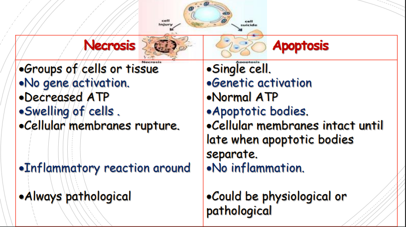
compare between necrosis and apoptosis
number ?
gene activation ?
atp ?
cellular membrane ?
inflammation ?
pathological or physiological ?
في نقطه مذكرتهاش في السؤال دور عليها :)
autophagy is ?
“self-eating” refers to lysosomal digestion of the cell’s own components.
autophagy causes (اشرح اللي بيحصل)
Nutrient deprivation, so that the starved cell can live by
eating its own contents and recycling these contents
to provide nutrients and energy.
enumerate the 6 types of tissue accumulation (deposits)
1-carbohydrates&glycoproteins
2-hyalinosis
3-amyloidosis
4-pathological calcification
5-pathological pigmentation
6-gout
glycogen tissue accumulation
etiology ?
shape under microscope ?
Etiology: due to abnormal glycogen metabolism
Microscopically: the cells are swollen with clear
cytoplasmic vacuole
mucin (mucoid or mucinous) is ?
shape under microscope ?
Glycoprotein Catarrhal inflammation, Mucoid carcinoma
Microscopically: cells distended with mucin (signet-ring cell). The cells may rupture with release of mucin (mucin lacks)
hyalinosis (hyaline deposits) what is present in the tissue ? (ايه اللي موجود يعني)
stain ?
Presence of:Glassy Refractile Homogenous Structureless Transparent material
Stains red with eosin
what are the 2 examples of hyalinosis ?
1-russell's bodies (in a disease named rhinoscleroma)
2-corpora amylacia (in benign prostatic hyperplasia)
amyloidosis is ?
Extracellular deposition ofabnormal fibrillar protein
(homogenous hyaline eosinophilic amyloid material).
amyloidosis are classified into two categories
localized and systemic
types of localized amyloidosis (3)
1-Nodular deposits: affect tongue, larynx, lung
2-Senile: cardiac or cerebral (Alzheimer dis.)
3-Endocrine amyloidosis in endocrine tumor
types of systemic (generalized) amyloidosis (3)
-1ry amyloidosis… plasma cell tumor called multiple myeloma
-2ry amyloidosis… reactive amyloid in conditions as in tuberculosis (TB)
-Heredofamilial amyloidosis
in amyloidosis diagnosis the tissue biopsy colour with each stain will be ? (3)
فيه 3 stains
هتقولهم وهتقول لون العينه في كل واحده منهم
Hx & E stain….esinophilic
• Congo red ……..Salmon pink
• By polarized light ……apple green
gout is ?
normal % of it is ?
what happens if it increases ?
Disturbance in purine metabolism with deposition of sodium urate in the tissue.
Normally 3 – 7 mg % in ♂. 2 – 6 mg% in ♀
when it increases in blood,urine & deposited in tissue as Na urates
what is the primary and secondary cause for gout ?
a) Primary:
familial, ♂ > ♀ at 40 years due to increased purine breakdown or decreased clearance .
b) Secondary:
increased cellular destruction as in Polycythaemia rubra ver
gout deposition site ?
NA urate deposited in: skin,kidney and joints affecting metatarsophalyngeal joint of big toe
pathological calcification is ??
structure as seen with naked eye ?
structure as seen under microscope ?
def. Deposition of Ca salts in tissue other than bone & teeth.
naked eye: as chalk, dull opaque white with finely granular surface & hard in consistency.
microscope: dark blue with Hx
what are the 3 types of pathological calcification
1-dystrophic calcification
( occurs in non viable tissue نسيج ميت ونسبه الكالسيوم في الدم طبيعيه)
2-metastatic calcification
(نسيج حي وواصله دم عادي بس نسبه الكالسيوم في النسيج عاليه
occurs in viable tissue with hypercalcemia)
3-stone formation
enumerate the 7 examples of dystrophic calcification
1- In fat necrosis
2= Following hyaline change
3- Wall of chronic abscess
4- Old scar
5- Dead bilharzial ova
6- fibrosed valve
7- Atheroma of large vessel
what are the 2 causes of metastatic calcification
الاسباب ليها ميكانيزم هو اسهل من اني اعمله كروت منفصله
a) Excess absorption of calcium from the intestine as in;
• Hypervitaminosis D (vitamin D intoxication)
• Milk-alkali syndrome: Excess milk and calcium carbonate
intake in patients treated for peptic ulcer.
b) Excess mobilization of calcium from bone as:
• Hyperparathyroidism, thyrotoxicosis, and Cushing syndrome
• Prolonged immobilization in bed
• Bone destruction by malignant tumors
enumerate the 4 sites of metastatic calcification
1. Renal tubular epithelium (Nephrocalinosis)
2. Mucosa of the stomach
3. Arteries
4. Lung alveoli
stone formation
precipitation of Ca salts in organic material in ducts of ? (3)
1- biliary tract
2- urinary tract
3- salivary gland
pathologic pigmentation means ?
types ? (2)
Pigments are colored substances that stain the
tissue. These could accumulate intracellularly.
Types: may be exogenous or endogenous.
exogenous routes of entry (pathologic pigments) are ? (3)
-Inhalation as anthracosis (carbon particles).
-Ingestion as in chronic lead poisoning (plumbism).
-Inoculation as in tattooing
enumerate the 3 types (الفئات يعني) of endogenous pigments (pathologic pigmentation)
1-melanin
2-lipochrome (lipofuscin)
3-hemosiderosis
melanin hyper and hypo pigmentation
localized and generalized for each
(مثال علي كل واحده كدا انا عايز اربع امثله)
melanin hyperpigmentation
localized: nevus, melanoma
generalized: prolonged exposure to sunlight
.
melanin hypopigmentation
localized: vetiligo
generalized: albinism
lipochrome (lipofuscin) def. ?
location ?
occurs due to ? (mechanism)
It is yellowish brown fat soluble pigment. normally in the heart,testis, seminal vesicles, corpus luteum and adrenal cortex.
Due to tear and wear →tissue breakdown →release of
phospholipids →phagocytosed by the healthy neighboring cells → intracellular accumulation
lipochrome (lipofuscin) causes ? (3)
1. Old age. (brown atrophy of heart)
2. Wasting diseases.
3. Cancer cachexia
endogenous hemosiderosis types ?
-localized
-general 1ry or 2ry
endogenous hemosiderosis
-localized due to ?
-general 1ry or 2ry due to ?
1-Localized hemosiderosis.
cause: due to localized haemorrhage
2-1ry hemosiderosis (due to inborn error of metabolism)
3- 2ry hemosiderosis in:
- Repeated blood transfusions.
- Hemolytic anemias.