Quiz #4 (Part #4, Electron Microscopy)
1/27
There's no tags or description
Looks like no tags are added yet.
Name | Mastery | Learn | Test | Matching | Spaced | Call with Kai |
|---|
No analytics yet
Send a link to your students to track their progress
28 Terms
Electrons
Charged, have rest mass, not visible to the eye
Photons
neutral, no rest mass, visible (400-760 nm)
Electrons shorter wavelength = higher resolution
Similarities between Electron and Light Microscopes? (EM vs LM)
Both have:
illumination system: generates and focuses beam/light
Specimen stage: holds the sample
Imaging system: lenses focus and magnify
Recording system: camera or screen to form imaging
Basically same radiation → specimen → lenses → image
Differences between Electron and Light Microscopes? (EM vs LM)
Optical lenses (glass, LM) vs magnetic lenses (coils + ferromagnet)
EM focal length can be changed by adjusting current through coils
Radiation source at top in EM (electrons come from top), LM can be top or bottom
EM must operate in a vacuum
Resolution
- LM ~ 200 nm
- EM = sub-nanometer
Magnification: higher and adjustable via current in EM
Biological samples must be dead and dehydrated for EM (since operated in a high vacuum, since the mean free path of electrons in air is very small)
Why are vacuums necessary in EM?
Vacuum necessary because electrons scatter in air
Resolution (X,Y)
R = λ/2NA
Limits of Resolution
Resolution is what limits light microscopes, which EM can overcome. We know light micropscopes can only resolve to ~ 200 nm. To achieve a better resolution → use shorter wavelengths like (X-rays, electrons). Electrons tiny wavelength gives EM its resolving power.
Types of Electron Microscopes
TEM (Transmission EM)
- Uses broad beam of electrons through a thin sample
- Similar to brightfield microscopy
SEM (Scanning EM) -
- Uses focused beam scanning the surface
- Creates a 3D surface image
STEM ( Scanning Transmission EM)
- Combines both; scans a thin sample like TEM, but pixel-by-pixel like SEM
- Often used for elemental analysis
Overall simple remembering of types of EM
TEM → thin samples
SEM → over surface (thick or thin)
SEM = confocal-like imaging (spot scanning)
Inside the TEM (Phillips/FEI CM10)
Electron gun emits electrons (heated tungsten or LaB6 crystal → electrons accelerate down a column toward specimen → electrons that pass through form the image → image captured by a fluorescent screen or CCD camera
What produces electrons?
Electron gun made of metal → heated → emits electrons
Brighter filament = more electrons (LaB6 more efficient → gives off more electrons)
Being cautious → X-rays generated during imaging
How do electrons interact with specimens in EM?
Transmitted electrons: pass through thin samples; used for TEM imaging
Scattered/backscattered electrons: reflect off atoms, used in SEM
X-rays: emitted when electrons hit atoms → used for elemental analysis (EDS/EDX)
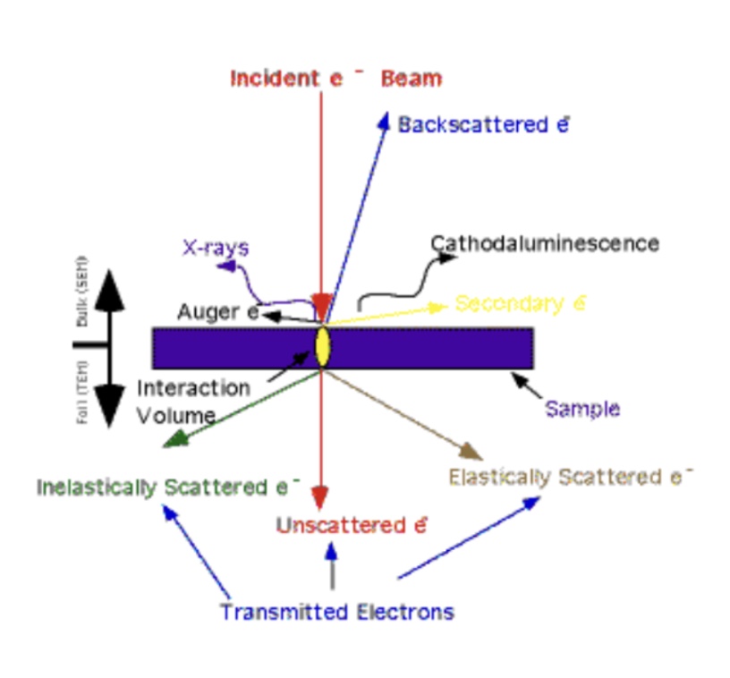
Electron - specimen interaction
electrons from top → sample (transmitted electrons) can either go out or travel back up
Transmitted Electrons
Only interact with the sample weakly, if at all.
Why do you have to stain your sample in order to see something?
Biological samples have low or no contrast so you have to stain your sample in order to see anything. Stains scatter electrons to enhance visibility.
Common stains
Osmium tetroxide → lipids
Uranyl Acetate → nucleic acids
Lead citrate → general contrast
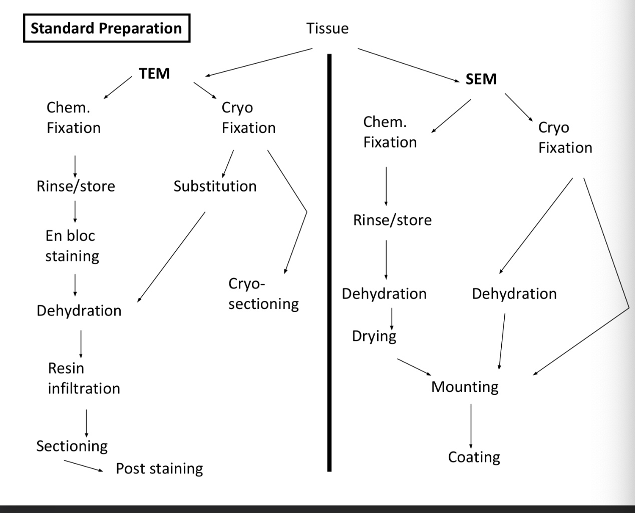
Goals of TEM specimen preperation?
Goals:
Preserve specimen’s natural state
Avoid artifacts (common artifacts: knife marks, wrinkles, and chatter)
Keep structure intact
Steps
Fixation (chemical or cryo) → dehydration → resin infiltration → sectioning (50-90 nm thin, with glass or diamond knife) → post staining (uranyl acetate, lead citrate) → mounting on copper grids (3 mm, grids act like a slide would = hold sample in place)
Image artifacts
Chatter → caused by knife “skipping”
Wrinkles → imperfections that can be smoothed out with heavy vapors
Knife marks → caused by knife imperfections
Stain contamination etc.
Immuno-EM
Immunoelectron microscopy, is a technique that uses antibodies tagged with electron-dense markers (like gold particles) to precisely locate specific molecules, such as proteins, within a cell or tissue at a very high resolution.
How does Immuno-EM work?
Detects specific molecules using antibody-gold labeling.
Steps:
Light fixation + sucrose cryoprotection → freeze in liquid nitrogen → section → grid → thaw → add primary antibody, then gold-conjugated secondary antibody → stain and view
Notes: can use different gold particle sizes for multiple labels
Problems: weak labeling or poor preservation
What is negative staining?
Provides quick visualization of small particles (proteins and viruses).
Uses heavy metal salts to surround, but not penetrate the sample → appears bright on dark background
Grid coated with formvar + carbon for specimen stability
factors affecting staining : stain concentration, pH of strain and time
SEM (scanning electron microscopy)
Type of electron microscope that uses a focused beam of electrons to scan the surface of a sample. Uses secondary electrons (low energy electrons ejected from surface of a material when an electron beam hits it) for topography. Producing high-resolution 3D-like images of surfaces.
Can also detect backscattered electrons (those that are deflected back out of the sample) (elemental contrast)
SEM sample prep
Steps: Fix (formaldehyde or glutaraldehyde) → dehydrate (ethanol or acetone) → critical point drying (sample is in vacuum so no liquids allowed) → replacing liquid (acetone) with CO2, then vent gas (prevents collapse, has vented without distorting the morphology of the sample)
Then coat the sample with gold (for a good source of secondary electrons) → view
SEM is for ___ samples allowed, and no need for ultra-___ sections
thicker, thin
Backscattered electrons
Created when incident electrons bounce back (180°) from dense atoms
Higher atomic number → brighter appearance
Used for elemental contrast (e.g, gold particles appear bright on E.coli)
All electron microscopy is
False coloring - all electron microscopy is black and white -psuedo-colored after the fact
Width of hair
Width = mλDwall / dmin
Demonstrates diffraction limit and resolution principles.
Critical points to be made
TEM → thin sections, internal detail.
SEM → surface topography.
STEM → scanning internal structure + composition.
Requires vacuum + fixation.
Sample prep is critical for accuracy.