introduction to the blood system
1/31
There's no tags or description
Looks like no tags are added yet.
Name | Mastery | Learn | Test | Matching | Spaced |
|---|
No study sessions yet.
32 Terms
blood
a liquid that fills the vascular compartment and serves to transport dissolved materials and blood cells throughout the body
the average human has 4-6L of blood
what are the constituent parts of blood
plasma (makes up approx 55% of total blood volume) - is a straw coloured liquid
WBC and platelets - <1% of the total volume - is called the buffy coat
Red blood cells ( approx 45% of whole blood volume)
Red blood cells and WBC and platelets together is called the haematocrit - but it often only refers to RBCs
Blood consists of plasma and cells - plasma 55%, cellular elements 45%
functions of blood
respiration - RBCs supply oxygen to tissues and remove CO2 from tissues and cells
transport of (in the plasma) -
nutrients to the tissues and cells
waste products from cells
messages such as hormones around the body
protection from infection (WBCs) - the immune system
repair (platelets) - repairing tissue damage
thermoregulation - plasma
thermoregulation
the maintenance of physiological core body temperature
extreme heat -
circulation diverts blood to the surface to cool the body
sweating
extreme cold -
circulation diverts blood to deep/core to maintain body heat
hair stands up
shivering
plasma
most water but also:
ions (such as Na+ etc)
plasma proteins (such as albumin etc.)
substances transported by blood (i.e. nutrients, waste products, hormones etc.)
Haematopoiesis
formation of blood
occurs in bone marrow (spongy tissue inside bones)
Red marrow in flat bones (such as pelvis, sternum or ribs) produce most blood cells#
yellow marrow in long bones produces some WBCs
Bone marrow consists of blood cells at various stages of development as well as supporting tissue called stroma
stroma is made up of fibroblasts macrophages, adipocytes, osteoblasts, osteoclasts and epithelial cells etc.
stroma produces growth factors that tell the stem cells what to become
haematopoiesis definition
production of blood cells
megakaryopoiesis or thrombopoiesis
production of platelets from megakaryocytes
erythropoiesis
production of red blood cells
granulopoiesis
production of granulocytes
monocytopoiesis
production of monocytes
lymphopoiesis
production of lymphocytes
totipotent
stem cells with the ability to become any cell in the body (e.g. cells in a fertilized ovum)
pluripotent
stem cells with the ability to become nearly all cells in the body (e.g. cells from the 3 germ layers)
multipotent
stem cells with the ability to become a number of different cells of the same family e.g multipotent haematopoietic stem cells in the bone marrow are able to become any of the different blood cells
oligopotent
stem cells with the ability to differentiate into a few different cell types e.g. common myeloid precursor can become any of the myeloid cells (platelets, RBCs
destruction of blood cells
spleen (an ovoid organ found in the upper left abdominal cavity) destroys red blood cells
the spleen is the largest ‘filter’ of blood in the body removing old or damaged blood cells (engulfed by phagocytes)
4 million blood cells are destroyed every second, 2.5 of these are RBCs
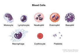
Red blood cells (erythrocytes)
biconcave disks
7-12um in diameter
hold approximately 90fl
doesn’t have a nucleus (is anucleate) - so can carry more oxygen
no mitochondria - as don’t want mitochondria using up oxygen to produce energy
RBC have a different energy
approx 4-6×10^12 RBCs/L
about 25 trillion total in the average human but it varies particularly between male and females
have a long lifespan of approx 120 days during which cells travel approx 300 miles
functions:
transport of oxygen and carbon dioxide around the body
critical role in respiration
why are mammalian RBCs anucleate
Humans are warm-blooded so have higher metabolic rates
By ejecting the nucleus, the RBCs have a higher O2 carrying efficiency
However this also means bone marrow has to work continuously
Haemoglobin (Hb)
responsible for red appearance of RBCs
about 250 million Hb molecules per RBC
There are 4 polypeptide chains each with a cofactor called a haem group that has an iron atom at the centre
Each iron atom binds one molecule of O2
co-operative binding - once one molecule of O2 has bound, it is easier to bind the 2nd and 3rd molecules but then it is harder to bind the 4th molecule. Likewise it is easier to offload the 2nd and 3rd molecules but harder to offload the 4th molecule
Hb binds to O2, CO2 and NO (can also bind CO) and transports these molecules around the body
Pulmonary circulation
blood vessels surrounding the lungs enable gas exchange (O2 and CO2)
pulmonary artery → heart → lungs (deoxygenated)
pulmonary vein → heart → body (oxygenated)
capillary bed surrounds the alveoli
Transport of oxygen
once in plasma, O2 is taken into RBCs and reversibly binds to haem group of Hb
oxyhaemoglobin (oxygenated state) - is bright red
deoxyhaemoglobin (deoxygenated state) - is dark red
Transport of CO2
CO2 follows the concentration gradient from the tissues into the interstitial fluid and then plasma
once in blood stream some CO2 binds to Hb but most reacts with the water in RBCs to become carbonic acid
HCO3- travels in the plasma and the H+ binds to Hb so the pH of the blood isn’t altered
In the lungs the reverse happens:
the HCO3- and the H+ recombine to carbonic acid and dissociates into CO2 and H2O
The CO2 follows the conc gradient out of the blood into the alveolar sacks and is inhaled
Anaemia
reduced haemoglobin conc in blood
there are several classes
it often causes low haematocrit and small RBCs
most common causes:
poor diet - iron deficiency
chronic blood loss i.e. internal bleeding, heavy periods etc
malabsorption of iron
pregnancy
signs and symptoms:
fatigue
pallor
tachycardia
shortness of breath
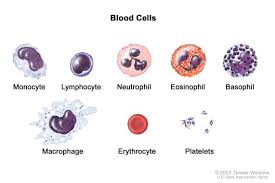
White blood cells
can be broken down into phagocytes (granulocytes and monocytes) and immunocytes (T-cells, B-cells, NK cells)
Neutrophil
approx twice the size of RBCs
multi-lobed nucleus
has neutral staining granules (myeloperoxidase)
2.5-7.5×10^9/L
life span - 6 hours to a few days
function:
vital role in protection from bacterial infections
phagocytosis of bacteria
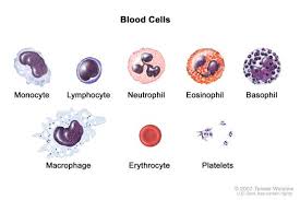
Eosinophils
approx same size as neutrophil
bi-lobed nucleus
large pink-staining granules (histamine, plasminogen, DNase, RNase) - stain with eosin
0.04-0.44×10^9/L
life span - 8-12 days
Functions:
immune-protection
phagocytosis of antibody-coated pathogens
attacks parasites
allergic responses
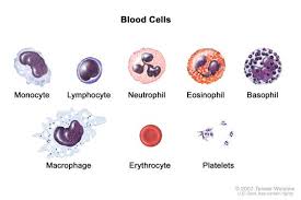
Basophils
slightly smaller than neutrophil
bi-lobed nucleus
large purple-staining granules (histamine, heparin, proteolytic enzymes) - stain with methylene blue
0.01-0.1×10^9/L
life span - a few hours to a few days
functions:
release of histamine during inflammation
are not phagocytes
mast cells are nor a type of basophil
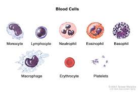
monocytes (blood)/macrophages (tissue)
large kidney shaped nucleus
larger than neutrophil
0.2-0.8×10^9/L
life span - lasts many months
function:
vital role in protection from infections
ingest bacteria, dead cells and cellular debris
phagocytosis
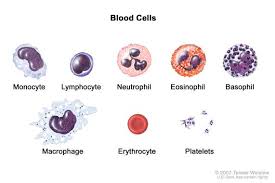
Lymphocytes
large and relatively round nucleus that fills the cytoplasm
slightly smaller than neutrophil
1.5-3.5×10^9/L
life span - can persist for many years
divided into two types of cell due to function: T cells and B cells
function:
central role in the immune system - protecting from infections especially viral infections
some lymphocytes attack pathogens directly (T-cells) and some produce antibodies (B-cells)
Natural killer cells
look similar to neutrophils
large round nucleus
purple-staining granules (perforin, proteolytic enzyme)
secrete cytokines (IFN gamma and TNF alpha)
very rare, not typically counted (elevated with physical exercise, massage etc.)
life span - 1-2 weeks
functions:
immunological surveillance
kill virus infected cells and some tumour cells non-specifically i.e are part of innate immunity
Platelets
small anucleate fragments of large precursor cells called megakaryocytes
2-4um diameter
appear in blood films as dark-staining granules
15—400×10^9/L
life span - 5-10 days
functions:
roles in blood clotting and prevention of blood loss (haemostasis)
activated platelets change from a smooth discoid shape to sending out filopodia and forming lamellipodia