Adult II Module 3: HEENT
 Call Kai
 Learn
Learn Practice Test
 Spaced Repetition
 Match
Match Flashcards
 Knowt Play
1/37

There's no tags or description
Looks like no tags are added yet.
Name | Mastery | Learn | Test | Matching | Spaced | Call with Kai | Chat |
|---|
No analytics yet
Send a link to your students to track their progress
38 Terms
Eye
• FUNDI: veins are larger & darker than arteries
• CONES: for color perception, 20/20 vision, sharp vision
• RODS: for detecting light and shadow, night vision
• MACULA: responsible for central vision; fovea contains large numbers of cones and is set in the middle; area of the eye that determines 20/20 vision
• PRESBYOPIA: Age-related visual change r/t a decreased ability of the eye to accommodate stiffening lenses; usually starts at the age of 40 yrs., near vision is affected w/decreased ability to read (especially small print)
• Legal Blindness defined as vision of 20/200

Cataracts
Opacity of the lens. Very common and a leading cause of blindness.
Vision is loss because the opacification prevents light from focusing
on the retina
Several Types
Nuclear cataract: the most common type of cataract, begins with a gradual hardening and yellowing of the central zone of the lens
Cortical cataract: Opacity occurs on the sides. Patients may c/o glare in dim illumination
Posterior subcapsular cataract: more rapid progression, occurs in younger patients
Cataracts can appear any age but aging is a risk factor for the most common form of cataract. Up to 20% of older adults are affected
Congenital cataracts (can be idopathic or related to infection or metabolic disease), traumatic cataracts, medication induced (e.g. corticosteroids). Diabetics, smokers at risk
Most common surgery in the U.S. is cataract surgery
ROS:
Non-painful
Progress over months to years
Difficulty with glare (example: headlights at night, sunlight)
Decreased night vision
Halos around lights
Blurred vision
PE:
Color of lens is white to gray
Use direct ophthalmoscopy
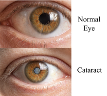
Cataract Management
Usually conservative at first: May warrant change in eyeglasses prescription, cessation of nighttime driving. Magnifiers, increased font size, and other visual aids may be helpful (remember this when giving written instructions, consents etc)
Discourage cigarette smoking, encourage glycemic control in diabetics, use appropriate eye protection
No medical treatments or topical drops available to reverse lens opacification
Surgery is indicated when the cataract has affected the patient’s vision to a point where ADLs/iADLs are impaired
The procedure consists of using an ultrasound device to “emulsify” the lens into microscopic fragments that are vacuumed out of the eye and replaced with an artificial lens
Aftercare: limit strenuous activities, protect the eye from irritants, avoid driving, do not let water directly enter eye
Red eye
Can be caused by other disorders. Examples of findings that support urgent opthalmologic referral include loss of visual acuity, photophobia, and ciliary flush (as pictured).

Allergic Conjunctivitis
Also known as Hay fever
Environmental allergens play a role
Results from the allergen causing an inflammatory response when it contacts the ocular surface
Vernal conjunctivitis and atopic conjunctivitis are considered to be more severe and chronic forms of allergic conjunctivitis. They are due to mast cell and lymphocyte-mediated immune processes
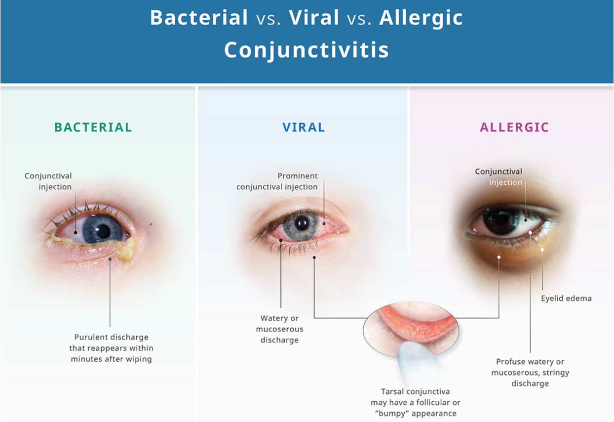
Viral Conjunctivitis
Spread by direct contact
Highly contagious
Assess for recent URI or sick contacts
S/S include acute onset of a red eye with excessive watery discharge. Usually starts in one eye then the other
3 forms:
Adenoviral conjunctivitis
Pharyngoconjunctival fever (more systemic disease: fever, HA, sore throat)
Epidemic keratoconjunctivitis (significant, bilateral conjunctival hyperemia and chemosis (sign of eye irritation). Subconjunctival hemorrhages may be present- refer to ophthalmology)
Bacterial Conjunctivitis
Thick, purulent discharge (“eyes glued shut”)
Acute conjunctivitis is the most common form of bacterial conjunctivitis. Symptoms manifest over days
In children, the causative agents include Haemophilus influenzae and Streptococcus pneumoniae.
S. aureus is more commonly seen in adults
Symptoms typically last for 7 to 10 days
>4 weeks is chronic bacterial conjunctivitis
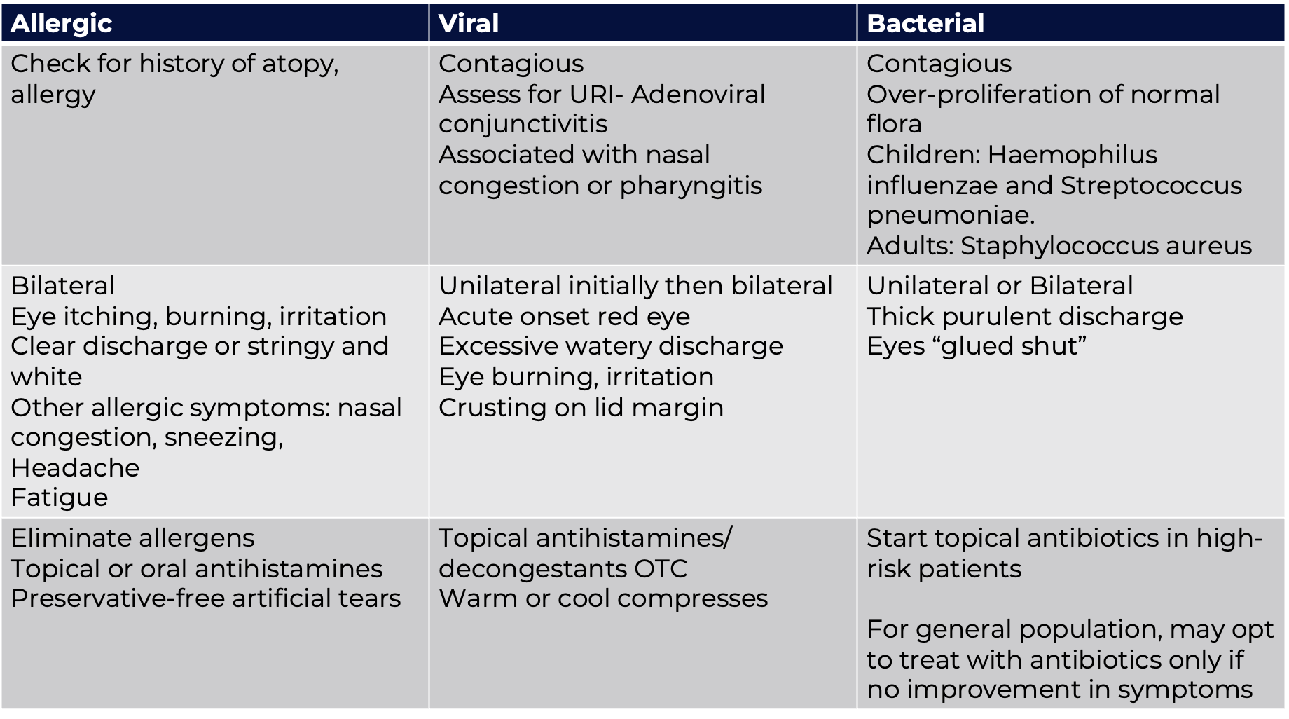
Bacterial conjunctivitis treatment

Glaucoma
Normally, the anterior chamber of the eye is moderately pressurized which helps the eye maintain its shape
When there is insufficient drainage of the anterior chamber of the eye (for example with aging which is associated with a loss of drainage cells) pressure begins to increase
Bottle-neck effect
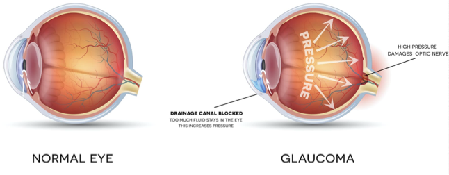
Primary open-angle glaucoma
Most common type of glaucoma (60-70%)
Gradual onset of increased IOP (intraocular pressure) due to blockage of aqueous humor inside the eye
IOP > 22 mmHg (normal IOP 8-21 mmHg), >30 mmHg is very high
The retina (CN II) undergoes ischemic changes which, if not treated, leads to permanent damage
No widespread screening recommendations but patients at risk for glaucoma, such as older adults, those with a family history of glaucoma, diabetics, may benefit from targeted screening
Older adult
Patients may be asymptomatic in the early stages
Gradual changes in peripheral vision (occurs first) that may be followed by central vision loss
Patient may report missing portions of words when reading
Fundoscopic exam shows cupping due to increased pressure
Assess visual fields
Tonometer checks IOP
Managed by ophthalmologist

Primary Open-Angle Glaucoma Treatment
Medications (eye drops)
Betimol (Timolol) 0.5% Beta Blocker (decreases aqueous production)
Latanoprost (Xalatan) topical prostaglandin (increases aqueous outflow)
Side Effects include bronchospasm, fatigue, depression, HF, & bradycardia
Contraindications: Asthma, emphysema, COPD, 2-3rd. Degree AV block, & HF
SURGERY
Selective Laser Trabeculoplasty (SLT)---Laser energy is applied to the drainage tissue in the eye. This results in better drainage of fluid through the drain and out of the eye. This eventually results in lowering of IOP.
Trabeculectomy---drainage hole in the sclera allowing fluid to flow out of the eye (bleb)
Acute Angle-Closure Glaucoma
Sudden blockage of aqueous humor results in an increased IOP resulting in ischemia and damage to the optic nerve (CN2)
Suspect acute angle-closure glaucoma if a patient presents with:
Acute onset of severe eye pain accompanied by
Headache
Decreased vision and/or blurry vision or halos around lights
N/V
Lacrimation
Exam Findings: Eyes: A mid-dilated (may be oval shaped), fixed pupil (4-6mm)
Pupil appears oval rather than round and reacts slowly w/ light
Corneal cloudiness
Conjunctival injection
Fundoscopic exam reveals cupping of the optic nerve
An ophthalmologic emergency! Treatment refer to ED- blindness can occur without intervention.
Tonometry: quickly measures the IOP. Usually done in ED
TREATMENT: Laser peripheral iridotomy is a microscopic opening that is made in the periphery of the iris which creates an area of outflow
Retinal Detachment
Sudden abrupt increase in floaters or a shower of floaters
Floaters associated with flashing lights
Associated w/ looking through a black curtain covering part of the visual field
Decreased vision
Refer patient to the ED
Treatment: Surgery

Hypertensive Retinopathy
Retinal vascular damage caused by hypertension, arteriosclerotic changes
Can result in retinal hemorrhage
Funduscopic examination can show arteriolar constriction, arteriovenous nicking, vascular wall changes, flame-shaped hemorrhages, cotton-wool spots, yellow hard exudates, and optic disk edema
Nicking, or indentation, of retinal veins is caused by stiff (arteriosclerotic) retinal arteries
Treatment: Control HTN
Encourage lifestyle changes

Diabetic Retinopathy
Non-proliferative diabetic retinopathy: Early stage
Tiny blood vessels leak, making the retina swell
Proliferative diabetic retinopathy: More advanced stage of diabetic eye disease.
The retina starts growing new blood vessels (neovascularization).
These fragile new vessels often bleed into the vitreous.
Ocular ischemia is a condition most commonly seen in association with proliferative diabetic retinopathy. Scar tissue forms
Patients may see dark floaters, blurry vision, vision loss
Perform a funduscopic examination. Assess for signs of arterial or venous disease. On exam you may see cotton wool spots (fluffy yellow white patches on retina)
For the evaluation and treatment of diabetic retinopathy: yearly visits to an ophthalmologist or an optometrist should begin at the time of diagnosis for all patients with diabetes
Annual screening: Dilated eye exam
Diabetic control is vital
Left untreated, diabetic retinopathy may progress to vision loss and blindness
Papilledema
Optic disc is swollen w/ blurred edges
Related to increased intracranial pressure (ICP)
Signs/symptoms of increased ICP may include:
Severe headache
Nausea, vomiting
Altered mental status
Somnolence
Diplopia
Pupillary changes (e.g., uneven pupil size, fixed dilated pupils)
Can be caused secondary to bleeding, brain tumor, abscess, pseudotumor cerebri (false brain tumor)

Optic Neuritis
Optic nerve inflammation
Variable vision loss; papilledema; pain on eye movement; sore globe
This may be alone or accompanied by nystagmus or other abnormal eye movements
Fundoscopic exam may reveal disc pallor
Assess for other neurological symptoms (i.e. Aphasia, paresthesias, abnormal gait, spasticity)
Refer to ophthalmology within 24-48 hours
Refer to neurology (h/o Multiple Sclerosis or may be first sign of Multiple Sclerosis)
Macular Degeneration
Age related macular degeneration (AMD)
Usually asymptomatic during early stages
Caused by gradual damage to the pigment of the macula (area of central vision). Metamorphopsia, or warped or distorted central vision, occurs
Results in severe vision loss to blindness. Straight lines appear distorted or curved (doors/windows)
Assess for family history of macular degeneration
Two types (Dry and Wet)
About 80% (8 out of 10) of people who have AMD have the dry form. Dry AMD is when parts of the macula get thinner with age and tiny clumps of protein called drusen grow. There is no way to treat dry AMD yet.
Wet form is less common but much more serious. Wet AMD is when new, abnormal blood vessels grow under the retina. These vessels may leak blood or other fluids causing scarring of the macula.
Vision loss is faster with wet AMD than with dry AMD
Macular Degeneration Treatment
Refer to ophthalmologist
Amsler grid: a simple square containing a grid pattern and a dot in the middle. This design can show problem spots in the field of vision
Dry Type:
Treated w/ ocular vitamins (lutein, zeaxanthin w/ zinc)
Preservision (to support cell structure & healthy pigment)
Wet Type:
Anti VEGF (vascular endothelial growth factor) is the most common treatment (intraocular injection: delivered to the eye through a very thin needle). Anti-VEGF treatment helps reduce the number of abnormal blood vessels in the retina. It also slows any leaking from blood vessels.
Slows progression of disease and may improve vision

Anterior Uveitis (Iritis)
Uveal tract of the eye: the iris, ciliary body, and choroid. Any or all of these structures may become inflamed. Uveitis is therefore an inflammation of any of these layers
Iritis is an inflammation of the anterior uveal structure, the iris
Eye pain (insidious onset)
Red eye
Injected conjunctiva (inflammation of iris & anterior chamber)
May occur in healthy individuals or in those w/ autoimmune disorders such as RA, Lupus, sarcoidosis, ankylosing spondylitis, syphilis
No purulent discharge as would be seen in bacterial conjunctivitis
Refer to ophthalmologist within 24 hrs.
May result in blindness
Treatment: Management of underlying condition, ocular steroids, and eye drops to dilate pupil (Medication Class: Mydriatics, i.e. cyclopentolate)
Otitis media (OM)
Otitis media (OM) is an acute infection of the middle ear cavity with bacteria pathogens due to mucus that becomes trapped in the middle ear
Severity and presentation may vary, and causative agents may be bacterial, fungal, or viral
Most often associated with upper respiratory tract infections or allergies: can occur due to a cold, sore throat, or respiratory infection
Very common complaint in primary care
OM is the most frequent childhood infectious illness, with the peak incidence at 6 to 15 months of age, but adults and older adults are also affected and is seen in all age groups
Usually unilateral but may involve both ears at times
Adult infections usually due to Streptococcus pneumoniae

Acute otitis media (AOM)
A bacterial or viral infection of the middle ear fluid
Rapid onset
Short duration
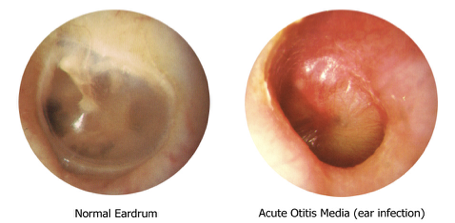
Bulging, opaque, erythematous tympanic membrane with reduced motility

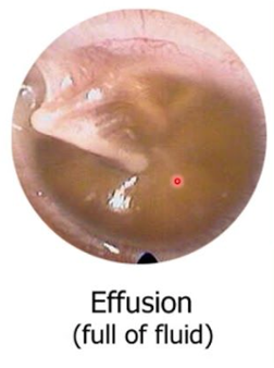
Middle ear effusion (MEE)
An accumulation of serous fluid in the middle ear
Can be associated with AOM, often persisting for weeks or months after an episode of AOM
Otitis media with effusion (OME)
Accumulation of serous fluid in the middle ear without acute inflammation
Can precede or follow AOM, but barotrauma or chronic allergic rhinitis also can precipitate an occurrence
Chronic effusion is characterized by hearing impairment and may persist for several months, with or without signs of infection
Tympanic membrane may bulge or retract but should not be red. May see yellow or clear fluid with air bubbles.
Oral decongestant
ROS
Recent URI? Exposure to smoke? Allergies? h/o ear infection
Presence of rapid-onset otalgia, worse in a prone position
Other symptoms include imbalance or vertigo, mild stuffiness, and a fullness or popping sensation in the ear
Assess for dizziness, changes in hearing,
Younger patients may have non-specific symptoms such as ear rubbing, rhinorrhea, vomiting, diarrhea, and fever
Assess for recent activities that involve barometric pressure changes
Clinical Presentation
Otalgia
Popping noise
Muffled hearing
Adult infections usually develop more slowly than in children
Generally afebrile
Assessment
Vital signs including temp
Level of alertness (especially in children)
Assess for signs of URI: oral, throat, nose, lymph nodes, mastoid, sinuses
Ear examination with otoscope
Diagnosis of AOM requires bulging of the TM with obscured landmarks or new onset of otorrhea not caused by acute otitis externa
Acute Otitis Media is primarily a clinical diagnosis
Treatment
Pain medication: patient specific (NSAIDs or acetaminophen)
First-line antibiotic therapy (indicated for adults with fever and if obvious significant discomfort is apparent)
Amoxicillin–clavulanate 875/125 mg PO every 12 hours for 5 to 7 days
For adults at high risk for severe infection (older than 65 years, immunocompromised, etc)
Amoxicillin–clavulanate 2000/125 mg PO every 12 hours for 10 days
If an adult is allergic to penicillin, azithromycin or doxycycline are options
Antihistamines, decongestants, and steroids are not beneficial for treatment of AOM
Treatment decisions should be reconsidered if symptoms worsen or fail to respond to initial antibiotic treatment within 48 to 72 hours
Vestibular Neuritis
An acute unilateral labyrinthine dysfunction
Viral inflammation of the vestibular nerve (8th cranial nerve), otitis media, acute suppurative labyrinthitis, chemical products associated with acute or chronic otitis media, as well as evidence also suggests an association w/ latent herpes simplex (type 1: infection of the vestibular ganglia)
CLINICAL PRESENTATION:
Severe vertigo, N&V, disequilibrium aggravated by head movement
Tinnitus may be present
Hearing remains intact
Severe symptoms typically resolve in 48-72 hrs. (may last for 4-5 days)
Most episodes resolve spontaneously
Up to 50% of patients will have symptoms of dizziness & equilibrium for several months
Vestibular Neuritis: ROS and PE
ROS:
Current medications
Vertigo (duration, nature & severity)
Any head trauma
Recent infections (esp. respiratory infections)
Precipitating factors (head turning, coughing, sneezing, etc..)
PHYSICAL EXAM and DIAGNOSTICS:
HEENT: Ears, Nose, Throat
Neurological exam (balance testing - Romberg)
Hearing test will reveal normal hearing
Spontaneous nystagmus, horizontal, or rotary is usually present and directed away from the affected ear
Abnormal findings on neurological examination suggests a central cause (refer to neurology)
Obtain a CBC w/diff. if a bacterial infection is suspected
If a tumor is suspected imaging should be done (MRI/CT)
Vestibular Neuritis: Treatment and Management
Alleviating vertigo, nausea & vomiting
Antihistamines, anticholinergics, benzodiazepines, or anti-emetics
Antihistamines and anticholinergics are preferred over benzodiazepines
Antiemetics may be used during the acute phase but stopped after 3 days (may hamper vestibular recovery)
Some studies have revealed improvement of symptoms w/ corticosteroids, more recent studies have shown little benefit
Still reasonable to start steroids during the acute phase (10-day tapering course of prednisone)
Once the severe symptoms have passed, patients may benefit from vestibular enhancement exercises, which can be obtained through physical therapy
When to Refer:
Otolaryngology referral when diagnosis is unclear
Severe bacterial infection
Does not resolve in 4-6 weeks
Severe dehydration

Meniere's Disease
Meniere’s disease is a chronic condition of the inner ear characterized by recurrent vertigo and hearing loss
It is a triad of symptoms that may or may not occur simultaneously:
Episodic vertigo (spinning or rocking), may be associated with nausea and vomiting
Hearing loss (chronic), unilateral, initially at lower frequencies, progresses to permanent hearing loss at all frequencies
Tinnitus (fluctuating or constant)
Meniere disease involves excess fluid and pressure in the labyrinth of the inner ear that episodically distends the structures of the labyrinth and damages the vestibular system (involved in balance) and cochlear hair cells (involved in hearing)
The majority of cases are likely caused by viral infections or immune system–mediated mechanisms
Up to 1/3 of all cases originate from an autoimmune process
Cause may be unknown
Usually unilateral presentation
No associated neuro signs

Meniere’s Disease: Clinical Presentation and Physical Exam
Clinical Presentation
Early signs: Vertigo lasting minutes to hours, associated nausea and vomiting
Pressure/fullness in ear
Low pitched tinnitus
Unilateral hearing loss
May have periods of remission
Physical Exam
Rule out other causes of vertigo & hearing loss
Head & neck exam to exclude infection
Neurological exam to exclude other causes
Weber test will show sensorineural hearing loss (usually low frequency)
Rinne test will show AC>BC
Spontaneous nystagmus during attack
Meniere’s Disease: Treatment and Management
Diagnostic criteria for Meniere disease include two episodes of spontaneous vertigo lasting at least 20 minutes each, audiometrically documented hearing loss, tinnitus or aural fullness, and the exclusion of other causes
Refer to otolaryngologist for testing and management
No Cure. Goals of care are directed managing vertigo & arresting the disease process
Lifestyle modifications which may include limiting salt, avoiding MSG, nicotine, caffeine
Steroid Therapy
A brief oral steroid course may provide temporary relief from vertigo for several weeks; however, longer-term efficacy has not been shown.
Intratympanic steroid injection into affected ear may produce somewhat longer relief for several months.
If autoimmune process is suspected, diagnosis is typically confirmed after a positive response to steroids
For Symptom Relief
Centrally acting antihistamines with anticholinergic effects can suppress the vestibular system while also providing antiemetic relief.
Options include dimenhydrinate, meclizine, and promethazine. Meclizine is the least sedating option and therefore the most common therapy.
Meniett device- (non-invasive treatment that generates low pressure pulses that may displace inner ear fluids)
Drop attacks (sudden falls)- Rarely (Approx. 6%) patients will develop this
Salt restriction- low salt diet has been suggested to reduce osmotic buildup of pressure in the endolymphatic compartment; long-term efficacy is not proven.
Hearing aids should be prescribed as required
Vestibular rehabilitation therapy
Persistent attacks: refer to ENT
Chronic Rhinosinusitis
Rhinosinusitis is related to nasal cavity and paranasal sinus inflammation
Chronic rhinosinusitis (CRS): symptoms of nasal congestion and discharge that are persistent
Criteria for CRS diagnosis:
Symptoms present 12 weeks or more
and two of the following symptoms: a diminished sense of smell, facial pain, nasal congestion, and mucopurulent drainage
Additional requirements for diagnosis of CRS include:
(1) confirmation of presence of edema or purulent mucous discharge in anterior ethmoid region or middle meatus
(2) nasal cavity or middle meatus polyps, and/or
(3) radiographic documentation of paranasal sinuses inflammation
Possible predisposing and associated factors for CRS include:
Dysfunctional cilia as seen in smokers
Cystic fibrosis
Allergy
Asthma
aspirin sensitivity
genetic factors
Immunodeficiency
infection
Pregnancy
Chronic Rhinosinusitis: Review of Systems
Common complaints can include cough, dental pain, facial discomfort, fatigue, fever, halitosis, headache, nasal blockage or discharge (anterior or posterior nasal drip), and reduction in or loss of smell
Symptoms can be chronic, worsening over time, and associated with asthma, allergic rhinitis, tonsillar hypertrophy, and recurrent otitis media
Obtain a detailed history: onset and timing of symptoms? location of congestion? associated symptoms? Triggers? h/o allergies? Asthma? family history of seasonal or environmental allergies? Smoking? Pollutants? Recreational drug use?
Obtain a detailed medication history (ask about chronic decongestant use)
Chronic Rhinosinusitis: Physical Exam
Observe for any asymmetry or deformity of the nasal structure. Assess for nasal obstruction
Use the otoscope with a wide speculum to examine each nostril. Nasal mucous membranes should be inspected for erythema, pallor, atrophy, edema, crusting, and discharge. Turbinates may be more erythematous in patients with chronic nasal congestion
The frontal and maxillary sinuses should be palpated for pain
Accurate diagnosis requires specialist consultation and diagnostic evaluation
Chronic Rhinosinusitis: Diagnosis and Treatment
Objective confirmation of CRS via nasal endoscopy by an otolaryngology specialist
Sinus CT
Saline irrigations and intranasal corticosteroids can be helpful for patients c/o nasal congestion
Daily saline irrigations and intranasal corticosteroids are the mainstay of treatment to minimize and control inflammation in CRS with and without nasal polyps; however, topical steroids may be less effective in patients who have CRS without nasal polyps
Patients who present with nasal congestion associated with acute bacterial rhinosinusitis without complications, “watchful waiting” before treating with antibiotic therapy is advised, as symptoms can resolve within a few days
For patients with nasal polyps, sinus surgery to remove polyps can be helpful for some, but recurrent polyps are possible
Antibiotics may be a consideration if an exacerbation of CRS results in acute rhinosinusitis
Patients with rebound nasal congestion related to topical decongestant use will have resolution 2 to 3 weeks after the offending medication is stopped
Referral to an otolaryngologist is necessary for patients with severe congestion refractory to treatment