Ovel Fetal Abnormalities quiz
1/49
There's no tags or description
Looks like no tags are added yet.
Name | Mastery | Learn | Test | Matching | Spaced | Call with Kai |
|---|
No analytics yet
Send a link to your students to track their progress
50 Terms
Echogenic debris within the fetal stomach is commonly associated with:
down syndrome
Demonstration of multiple unilateral renal cysts is most suspicious for:
multicystic dysplastic kidney
“Double bubble” is a sonographic sign associated with:
duodenal atresia
Which of the following sonographic findings helps to differentiate Dandy-Walker syndrome from an arachnoid cyst?
presence of a normal vermis
Dilation of the third ventricle is a sonographic finding associated with:
agensis of the corpus callosum
Maternal alpha-fetoprotein levels in a pregnancy with gastroschisis will:
markedly increase
Which skeletal abnormality is most likely to demonstrate a cloverleaf skull?
thanatophoric dysplasia
A crescent-shaped appearance to the cerebellum should signal the sonographer to give additional attention to which of the following fetal structures?
spine
Peritoneal calcifications with associated dilated loops of bowel and polyhydramnios visualized in a 30-week fetus most likely represent:
meconium peritonitis
Which of the following abnormalities is the most common neural tube defect?
anencephaly
Which of the following conditions is most likely associated with frontal bossing?
hydrocephalus
Which of the following conditions is most likely associated with frontal bossing?
encephalocele
Which of the following is a common sonographic finding with fetal facial abnormalities?
polyhydramnios
Demonstration of fetal bone fractures raises suspicion for which skeletal abnormality?
osteogenesis imperfecta
A large single ventricular cavity is most suspicious for:
holoproencephaly
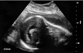
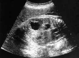
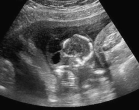
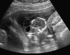
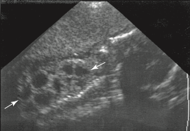
The sonographic finding in this image is most suspicious for:
ventriculomegaly
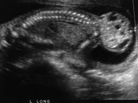
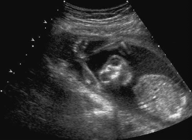
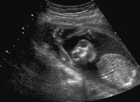
An asymptomatic patient arrives for a second- trimester fetal surveillance examination. A sagittal image of the fetal body is most suspicious for:
sacrococcygeal teratoma
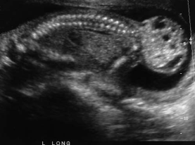
Associated findings with this abnormality include:
hydronephrosis
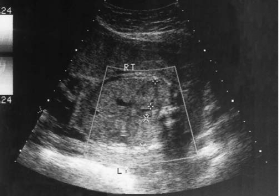
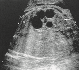
During a late second-trimester screening examination, what does this image of the fetal abdomen most likely show:
infantile polycystic disease
Which of the following conditions will likely occur because of this abnormality?
oligohydramnios
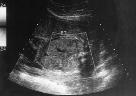
A sagittal image of the fetal abdomen most likely demonstrates:
hydronephrosis
A patient arrives for an early second-trimester sonogram for gestational dating. An endovaginal image demonstrates a fetal abnormality that is most suspicious for:
acrania
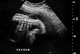
A sagittal image of the lower spine is most suspicious for:
spina bifida
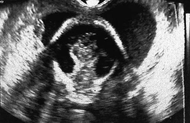
What abnormality is most likely present in this cross-sectional image of the cranium?
cystic hygroma
The etiology of this abnormality is typically:
chromosomal
A patient presents for an ultrasound to determine gestational age. An image of this early second- trimester fetus is most suspicious for:
anencephaly
Which of the following is most likely associated with this finding?
elevated maternal alpha-fetoprotein
A patient arrives for a second-trimester screening sonogram. A sagittal image of the fetus is most suspicious for which of the following pathologies?
cystic adenomatoid malformation
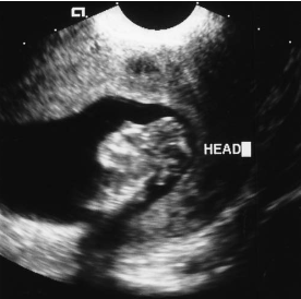
This sonogram of an early second-trimester cranium is most suspicious for:
holoprosencephaly
An oblique sonogram of the fetal abdomen most likely demonstrates:
multicystic dysplastic kidney
Lateral ventricular enlargement becomes ventric- ulomegaly after the diameter exceeds:
10 mm
Caudal regression syndrome is more commonly found in patients with:
diabetes mellitus
Which of the following is the most common fetal neck mass?
cystic hygroma
Which of the following abnormalities is more commonly associated with proboscis?
holoprosencephaly
Which of the following abnormalities is not asso- ciated with pulmonary hypoplasia?
duodenal atresia
A diagnosis of clubfoot may be made with persis- tent abnormal inversion of the:
foot perpendicular to the lower leg
Opening in the layers of the abdominal wall with evisceration of the bowel describes which of the following abnormalities:
gastroschisis
Which of the following is the most common nonlethal skeletal dysplasia?
achondorplasia
Hydronephrosis in utero is most commonly caused by an obstruction:
at the ureteropelvic junction
Herniated contents of an omphalocele are covered by a membrane consisting of:
amnion and peritoneum
The presence of a posterior fossa cyst and agenesis of the cerebellar vermis are characteristic findings of:
Dandy-Walker malformation
Which of the following is not associated with hydrocephalus?
choroid plexus cysts
Anechoic regions within brain tissue are most suspicious for:
hydranencephaly
Outward angling of the frontal and lateral horn of the lateral ventricles is a sonographic finding in:
agenesis of the corpus callosum
The renal pelvis in a third-trimester fetus demonstrates an anterior–posterior diameter of 10 mm. This is considered:
mild hydronephrosis
In the late second trimester, which sonographic finding consistently displays with renal agenesis?
oligohydramnios
The most common sonographic finding associated with multicystic renal dysplasia is:
unilateral multicystic kidney
Sonographic findings associated with osteogene- sis imperfecta may not be apparent before:
24 weeks’ gestation
Which classification of osteogenesis imperfecta is the most severe?
type II
A consistently small fetal stomach on serial sono- grams is most suspicious for which abnormality?
esophageal atresia