clinical immunology
 Call Kai
 Learn
Learn Practice Test
 Spaced Repetition
 Match
Match Flashcards
 Knowt Play
1/81

There's no tags or description
Looks like no tags are added yet.
Name | Mastery | Learn | Test | Matching | Spaced | Call with Kai | Chat |
|---|
No analytics yet
Send a link to your students to track their progress
82 Terms
innate immune response
The response consists of two phases (Figure 1.6). First is recognition that a
pathogen is present. This involves soluble proteins and cell-surface receptors
that bind either to the pathogen or to human cells and plasma proteins that
have been altered by the pathogen's presence. Once the pathogen has been
recognized, the second phase of the response recruits effector mechanisms
that kill or expel the pathogen. Mediating the effector mechanisms are effector
cells that engulf bacteria, kill virus-infected cells, and attack protozoa.
Supporting the effector cells is complement, a system of plasma proteins
that assists the effector cells by tagging pathogens with molecular flags.
Complement proteins can also kill pathogens without assistance from effector
cells by perturbing the integrity of the pathogens' membranes. Collectively,
these defenses comprise.
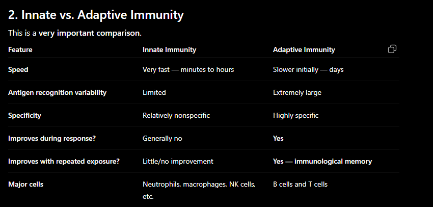
The body's immediate, nonspecific defense against pathogens.
Present from birth; cells include neutrophils, macrophages, NK cells, etc.
Rapid response within hours
Fixed
Limited number of specificities
Constant during the course of response
antigen
A substance that can be specifically recognized by immune receptors, especially B-cell receptors, antibodies, or T-cell receptors
Can be from microbes, toxins, proteins, etc.
Adaptive immunity
Slow response in days to weeks
Variable
Numerous highly selective specificities
Improves during the course of response
Antigen-specific immune defense that develops after exposure
Innate Immunity vs Adaptive Immunity
The receptors of innate immunity are structurally of many different types. Each receptor recognizes molecular features that are common to groups of pathogens, and none isspecific for a particular pathogen. Conversely, the lymphocytes of adaptive immunity recognize pathogens using only one type of cell-surface receptor, but this receptor is made in billions of different versions.
Main Advantages of Adaptive Immunity
High specificity
Can distinguish between many different antigens.
Huge recognition diversity
The adaptive immune system can recognize an enormous variety of different antigens.
Immunological memory
After the first exposure, memory cells remain.
A second exposure can produce a faster and stronger response.
Improved response with repeated exposure
Adaptive responses can become more effective after repeated encounters with the same antigen.
Mast cell
Expulsion of parasites from the body by release of granules
containing histamine and other active agents
CD markers that can be used to identify T lymphocytes
CD3
Helper T → CD4
Cytotoxic T → CD8
CD markers that can be used to identify B lymphocytes
CD19, CD20
List the mature immune cell types that can arise from the common lymphoid precursor
B lymphocytes
T lymphocytes
NK cells
Lymphoid → B, T, NK
List the mature immune cell types that can arise from the common myeloid precursor
granulocytes, (which comprise neutrophils, eosinophils, and basophils)
Dendritic cells
Megakaryocytes → platelets
Erythrocytes (red blood cells)
Monocytes → macrophages
Myeloid → monocytes/macrophages + granulocytes
granulocytes (polymorphonuclear leukocytes)
Comprises neutrophils, eosinophils, and basophils. These effector cells all have cytoplasmic granules fill which is why they are granulocytes.
Neutrophils
polymorphonuclear (usually 2-5 lobes) granulocyte with tiny pink/blue cytoplasmic granules
Important in acute inflammation and bacterial defense
lymphocytes
mononuclear cells with round nucleus, high nucleus to cytoplasmic ratio (big nucleus, very little cytoplasm around it) rare cytoplasmic granules. B cells t cells, and NK cells are all in this group because they cannot be distinguished morphologically using a microscope.
eosinophils
usually, bilobed nucleus (like basophils) with large orange pink (salmon) colored cytoplasmic granules
Important in parasite defense and allergic responses
basophils
rare cells; usually bio\lobed nuclesus (like eosinophils) obscured by large purple purple black cytoplasmic granules.
Important in inflammatory/allergic responses
Explain the relationship between monocytes and macrophages
Monocyte
Found primarily in peripheral blood
Circulating leukocyte
Can leave the bloodstream and enter tissues
Macrophage
A differentiated tissue phagocyte
Develops from monocytes that migrate into tissues
Engulfs pathogens, dead cells, and debris
Can present antigen to T cells
Easy sequence:
Bone marrow → monocyte → blood → tissue → macrophage
List the 2 tissues/organs that represent the primary lymphoid tissues
1. Bone marrow
Site of blood-cell production
B-cell development/maturation
2. Thymus
Site of T-cell maturation
Memory trick:
B = Bone marrow
T = Thymus
Describe how a naïve lymphocyte enters a lymph node, and how it would exit if it did not
get stimulated by antigen
enter via arterioles (blood vessel), exit via efferent lymph
Describe how antigen enters a lymph node
anitgens enters lymph node via fluid drainijng from tissues (in afferent lymph vassals)
List where T lymphocytes and B lymphocytes are primarily located in the lymph nodes
and the white pulp of the spleen.
Lymph node
B cell zone → Outer (follicles)
T cell zone→ Middle
Spleen
B → follicles
T → PALS (periarteriolar lymphoid sheath)
CD antigen/marker
A cell-surface molecule identified by a "CD" (cluster of differentiation) number
Used to identify/classify immune cells
Primary lymphoid tissue
Where lymphocytes develop and mature
Bone marrow and thymus
Secondary lymphoid tissue
Where mature lymphocytes encounter antigen and become activated
Lymph nodes, spleen, MALT, etc.
Lymph
Fluid collected from tissues that travels through lymphatic vessels
Eventually e-mph nodes and returns to blood
PALS
Periarteriolar lymphoid sheath
t-cell region of the spleen's white pulp
GALT
Gut-associated lymphoid tissue
Lymphoid tissue associated with the gastrointestinal tract
BALT
Bronchus-associated lymphoid tissue |
Lymphoid tissue associated with the respiratory tract |
MALT
Mucosa-associated lymphoid tissue
Lymphoid tissue associated with mucosal surfaces
M cell
Specialized epithelial cell that transports material from the lumen to underlying immune cells
Important in mucosal immune surveillance, especially GALT
Describe how plasma proteins limit the spread of infection throughout the body
Plasma contains several proteins that help prevent an infection from spreading.
Major mechanisms:
1. Complement proteins
Bind to microbes.
Can directly damage microbial membranes.
Help mark microbes for destruction (opsonization).
Promote inflammation and recruitment of immune cells.
2. Pentraxins
Bind to structures on microbes.
Help identify microbes for phagocytosis.
Can activate complement.
3. Acute-phase proteins
Produced mainly by the liver in response to inflammatory cytokines.
Circulate throughout the body.
Help recognize and contain pathogens
phagocytosis
cellular process where cells engulf and digest large particles, such as pathogens and cellular debris, playing a crucial role in the immune response and maintaining tissue homeostasis
Describe how pentraxins and defensins function in innate immune system protection of
the body
Pentraxins
Circulating proteins that bind to pathogens and effector molecules
target for destruction
Similar role to antibodies of the adaptive immune response
Form a bridge between pathogen and phagocytes.
Promote phagocytosis.
Pentraxins = "tag the pathogen”
Defensins
Defensins are antimicrobial peptides.
Insert into microbial membranes.
Disrupt membrane integrity.
Kill microbes.
Defensins = "damage the pathogan”
Identify the major macrophage cytokine that triggers the “acute phase” response and the proteins that are produced as a part of the acute phase response.
A major macrophage cytokine that triggers the acute-phase response is:
IL-6
Macrophages release IL-6, which acts primarily on the liver.
The liver then produces acute-phase proteins.
Important examples:
C-reactive protein (CRP)
Serum amyloid A (SAA)
Fibrinogen
Surfactant proteins
Pathway to memorize:
Macrophage → IL-6 → liver → acute-phase proteins
The acute-phase response is a systemic response to local infection/inflammation.
Describe the main functions of the inflammatory response
Earliest and most notable immune response:
Signs: redness, swelling, heat, pain, loss of function.
Inflammatory response:
Recruitment of leukocytes to the site of infection via soluble and cellular receptors
Reduced integrity of blood vessels allows for leukocytes to enter infected tissues
Macrophage activation
Release of cytokines (pro/inflammatory)
Helps induce adaptive immune response if necessary.
THINK: Inflammation gets immune cells and immune proteins from the blood to the site where they're needed.
Identify the cytokines and chemokines released by macrophages that cause beneficial
LOCAL immune system effects and the specific effects that each of the cytokines is
responsible for.
at the site of infection, activated resident macrophages secrete inflammatory cytokines.
TNF-a (cytokine) induces blood vessels to be more permeable, enabling cells, fluid, and soluble effectors to enter infected tissue.
IL-6 (cytokine) induces fat and muscle cells to metabolize, generate heat, and raise temperature in the infected tissue.
IL-12 (cytokine) recruits and activates natural killer cells to secrete cytokines that strengthen macrophages’ response to infection
CXCL8 (chemokine) recruits neutrophils from the blood and directs them to the infected tissue
CCL2 (Chemokine) recruits monocytes from the blood and directs them to the infected tissue.
cytokines
Cytokines are small signaling proteins that allow cells to communicate with one another
Cytokines that start the immune response are pro-inflammatory
Cytokines that limit the immune response are anti-inflammatory
chemokines
attract effecter cells into infected tissues.
What are Pattern Recognition Receptors? Differentiate between PAMPs and DAMPs
PRRs are receptors used by innate immune cells to recognize common molecular patterns associated with infection or tissue damage.
Receptors on all leukocytes, as well as some epithelial and endothelial cells.
PAMP = Pathogen-Associated Molecular Pattern
These are any molecular structures associated with microbes recognized by PRR
PAMP = Pathogen
DAMP = Damage-Associated Molecular Pattern
These are molecules released or exposed by damaged or stressed cells/tissues. They signal that tissue injury has occurred. DAMP = Damage
Describe the process of leukocyte recruitment from blood vessels into surrounding
tissues
When tissue becomes infected or damaged, leukocytes must leave the bloodstream and enter the tissue.
The sequence:
1. Rolling
Leukocytes temporarily attach to endothelial cells using selectins.
⬇
2. Activation
Chemokines activate leukocytes.
⬇
3. Firm adhesion
Leukocyte integrins become activated and bind strongly to endothelial adhesion molecules.
⬇
4. Transmigration / diapedesis
The leukocyte moves between endothelial cells and leaves the blood vessel.
⬇
5. Chemotaxis
The leukocyte follows a chemical gradient toward the site of infection.
Memorize:
ROLL → ACTIVATE → ADHERE → TRANSMIGRATE → CHEMOTAXIS
Or:
"Rolling → Activation → Adhesion → Exit → Follow the signal."
What are the three systemic inflammatory cytokines? What tissues do they act on, and
what kind of response is produced?
Systemic effects to know
Cytokine | Tissue/organ | Main response |
|---|---|---|
TNF-α | Muscle + fat | Changes metabolism; promotes breakdown of energy stores → (decreased viral and bacterial infections) |
IL-1 | Hypothalamus | Increased body temperature → decreased viral and bacterial infections |
IL-6 | Liver | Acute-phase protein production → activates complement opsonization |
TNF-α + IL-1 | Bone marrow | Increased leukocyte production/release → phagocytosis |
What are the mechanisms of systemic shock?
1. Widespread vasodilation (due to TNF-a)
Blood vessels become more dilated.
2. Increased vascular permeability
Fluid leaves blood vessels and enters tissues.
3. Decreased blood volume/pressure
Less effective circulating blood volume remains in the vascular system.
4. Poor tissue perfusion
Organs don't receive enough blood and oxygen.
This can result in:
Systemic inflammatory shock
Simple chain:
Cytokines (TNF-a) → vasodilation + vascular leakage → low blood pressure → inadequate tissue perfusion → organ dysfunction
Differentiate autoinflammation from (allo) inflammation
Autoinflammation
Inflammation caused by activation of the innate immune system (not adaptive immunity) in response to abnormal/damaged components of the person's own body.
It does not primarily require recognition of a foreign antigen by adaptive immunity.
AUTO = self
not adaptive immunity
Alloinflammation
Inflammation caused by recognition of genetically different cells/tissues from another individual of the same species.
This is particularly important in situations such as:
Transplantation
Transfusion-related immune responses
ALLO = another individual
involves adaptive immunity and innate
Neutrophils vs. Macrophages
Both are phagocytes, but they behave very differently.
Feature | Neutrophils | Macrophages |
|---|---|---|
Lifespan | Short — usually hours to a few days | Much longer — weeks to years depending on tissue |
Location | Mostly blood until recruited to tissues | Primarily tissues |
Main role | Rapid response to infection | Long-term defense, cleanup, regulation, repair |
Phagocytosis | Very strong | Very strong |
Repeated use | Generally die after intense activity | Can survive and continue functioning |
Antigen presentation | Limited | Yes — can present antigen to T cells |
Tissue repair | Limited | Important |
Can produce cytokines | Yes | Yes |
Neutrophil = emergency responder
Macrophage = long-term resident
Neutrophils arrive quickly and kill microbes, but they have a relatively short lifespan.
Macrophages can remain in tissues much longer and can phagocytose, secrete cytokines, present antigen, and help with tissue repair.
Differentiate M1 and M2 macrophages
M1 = Intracellular infections
M2 = wound healing, antiparasitic infections
List the different PRRs activated dendritic cells use to traffic molecules to the lymph nodes, and what cell(s) respond to each type of PRR activation
PRR activation tells dendritic cells that danger is present and promotes their migration to lymph nodes, where they can activate T cells.
Identify the main function of plasmacytoid dendritic cells.
The main function of plasmacytoid dendritic cells (pDCs) is:
Producing large amounts of Type I interferons
Especially:
IFN-α
IFN-β
They are particularly important in antiviral innate immunity.
Memorize:
pDC → Type I IFN → antiviral defense
Explain what is meant by “respiratory burst” regarding phagocytes
The respiratory burst is the rapid production of reactive oxygen species (ROS) by activated phagocytes.
It happens after a phagocyte has engulfed a pathogen.
Simplified process:
Phagocytosis
↓
NADPH oxidase activated
↓
O₂ → reactive oxygen species
↓
ROS help kill the pathogen
Important ROS include:
Superoxide
Hydrogen peroxide
Other reactive oxygen products
The respiratory burst is an important mechanism for killing phagocytosed microbes
Identify the immune system defect that causes Chronic Granulomatous Disease
CGD is caused by a defect in:
NADPH oxidase
Remember:
NADPH oxidase → respiratory burst
Therefore:
Defective NADPH oxidase → defective respiratory burst → impaired killing of certain phagocytosed microbes
This makes patients particularly susceptible to certain bacterial and fungal infections.
Exam connection:
CGD = NADPH oxidase defect = respiratory burst defect
phagocytosis, netosis, apoptosis, pyroptosis, and trogocytosis
Process | What happens? | Common cell types | Inflammation? |
|---|
Phagocytosis | Cell engulfs and digests target | Neutrophils, macrophages, dendritic cells | Usually can promote inflammation depending on context |
NETosis | Neutrophils release DNA/proteins that form NETs to trap microbes | Neutrophils | Yes, generally |
Apoptosis | Programmed cell death; cell is removed in an orderly manner | Most nucleated cells | Usually no |
Pyroptosis | Inflammatory programmed cell death with membrane disruption | Macrophages, monocytes, other innate cells | Yes |
Trogocytosis | Cell takes small pieces of membrane/material from another cell | Immune cells, including phagocytes | Can contribute to immune activation depending on context |
Identify the two main functions of NK cells
1. Kill infected or abnormal cells
NK cells can kill:
Virus-infected cells
Some tumor/abnormal cells
They release cytotoxic molecules that cause target-cell death.
2. Produce cytokines
Especially:
IFN-γ
IFN-γ activates macrophages and helps promote antimicrobial immune responses.
Memorize:
NK = Kill + IFN-γ
Compare the three known ILCs based on what type of immunity they are associated with
ILC | Immunity | Major cytokines | Main association |
|---|---|---|---|
ILC1 | Type 1 | IFN-γ | Intracellular microbes |
ILC2 | Type 2 | IL-4, IL-5, IL-13 | Parasites/allergy |
ILC3 | Type 3 | IL-17, IL-22 | Extracellular bacteria/fungi |
Discuss how interferons (Type I and Type II together) activate NK cells to
combat/counteract infections
Type I IFNs (IFN-α/β) are produced during viral infections and activate NK cells. NK cells then kill infected cells and produce Type II IFN (IFN-γ), which activates macrophages and increases their ability to kill intracellular microbes.
Easy pathway: Virus → IFN-α/β → NK cell → IFN-γ → Macrophage activation
Describe the interaction between macrophages and NK cells, including the cytokines and
interferons involved
Macrophage
↓ releases IL-12 + IL-18
NK cell
↓ produces IFN-γ
Macrophage
↓ becomes more activated
Better killing of intracellular microbes
Describe the process of immunological (NK-cell) synapse, and the responses to activating
and inhibitory signals.
The NK-cell synapse is the organized contact between an NK cell and its target cell.
NK cells receive both:
Inhibitory signal
Healthy cells typically express MHC class I. NK-cell inhibitory receptors recognize MHC I. This tells the NK cell: "This is a normal self cell — don't kill it."
Activating signal
Stress or infection can cause a cell to express molecules recognized by activating NK receptors. If activating signals outweigh inhibitory signals, the NK cell kills the target.
The basic decision:
Inhibitory signals dominate → DON'T KILL
Activating signal dominates → KILL
Identify the general role of Pattern Recognition Receptors in innate immune responses
Pattern recognition allows innate immune cells to detect nonself and altered self
Soluble and cellular receptors that detect the presence of infecting organisms and recruit leukocytes to the infected tissue. These receptors operate using pattern recognition, the mechanism of examining molecules to determine if the motif present belongs to the host or to an outside entity, and among host cells, to distinguish between healthy cells and altered unhealthy cells.
List and describe each of the TLR subfamilies: Type I, II, III, IV (not each individual TLR), including general type of ligands, microbe type recognized, cells on which they appear (immune or other), and cellular location of receptor.
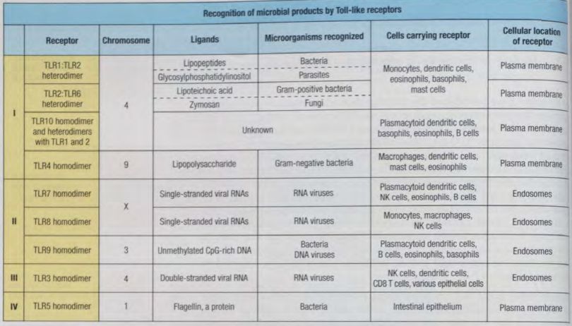
Describe how TLR’s main cell location relates to their target antigen type
Location | What TLRs mainly detect |
|---|---|
Plasma membrane | lipids, lipoproteins, cell-wall components, carbohydrates |
Endosome | Microbial nucleic acids → viral RNA/DNA or intracellular bacterial infections. |
Describe the function of MyD88, and which TLR’s utilize this protein
MyD88 is an adaptor protein that brings two signaling proteins together.
Most TLRs use MyD88 except TLR3
Identify which of the interferons (IFN-α, IFN-β, or IFN-γ) are “Type I interferons” and the type of microbial infection they combat (ie, Bacterial? Viral? Fungal? Parasitic?)
Interferon | Type | Major role |
|---|---|---|
IFN-α | Type I | Antiviral |
IFN-β | Type I | Antiviral |
IFN-γ | Type II | Activates immune responses, especially macrophages |
IFN-α and IFN-β = Type I interferons
Describe the function of NFκB, IRF3, and IRF7 transcription factors and their cytokine/interferon production in response to cell surface (plasma membrane) or endosomal TLR binding
NF-κB, IRF3, and IRF7
Transcription Factor | Main Function | Activated By / Pathway | Main Products | Main Result |
|---|---|---|---|---|
NF-κB | Promotes inflammation | TLR signaling, especially through MyD88 | TNF, IL-1, IL-6 | Inflammatory response |
IRF3 | Promotes antiviral responses | TLR signaling and RIG-I/MAVS pathways | Mainly IFN-β | Antiviral response |
IRF7 | Promotes and amplifies Type I interferon production | Endosomal TLR signaling and interferon signaling | Mainly IFN-α | Antiviral response |
Identify the outcome of RIG-1 binding and activation of MAVS receptors
RIG-I is a cytosolic pattern-recognition receptor.
It detects viral RNA inside the cytoplasm.
Leads to the synthesis and secretion of type 1 interferons
On what type of leukocyte are scavenger receptors found? What is the function of scavenger receptors, and what kinds of ligands do they bind to
PRRs on tissue-resident macrophages
Scavenger receptors bind a broad range of molecules and help cells:
Recognize material
Internalize material
Remove potentially harmful substances
Participate in microbial recognition
Types of ligands
They can recognize:
Microbial components
Modified lipids
Damaged cellular material
Other altered molecules
Describe the importance of the cGAS-STING pathway in the innate immune response
cGAS–STING Pathway
Step | What happens |
|---|---|
cGAS | Detects DNA in cytosol |
STING | Activates signaling |
IRF3 | Turns on Type I interferons |
Result | Antiviral immune response |
Remember: cGAS = DNA sensor → STING → Type I IFN.
memorize → produce type 1 interferons (a,b)
Detail the structure and function of the inflammasome
Part | Function |
|---|
Sensor | Detects microbes, cell damage, or danger signals |
ASC | Adaptor protein that connects the sensor to caspase-1 |
Caspase-1 | Processes IL-1β and IL-18 into active cytokines |
IL-1β | Promotes inflammation |
IL-18 | Helps activate immune responses |
Pyroptosis | Inflammatory form of programmed cell death |
Overall result | Strong inflammatory immune response |
The inflammasome is a cytosolic multiprotein complex that detects danger and activates inflammatory responses.
Complement (C′)
A group of plasma and membrane proteins that help destroy pathogens, promote inflammation, and enhance phagocytosis.
Opsonin
A molecule that coats a pathogen and makes it easier for phagocytes to recognize and ingest it. C3b is an important complement opsonin.
Anaphylatoxin
Small complement fragments, mainly C3a and C5a, that promote inflammation and activate/recruit immune cells.
Chemoattractant
A substance that causes immune cells to move toward a site of infection or inflammation. C5a is an important example.
Complement fixation
Activation of complement after recognition of a target, resulting in complement proteins binding to/depositing on the target.
CR# (Complement Receptor)
Receptors on immune cells that recognize complement fragments attached to pathogens or immune complexes
Opsonization vs. Complement Fixation
Opsonization | Complement Fixation |
|---|---|
Coats a pathogen to make it easier to phagocytose | Complement proteins become activated and deposited on a target |
Important opsonin: C3b | Can lead to opsonization, inflammation, and MAC formation |
Easy memory:
Opsonization = “tag it for eating.”
Complement Cascade
Complement works as a cascade:
One protein activates → many molecules of the next protein → even more molecules downstream.
Each activated enzyme can activate many molecules of the next component, producing amplification
Advantage of Complement vs. Antibody
Complement can rapidly amplify and coat a pathogen with C3b without requiring a large amount of antibody already attached to the pathogen.
This is especially important for the innate immune response
Four Main Functions of Complement
Function | What it does |
|---|
Opsonization | C3b coats pathogens → promotes phagocytosis |
Inflammation | C3a/C5a promote inflammatory responses |
Chemotaxis | C5a attracts immune cells to infection |
Cell lysis | MAC (C5b–9) can create pores and lyse target cells |
Three Complement Pathways
Pathway | How it starts | First Recognition Component |
|---|
Classical | Antibody bound to antigen | C1q |
Lectin | Recognizes microbial sugars such as mannose | MBL |
Alternative | Spontaneous C3 activation and recognition of pathogen surfaces | C3/C3b |
C3 and C5 Convertases
Pathway | C3 Convertase | C5 Convertase |
|---|---|---|
Classical | C4b2a | C4b2a3b |
Alternative | C3bBb | C3bBb3b |
Lectin | C4b2a | C4b2a3b |
Note: Some textbooks use different naming for the C2 fragments. Follow the notation used in your Parham course materials. The functional complex is commonly written C4b2a.
iC3 vs. iC3b and iC3Bb vs. C3bBb
Molecule | Status | Meaning |
|---|
C3b | ✅ Active | Functional complement fragment |
iC3b | ❌ Inactivated | C3b after regulatory cleavage |
C3bBb | ✅ Active | Alternative pathway C3 convertase |
iC3Bb | ❌ Inactivated | Inactivated form of the alternative C3 convertase |
Key idea:
“i” = inactive/inactivated.
Factor I helps cleave C3b into inactive forms, helping stop complement activation.
Greatest Amplification
C3b
C3b provides major amplification because it can form the alternative pathway C3 convertase (C3bBb).
That convertase produces more C3b, which produces more convertase:
C3b → C3bBb → more C3b → more C3bBb
This creates a powerful positive-feedback amplification loop
Membrane Attack Complex (MAC)
MAC components:
C5b + C6 + C7 + C8 + C9
Written as:
C5b–9
C9 is the component that polymerizes to form the pore in the target membrane.
Memory:
C5b → C6 → C7 → C8 → C9
Complement Amplification Protein
Properdin (Factor P)
Question | Answer |
|---|---|
What protein amplifies complement? | Properdin |
Which pathway? | Alternative pathway |
How? | Stabilizes C3bBb, the alternative C3 convertase |
Result | More C3 is cleaved → more C3b |
Properdin stabilizes the alternative pathway C3 convertase, extending its activity and increasing complement amplification.
Complement Inhibition
Complement can be inhibited at important points in the cascade, including:
C3 convertase formation/activity
C3b activity/deposition
Factor I
Factor I cleaves C3b → iC3b
C3b → iC3b
This helps prevent continued complement activation on host cells
three Pathways — Big Picture
Pathway | Initiated By | Early Components | C3 Convertase |
|---|---|---|---|
Classical | Antibody–antigen complex | C1q → C1r/C1s → C4 + C2 | C4b2a |
Lectin | MBL binding microbial sugars | MBL → MASPs → C4 + C2 | C4b2a |
Alternative | Spontaneous C3 activation on pathogen surface | C3b + Factor B + Factor D | C3bBb |
Where do they converge?
All three pathways converge at:
C3
The C3 convertases cleave:
C3 → C3a + C3b
This is important because C3b provides opsonization and amplifies complement activation, while the pathway continues toward C5 activation and the MAC.