Kidneys T2 W8
1/23
Earn XP
Description and Tags
Flashcards about the Kidneys Macroscopic & Microscopic Structure, including the functions and structures of the kidneys and nephrons.
Name | Mastery | Learn | Test | Matching | Spaced |
|---|
No study sessions yet.
24 Terms
Kidneys structure
Reddish-brown, bean-shaped organs, approximately 11cm long, 6cm wide, and 3cm thick, located in the posterior abdominal cavity.
Role of Kidneys
Filter nearly 200L of fluid from our bloodstream daily, removing toxins, metabolic wastes, and excess ions while returning needed substances to the blood.
Kidneys Function
Regulating body fluids by controlling the total volume of water in the body and the total concentration of solutes in that water.
Ensuring long-term acid-base balance of the blood by maintaining the pH of the blood (pH 7.4).
Excreting metabolic wastes, including nitrogenous wastes, and foreign substances such as drugs and toxins from the blood.
Macroscopic structure
⚬What can be seen with the naked eye
⚬3 major regions
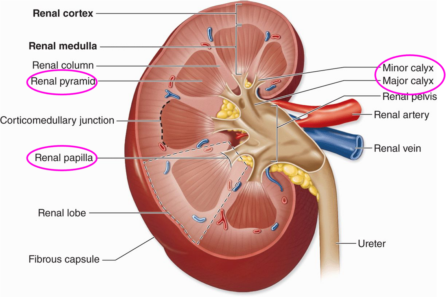
Renal capsule, renal cortex, renal medulla, renal pyramids, renal pelvis, renal artery + renal vein.
Microscopic structure
⚬What can only be seen under the microscope
⚬Filtering units of the kidney that produce urine – the nephrons
Renal capsule
•A layer of fibrous connective tissue that surrounds each kidney.
•Helps support the kidney and protects it from injury.
Renal cortex
•Outer part of kidney that has a grainy appearance & is lighter reddish-brown in colour.
•Contains the glomerulus of the nephrons.
Renal medulla
•Middle part of kidney containing renal pyramids, is darker reddish-brown in colour & is striated in appearance.
•Contains capillary networks that loop around the long renal tubules of the nephrons.
Renal pyramids
•Cone-shaped structures in the medulla. The apex of each renal pyramid is called the papilla.
Renal pelvis
•White in colour & has a funnel-shaped cavity.
•Branching extensions of the renal pelvis form cup-shaped areas:
⚬2-3 major calyces (singular calyx)
⚬Subdivide to form minor calyces, that enclose the papillae of each renal pyramids.
⚬The calyces collect urine, which drains continuously from the papillae, and empty into the renal pelvis.
⚬Urine then flows through the renal pelvis into the ureter.
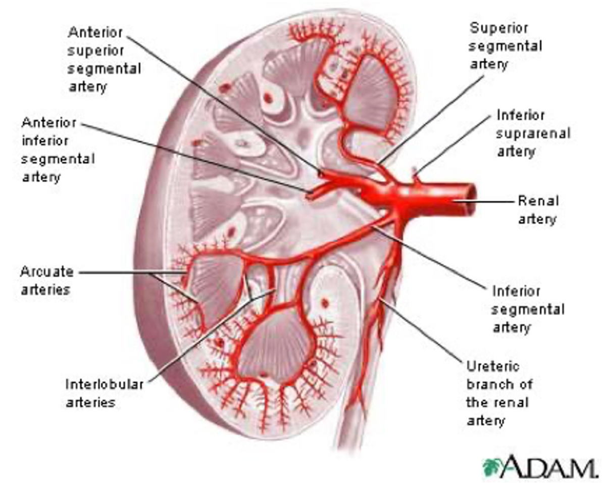
Renal artery
•Supplies blood to the kidneys from the aorta.
Renal vein
•Carries blood from the kidneys & empties into the inferior vena cava.
•Since the kidneys must continuously cleanse the blood and adjust its composition, it is not surprising that they have a very rich supply of blood.
Functional unit of kidney
A nephron
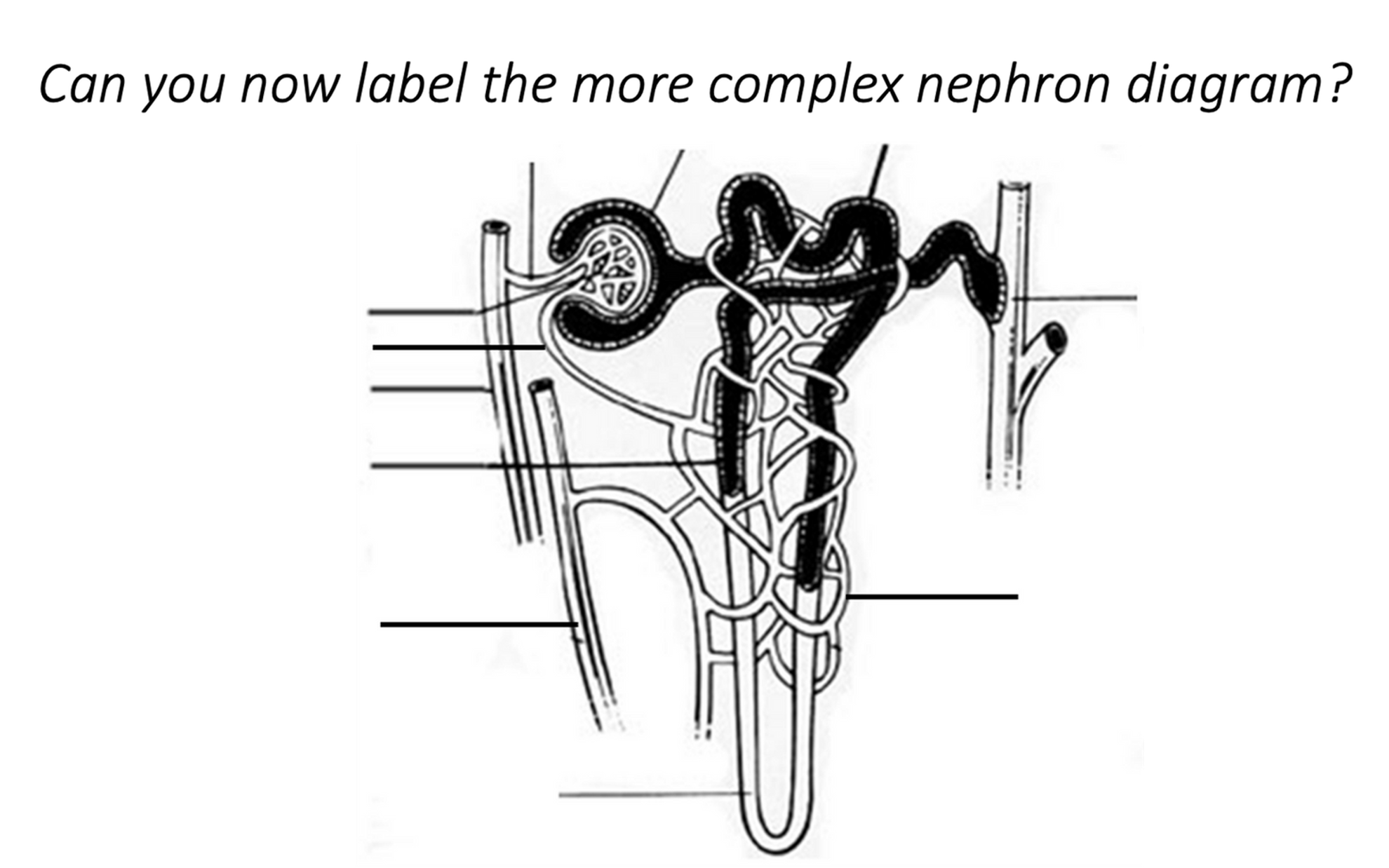
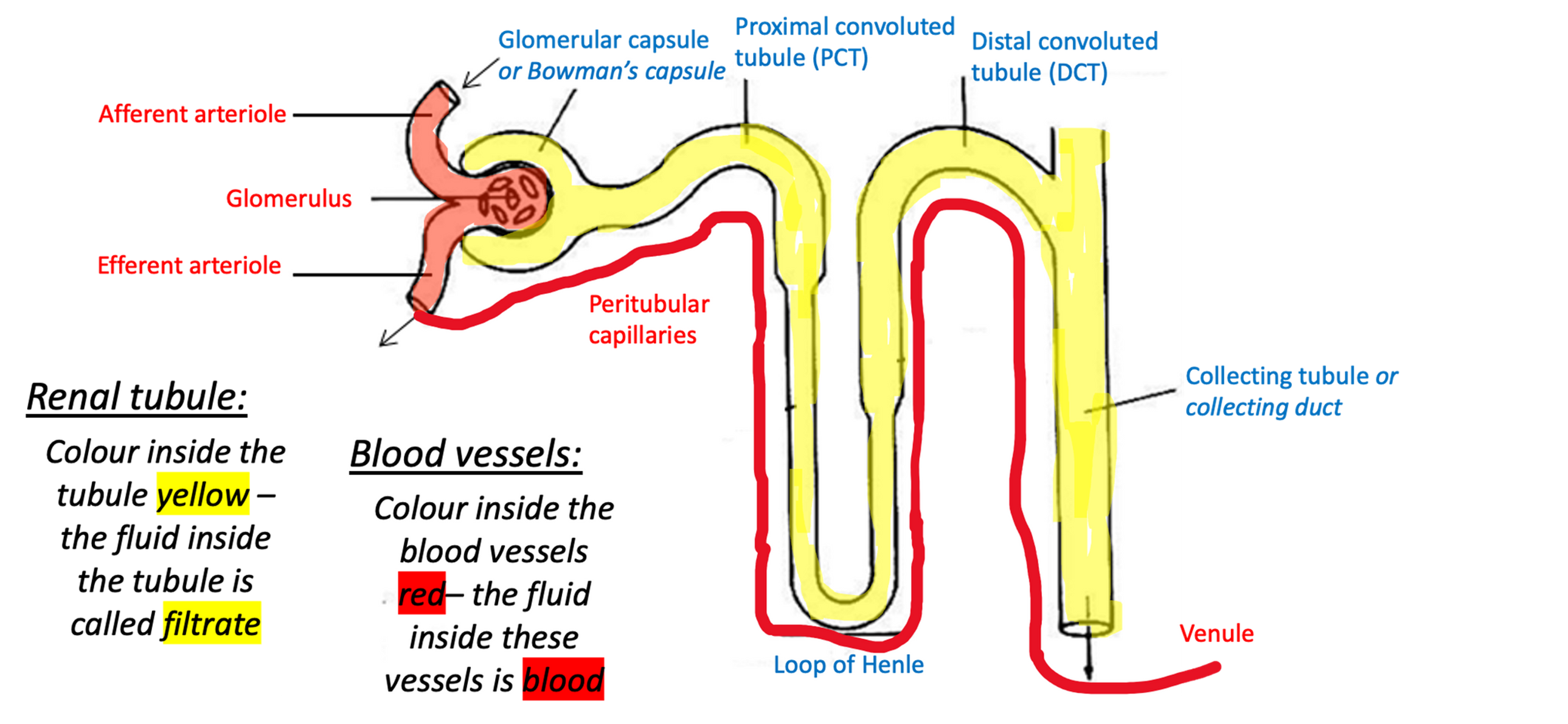
Nephron
•The nephron is where urine is produced.
•There are an estimated 1 million nephrons in each human kidney, and each is surrounded by a complex capillary network.
•Highly folded tubule + complex capillary network.
Renal tubular
structures involved in urine formation. Fluid inside it is called filtrate.
•About 3cm long and has 3 major parts.
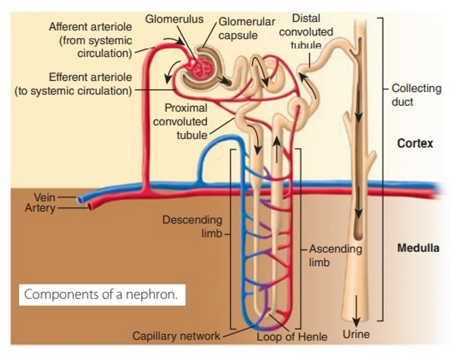
•The renal tubule leaves the glomerular capsule coiling to form the proximal convoluted tubule (PCT), drops into a hairpin loop called the loop of Henle and twists again as the distal convoluted tubule (DCT) before emptying into the collecting duct (CD).
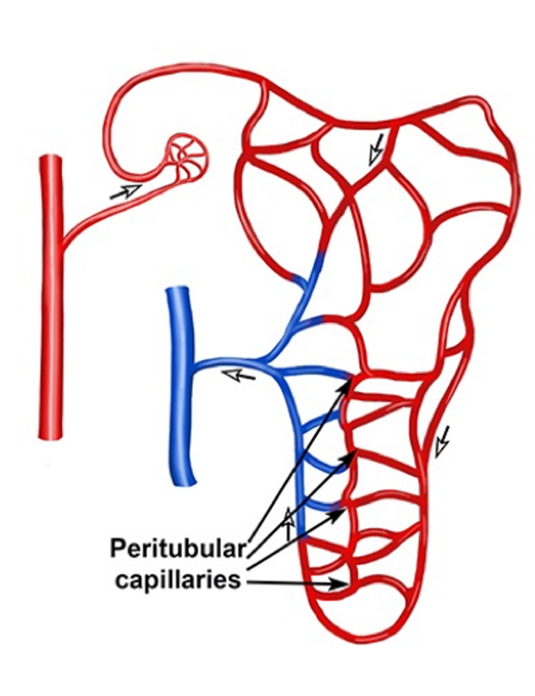
How kidney blood supply works
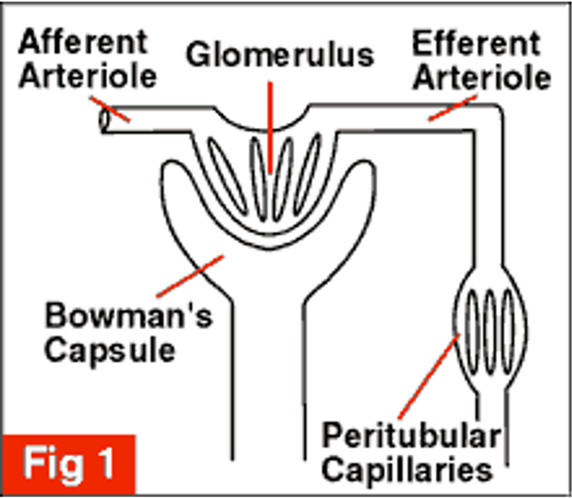
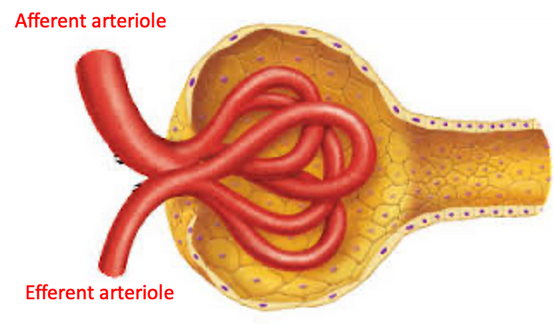
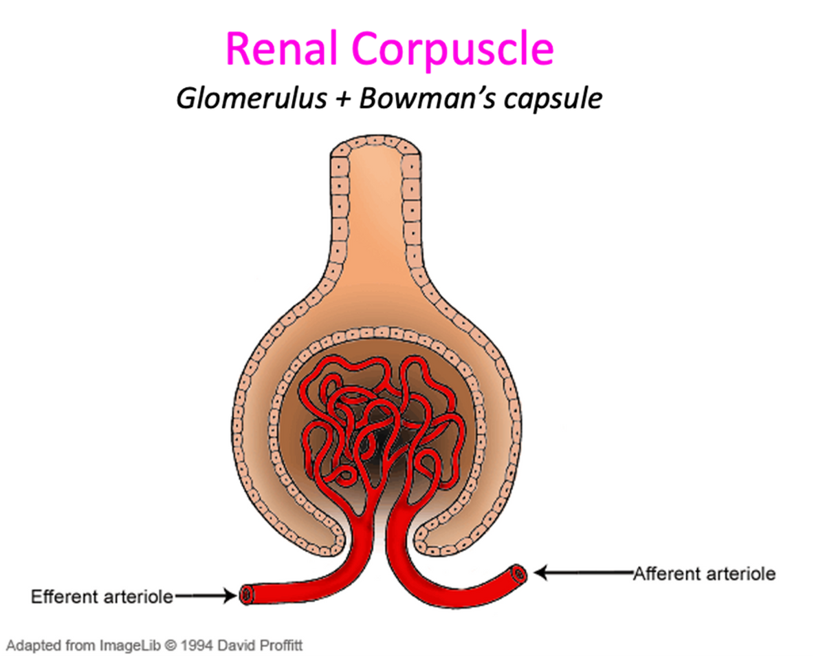
•The afferent arteriole takes blood into the glomerulus.
•The first capillary network is called the glomerulus.
•The efferent arteriole drains blood from the glomerulus and empties into the second capillary network.
•The second capillary network are called the peritubular capillaries.
Glomerulus
•The glomerulus differs from other capillary beds in that blood moves into the capillaries via the afferent arteriole, but then leaves via the efferent arteriole (not a venule).
•The efferent arteriole has a narrower diameter than the afferent arteriole.
•This narrowing effect increases resistance to blood flow, and thus increases the blood pressure in the glomerular capillaries - this is needed for the process of glomerular filtration (we will look into this later).
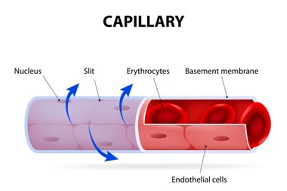
Peritubular capillaries
•These blood vessels cling closely to the renal tubules.
•Empty into nearly venules.
•They have a thin wall and a large surface area.
•As they arise from the efferent arterioles, which have high resistance to blood flow, they have low blood pressure.
•They help filter waste out of the blood, which leaves the body via urine.
Renal corpuscle
•Each renal corpuscle consists of a ball of capillaries called a glomerulus (glom = ball of wool) and a cup-shaped hollow structure called the glomerular capsule (or Bowman’s capsule).
•This is the initial blood-filtering component of the nephron.
Blood flow in nephron (1)
•Via the renal arteries, which branches directly off the abdominal aorta.
•This is very important as it means that the blood entering each kidney is under high blood pressure.
•The renal artery then branches into a series of minor arteries inside each kidney.
Blood flow in nephron (2)
•In the renal cortex, an afferent arteriole supplies blood to the renal corpuscle.
•This branches into a capillary network – the glomerulus.
•The glomerular capillaries eventually unite to form the efferent arteriole.
Blood flow in nephron (3)
•The efferent arteriole, once leaving the renal corpuscle, will now branch again to form a second capillary network called the peritubular capillaries.
•These peritubular capillaries surround the PCT, loop of Henle, DCT and CD.

Blood flow in nephron (4)
•Venous blood drains away from the peritubular capillaries and empties into a venule.
•These smaller veins eventually form larger and larger blood vessels, until the blood empties into the renal vein (which connects to the inferior vena cava).
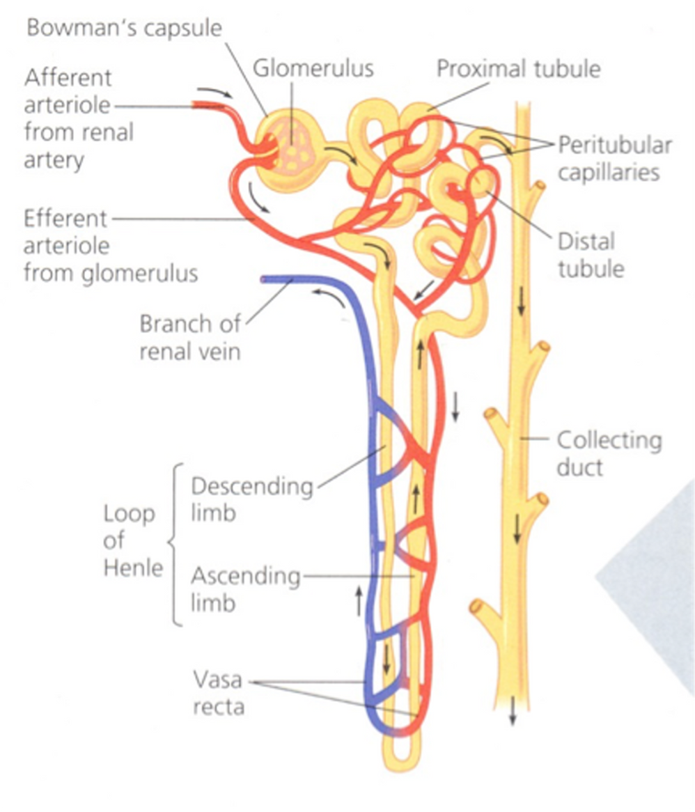
Blood flow in nephron (5)
•Blood flow through the kidney has two unusual features:
•Blood flows through two capillary networks.
•The efferent arteriole has a narrower diameter than the afferent arteriole.