Ch 4- Pt 3: Muscle, Nervous Tissue, Body Membranes, and Tissue Repair
 Note
Note Call Kai
 Learn
Learn Practice Test
 Spaced Repetition
 Match
Match Flashcards
 Knowt Play
1/27

Earn XP
Description and Tags
Vocabulary flashcards identifying tissue types, specialized cell structures, body membrane classifications, and tissue repair stages based on lecture slides.
Name | Mastery | Learn | Test | Matching | Spaced | Call with Kai | Chat |
|---|
No analytics yet
Send a link to your students to track their progress
28 Terms
Myofilaments
Structures in muscle cells made up of actin and myosin proteins that bring about contraction.

Skeletal muscle

Cardiac muscle
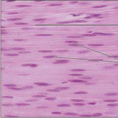
Smooth muscle
Neurons
Specialized branching cells of nervous tissue that transmit electrical signals from sensory receptors to effectors.
Dendrites
Neuron cell processes that respond to stimuli.
Axons
Long cell processes of neurons that transmit electrical signals.
Glial cells (Neuroglia)
Nonexcitable supporting cells in nervous tissue that support, insulate, and protect neurons.
Membranes
Continuous epithelial and connective tissue sheets that cover body surfaces, line internal cavities, and wrap visceral organs
Cutaneous membrane
The skin; a dry membrane made of keratinized stratified squamous epithelium (epidermis) attached to a thick layer of connective tissue (dermis).
Mucous membranes (Mucosae)
Moist membranes that line body cavities open to the exterior, including the hollow organs of the digestive, respiratory, and urogenital tracts.
Lamina propria
The layer of loose connective tissue directly underlying the epithelial sheet in a mucous membrane.
Serous membranes (Serosae)
Moist membranes lining closed ventral body cavities, constructed from mesothelium resting on thin areolar connective tissue.
Mesothelium
The simple squamous epithelium that forms part of a serous membrane.
Parietal serosae
Serous membrane layers that line internal body cavity walls.
Visceral serosae
Serous membrane layers that directly cover internal organs.
Pleurae
Specific serous membranes surrounding the lungs.
Pericardium
Specific serous membrane surrounding the heart.
Peritoneum
Specific serous membranes surrounding organs in the abdominopelvic cavity.
Granulation tissue
Fragile capillary-rich tissue that grows into a wound area during organization to replace the blood clot and restore vascular supply.

Nervous tissue
Three stages of tissue repair
Inflammation sets the stage: Trauma triggers release of inflammatory chemicals, making blood vessels leaky to allow white blood cells, fluid, and clotting proteins to form a clot and scab.
Organization restores the blood supply: Granulation tissue (fragile capillaries) replaces the clot, fibroblasts produce collagen fibers to bridge the gap, and macrophages clean debris.
Regeneration and fibrosis effect permanent repair: The fibrosed area contracts, pulling wound margins together, and surface epithelium thickens and regenerates over underlying scar tissue.
What characterizes muscle cells?
Highly vascularized and are responsible for most types of movement
Skeletal muscle
Description: Highly vascularized muscle tissue, Long, cylindrical, multinucleate cells with obvious striations.
Function: Voluntary movement, locomotion, manipulation of the environment, facial expression, and voluntary control.
Location: Attached to bones or occasionally to skin.
Cardiac muscle
Description: Branching, striated, generally uninucleate cells that connect at specialized junctions (intercalated discs).
Function: Contracts to propel blood into the circulation under involuntary control.
Location: Walls of the heart.
Smooth muscle
Description: Spindle-shaped cells with central nuclei and no striations, arranged closely to form sheets.
Function: Propels substances or objects (foodstuffs, urine, a baby) along internal passageways under involuntary control.
Location: Mostly in the walls of hollow organs.
Nervous tissue
Definition: Main component of the nervous system composed of branching neurons (with cell bodies, dendrites that respond to stimuli, and long axons that transmit signals) and nonexcitable glial cells (neuroglia) that support, insulate, and protect neurons.
Function: Regulates and controls body functions by transmitting electrical signals from sensory receptors to effectors.
Location: Brain, spinal cord, and nerves
Types of muscle tissue
cardiac muscle, smooth muscle, and skeletal muscle