Cell Divisions
1/84
There's no tags or description
Looks like no tags are added yet.
Name | Mastery | Learn | Test | Matching | Spaced |
|---|
No study sessions yet.
85 Terms
Why do cells divide? x4
Growth
To replace cells
To repair tissues
Asexual reproduction
Why is it important to replicate all the genetic material?
For inheritance of genetic traits so that the genetic material is identical and both daughter cells receive a full set of chromosomes.
Why do daughter cells need to be genetically identical to the parent cell?
To enable them to carry out the same functions as the parent cell.
What is the Cell Cycle?
A highly ordered sequence of events that takes place in a cell, resulting in the formation of two genetically identical daughter cells.
In Eukaryotic cells what are the 2 main stages of the cell cycle?
interphase
miotic (division) phase
What is interphase?
When the cell is actively continuing its function while also preparing for cell division. (longest phase)
What occurs during interphase? x5
DNA is replicated and checked for errors in the nucleus
protein synthesis occurs in the cytoplasm
mitochondria grow and divide, increasing in number in the cytoplasm
chloroplasts grow and divide in plant and algal cell cytoplasm, increasing in number
the normal metabolic processes of the cell.
What are the 3 substages of interphase?
G1 - first growth phase
S - synthesis phase
G2 - second growth phase
What occurs in G1?
proteins from which organelles are synthesised are produced and organelles replicate.
the cell increases in size.
What occurs in S?
DNA is replicated in the nucleus.
What occurs in G2?
the cell continues to increase in size, energy stores are increased and the duplicated DNA is checked for errors.
What occurs in mitosis (short answer)?
the nucleus divides
What occurs in cytokinesis (short answer)?
the cytoplasm divides and two cells are produced.
What are the checkpoints in the cell cycle?
the control mechanisms of the cell cycle
they monitor and verify whether the processes at each phase of the cell cycle have been accurately completed before the cello is allowed to progress into the next phase. - therefore ensuring that daughter cells are exact duplicates of parent cells.
What is being checked at the G1 checkpoint?
Where is it located?
What happens if it is passed?
this checkpoint is at the end of the G1 phase, before entry into the S phase. If the cell satisfies the requirements of this checkpoint it is triggered to begin DNA replication. If not it enters the G0 resting state.
- what is being checked:
cell size (needs to be large enough)
contain enough nutrients for DNA synthesis/cell growth
contain enough growth factors
have no DNA damage
What is being checked at the G2 checkpoint?
Where is it located?
What happens if an error is detected?
What happens if it is passed?
this checkpoint is at the end of the G2 phase, before the start of the biotic phase. If this checkpoint is passed, the cell initiates the molecular processes that signal the beginning of mitosis.
cell size
DNA replication/ damage (otherwise daughter cells won’t receive identical genetic information, and proteins may not be made or function properly/correctly.
If an error is detected the cell cycle stops and the cell attempts to repair the damaged DNA. If the damage cannot be repaired the cell will be destroyed by apoptosis - programmed cell death.
What is being checked at the Spindle Assembly checkpoint?
What is it also called?
Where is it located?
What cannot proceed until this checkpoint is passed?
also called metaphase checkpoint, it is at the point in mitosis where all the chromosomes should be attached to spindles and have aligned. Mitosis cannot proceed until this checkpoint is passed. It checks for chromosome attachment to spindles.
What is G0?
The phase when the cell leaves the cycle either temporarily or permanently during G1.
What are the reasons for leaving the cell cycle?
Differentiation- a cell that becomes specialised to carry out a specific function is no longer able to divide- it cannot enter the cell cycle again.
the DNA of the cell may be damaged- no longer viable and cannot divide, enters a period of permanent cell arrest- and becomes senescent.
As you age the number of these cells in your body increases causing age-related diseases like cancer or arthritis.
What is an example of a cell that can be stimulated to return to the cell cycle and start dividing again?
Lymphocytes in an immune response
what are the time periods of the cell cycle
24 hrs
specialised cells generally have a longer cell cycle
eg embryo cells: 8-60 mins; yeast cells: 1.5-3 hours; intestinal epithelial: 12 hours; stomach epithelial: 24 hours
the G1 phase might last about 11 hours, S phase about 8 hours, G2about 4 hours, and M about 1 hour
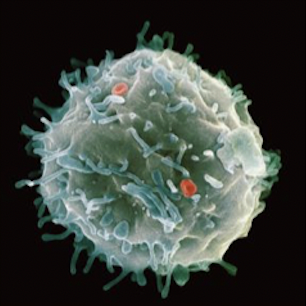
What is a stem cell?
the undifferentiated cell that all cells begin as. They are not adapted to any function (unspecialised) and they have the potential to differentiate to become any one of the specialised cells in an organism.
Stem cells are the source of new cells necessary for growth, development and tissue repair- they can undergo cell division again and again.
Why does the activity of stem cells have to be strictly controlled?
if they do not divide fast enough then tissues are not efficiently replaced, leading to ageing
if there is uncontrolled division then they form masses of cells called tumours- which can lead to the development of cancer.
What happens to stem cells once they become specialised?
they lose their ability to divide
they enter the G0 phase of the cell cycle
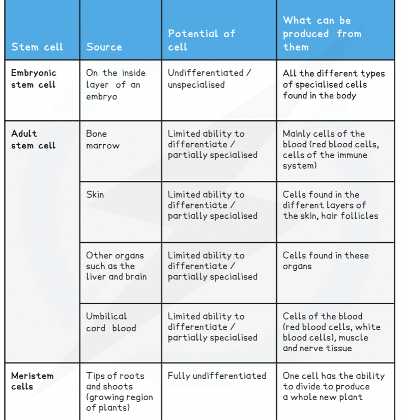
what is a stem cell’s potency?
a stem cell’s ability to differentiate into different cell types
Define Totipotent?
these stem cells can differentiate into any type of cell.
Examples: fertilised egg, zygote, the 8 or 16 cells from its first few miotic divisions are totipotent- they are destined to eventually produce a whole organism. They can also differentiate into extra- embryonic tissues like the amnion and umbilicus.
Define pluripotent
stem cells that can form all tissue types but not whole organisms.
Examples: They are present in early embryos- the origin of the different types of tissue within an organism.
Define multipotent
these stem cells can only form a range of cells within a certain type of tissue.
examples: haematopoetic stem cells in bone marrow- they give rise to the various types oof blood cell.
Where do all blood cells derive from?
stem cells in the bone marrow
Define unipotent?
can only develop into one type of cell
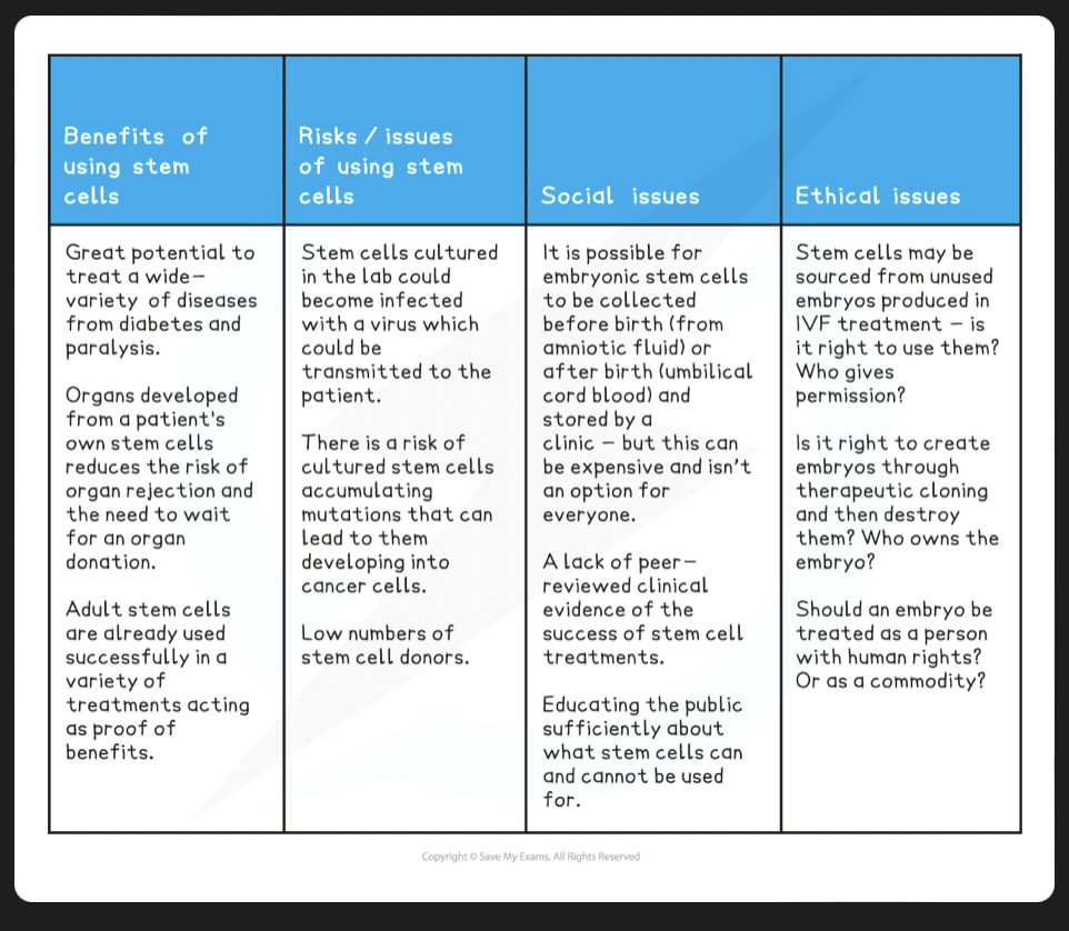
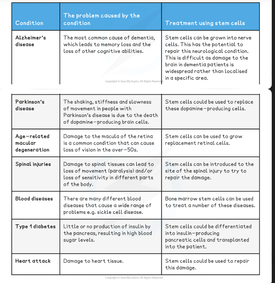
suggest some uses of stem cells
repair of damaged tissue e.g. cardiomyocytes after myocardial infarction
drug testing on artificially grown tissues
treating neurological diseases e.g. Alzheimer’s and Parkinson’s
Researching developmental biology e.g. formation of organs, embryos
Define differentiation
the process by which a cell specialises to carry out a particular function.
Describe the two groups of specialised cells in blood
erythrocytes are red blood cells- they are biconcave, no nucleus, lots of haemoglobin (because of the lack of organelles) to carry oxygen- short lifespan of around 120 days. Die quickly so must be replaced daily in the billions. elastic membrane- can squeeze through narrow capillaries
leucocytes are white blood cells- lymphocytes, eosinophils and neutrophils (live for 6 hours- the figure of new cells produced daily increases during infection) - engulf foreign material - monocytes
Neutrophils are the first white blood cells to arrive at an infection site on or in the body
They exit the blood through the tiny gaps in capillary walls and collect around foreign bodies (e.g. pathogens)
They then destroy these by engulfing them (phagocytosis) and digesting them using their hydrolytic enzymes
how do specialised cells in blood form?
Multipotent adult stem cells in the bone marrow differentiate into:
erythrocytes, which have a short lifespan and cannot undergo mitosis since they have no nucleus - process known as erythropoiesis
leucocytes, including neutrophils
Where are stem cells sourced from in plants?
Xylem vessels and phloem sieve tubes form the transport systems of plants and are found throughout their roots and stems
The xylem and phloem are formed from stem cells that are found in the tissue between them
This tissue is known as the cambium
The cambium is a meristem, which is the term given to any undifferentiated tissue in a plant that has the ability to give rise to new cells
For example, there are also meristems located at the tips of shoots and roots that provide new cells to these growing parts of the plant
In the roots and stems of plants, the stem cells at the inner edge of the cambium differentiate into xylem cells and the stem cells at the outer edge of the cambium differentiate into phloem cells
Cambium cells that differentiate to form the xylem lose their cytoplasm, deposit lignin in their cell walls and lose their end cell walls
Cambium cells that differentiate to form the phloem lose some of their cytoplasm and organelles and develop sieve plates (located at the ends of the cells)
This cell differentiation is stimulated by hormones (the balance of different hormones can determine whether xylem or phloem tissue is produced)
Why is mitosis important?
It ensures that both daughter cells produced when a parent cell divides are genetically identical- this is vital during growth, replacement and repair of tissues and asexual reproduction.
What is mitosis? short answer
the term used to describe the entire process of cell division in eukaryotic cells, it mainly the nuclear division.
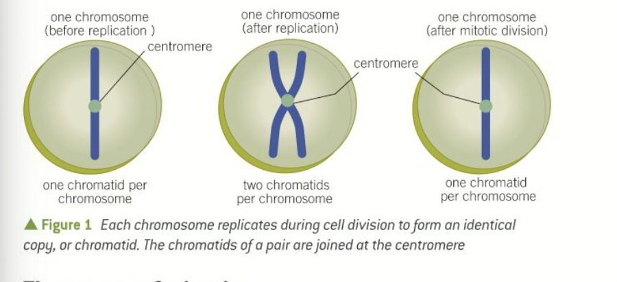
what happens to the chromosomes in interphase before mitosis can begin?
all of the DNA in the nucleus is replicated
each chromosome is converted into two identical DNA molecules, called chromatids
the two chromatids are joined together at a region called the centromere
it is necessary to keep the chromatids together during mitosis so they can be precisely manoeuvred and segregated equally.
what are the four stages of mitosis and what can you remember them by?
I picked many apples today
(interphase)
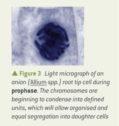
prophase
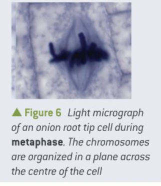
metaphase
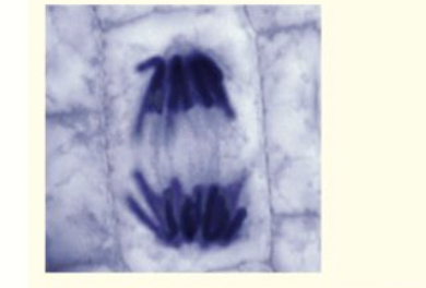
anaphase
telophase
what sort of microscope is needed to observe each phase
light microscope
What occurs during prophase and what does it look like?
chromatin fibres (made of various proteins, RNA, DNA, complex)- begin to coil and condense to form chromosomes that will take up stain to become visible under the light microscope - nucleus disappears - nuclear membrane begins to break down.
protein microtubules form spindle-shaped structures linking the poles of the cell. The fibres forming the spindle are necessary to move the chromosomes into the correct positions before division.
In animal and some plant cells, two centrioles (cylindrical bundles of proteins that help in the formation of the spindle) migrate to opposite poles of the cell.
the spindle fibres attach to specific areas on the centromeres and start to move the chromosomes to the centre of the cell.
By the end of the prophase the nuclear envelope has disappeared.
Describe Martin the metaphase
the chromosomes are moved by the spindle fibres to form a plane in the centre of the cell, called the metaphase plate, and then held in position.
describe anaphase
the centromeres holding together the pairs of chromatids in each chromosome divide during anaphase
the chromatids are separated - pulled to opposite poles of the cell by the shortening spindle fibres
the characteristic v shape of the chromatids moving towards the poles is a result go them being dragged by their centromeres through the liquid cytosol.
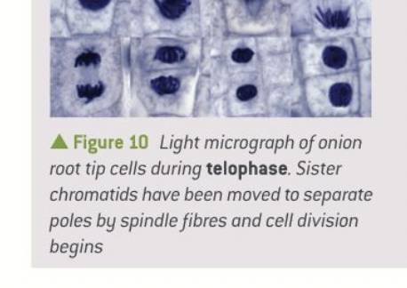
describe telophase
the chromatids have reached the poles and are now called chromosomes again yay!
the two new sets of chromosomes assemble at each pole and the nuclear envelope reforms around them.
the chromosomes start to uncoil and the nucleus is formed
cytokinesis begins….
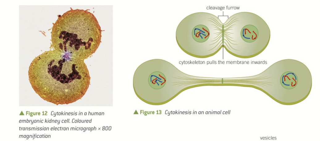
define cytokinesis
the division of the cell into two separate cells
describe cytokinesis in animal cells
a cleavage furrow forms around the middle of the cell
the cell-surface membrane is pulled inwards by the cytoskeleton until it is close enough to fuse around the middle forming two cells
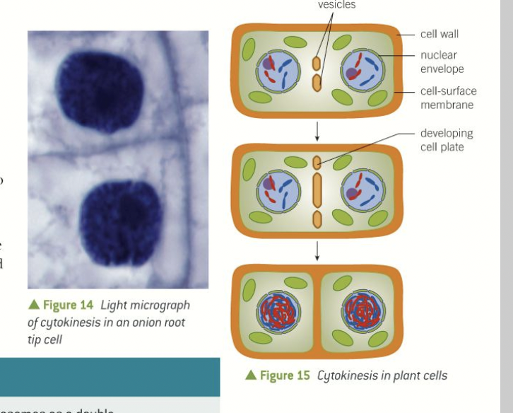
describe cytokinesis in plant cells
have a cell wall so cleavage furrow cannot be formed
vesicles from the Golgi apparatus begin to assemble in the same place as where the metaphase plate was formed
the vesicles fuse with each other and the cell surface membrane- dividing the cell into two
new sections of the cell wall then form alongside the new sections of the membrane.
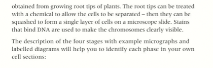
explain why plant root tips are a good source of cells to examine for mitosis
What its a diploid cell
a cell with two chromosomes of each type- one inherited from each parent - mitosis results in two genetically identical diploid daughter cells.
what are sex cells called and what do they fuse to produce?
gametes
fertilised egg (zygote)
how are gametes formed?
another form of cell division known as meiosis
the nucleus divides twice to produce 4 daughter cells which each contain half of the chromosome number of the parent - haploid - known as reduction division
what is a homologous chromosome?
each nucleus contains matching sets of chromosomes - they have the same genes at the same loci
- exactly the same length
- centromere in the same position
- same genes (but maybe different alleles) and same number of genes
- genes arranged in the same linear order
what are alleles?
different versions of the same gene (variants)
why is meiosis important?
in the formation of haploid gametes (sperm and ova) in animals in the production of spores in most flowering plants. produces genetic variation
what is meiosis I ?
separates homologous pairs of chromosomes into two cells.
what is meiosis II ?
separation of sister chromatids (chromatin pairs are not the same) two more cells formed - four haploid daughter cells in total
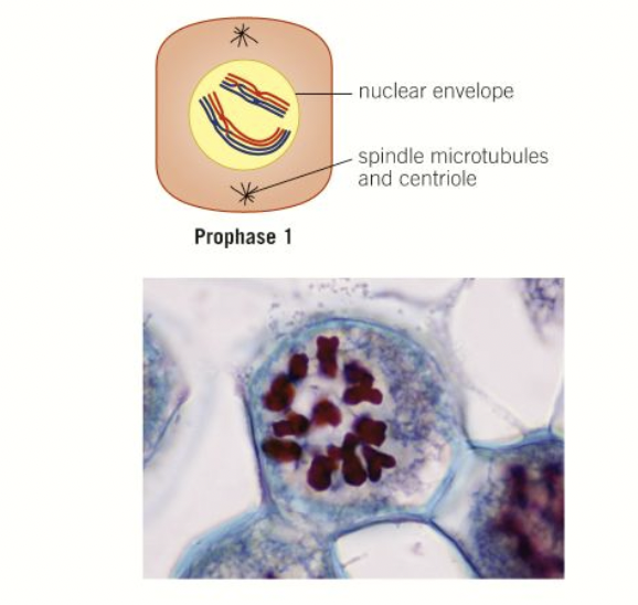
Describe prophase 1
chromosomes condense
nuclear envelope disintegrates
nucleolus disappears
spindle formation begins
homologous chromosomes pair up, forming bivalents
the chromosomes moving through the liquid cytoplasm as they are brought together results in the chromatids entangling- crossing over
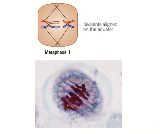
Describe Metaphase 1
homologous pairs of chromosomes assemble along the metaphase plate
orientation of them is random and independent - independent assortment - can result in many different combinations of alleles facing the poles- results in genetic variation.
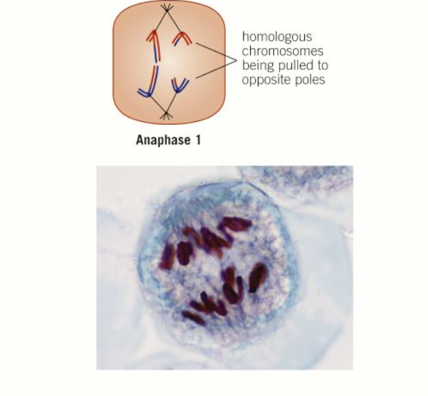
Describe anaphase 1
homologous chromosomes are pulled to the opposite poles and the chromatids stay joined to each other
sections of DNA on sister chromatids which became entangled during crossing over, now break off and rejoin- sometimes resulting in an exchange of DNA- the points at which the chromatids break and rejoin are called chiasmata.
when exchange occurs this forms recombinant chromatids - with genes being exchanged- the new combination of alleles gives rise to genetic variation - sister chromatids no longer identical
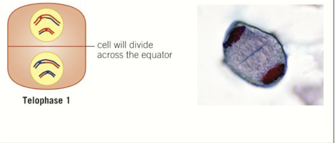
describe telophase 1
the chromosomes assemble at each pole
the nuclear membrane reforms
chromosomes uncoil
cell undergoes cytokinesis and divides into two haploid cells
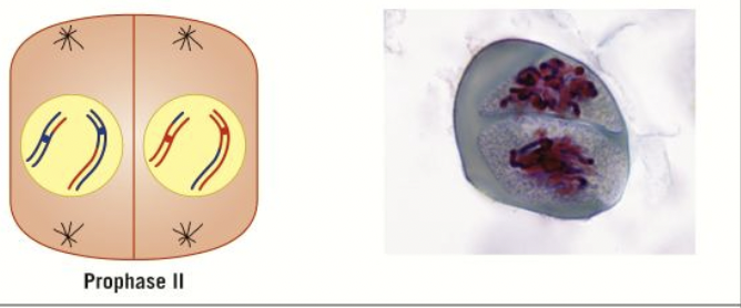
describe prophase 2
the chromosomes- which still consist of 2 chromatids condense and become visible again
nuclear envelope breaks down
spindle formation begins
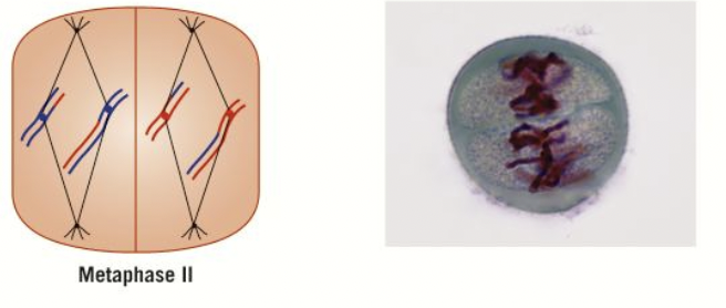
describe metaphase 2
the individual chromosomes assemble on the metaphase plate (like in mitosis)
because of crossing over- chromatids no longer identical - independent assortment, more genetic variation
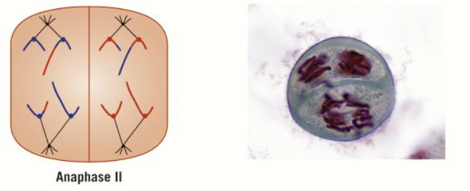
describe anaphase 2
the chromatids of individual chromosomes are pulled to opposite poles- after division of centromeres (same as mitosis)
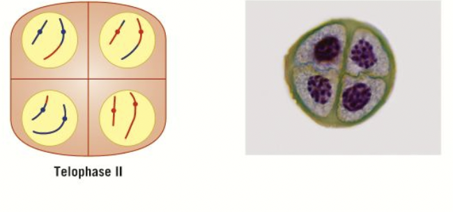
describe telophase 2
chromatids assemble at the poles
chromosomes uncoil and form chromatin again
nuclear envelope reforms
nucleolus becomes visible
cytokinesis results in a division of the cells forming four daughter cells in total
haploid due to reduction division - genetically different from each other and the parent cell.
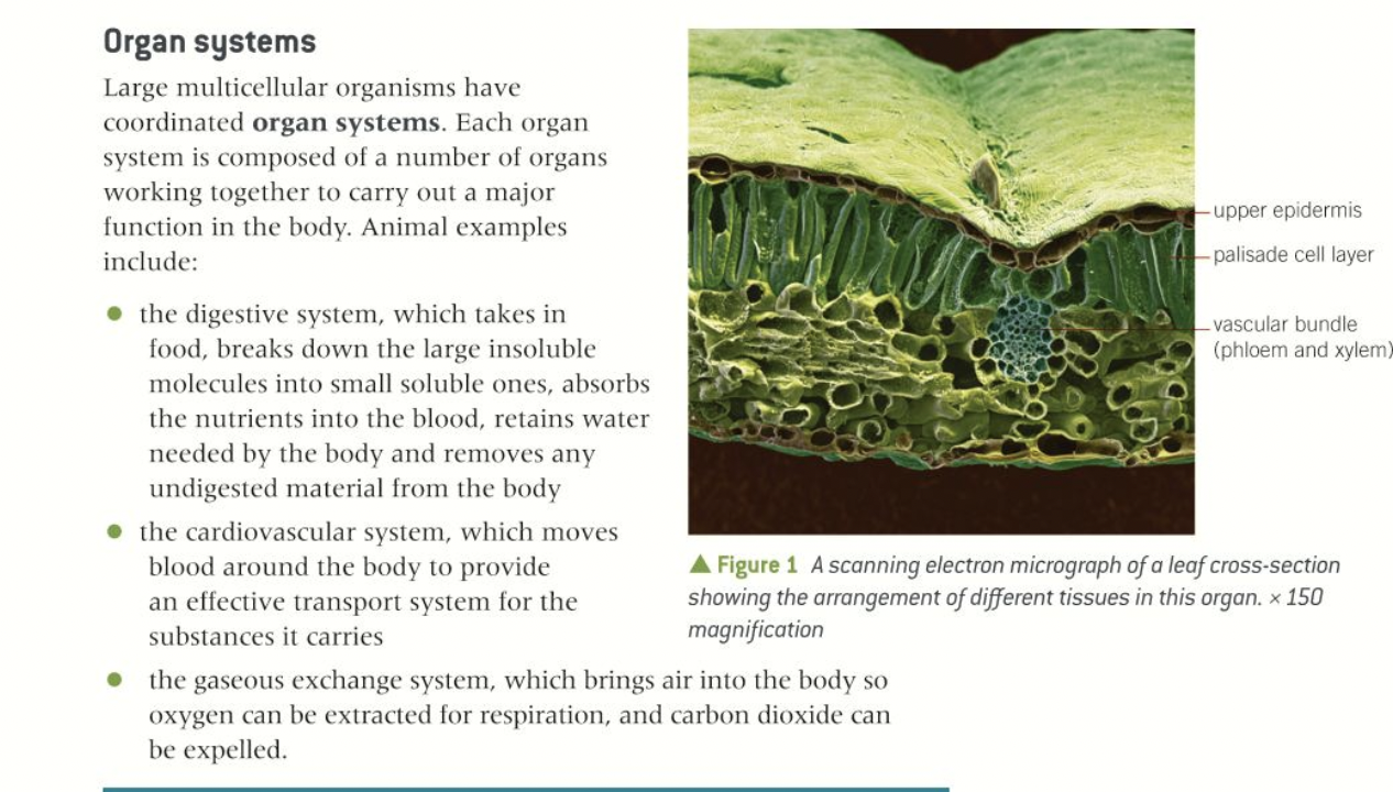
explain the levels of organisation in multicellular organisms

Sperm cells
male gametes
function: deliver genetic information to the female gamete- the ovum or egg
capable of movement
have a flagellum
contain many mitochondria
acrosome- contains digestive enzymes- released to digest the protective layers around the ovum- penetration > fertilisation

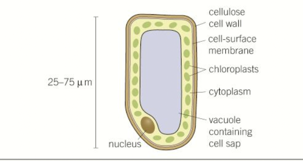
Palisade cells
present in the mesophyll
contain chloroplasts for light absorbency for photosynthesis - can move within cytoplasm
rectangular boxes- closely packed- continuous layer
thin cell walls
large vacuole maintains tugor pressure
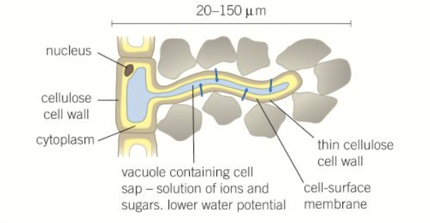
root hair cells
present at surfaces of roots near the growing tips
have long extensions called root hairs- increase sa - maximises water and mineral uptake
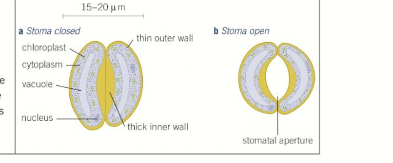
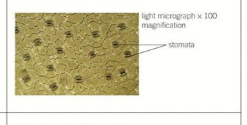
guard cells
pairs on leaf surfaces form small openings called stomata
necessary for co2 to enter
when they lose water and become less swollen as a result of osmotic forces they change shape and the stoma closes to prevent further water loss
cell wall thicker on one side- symmetrically doesn’t change
what is a tissue?
a collection of differentiated cells that have a specialised function (s)
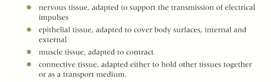
what are the four main categories of animal tissues?
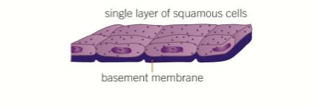
Squamous epithelium
Consists of a single layer of flattened cells on a basement membrane
- layer of cells forms, a thin cross-section which shortens diffusion distance
- permeable so easy diffusion of gases
- provides a surface cover up.
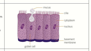
Ciliated epithelium
- moving substances across the surface of a tissue. - has cilia which beat in a coordinated way to shift material
- goblet cells secrete mucus to trap dust, dirt etc
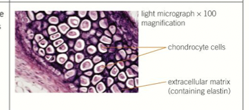
Cartilage
- connective tissue found in outer ear, nose and at ends of bones
- contains fibres of proteins elastin and collagen
- firm and flexible tissue composed go chondrocyte cells in extracellular matrix
- prevents bones from rubbing together and causing damage
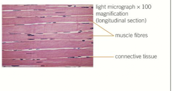
Muscle
- contraction for movement
- layers of protein filaments that can slide over each other and cause muscle contractions
- high density of mitochondria
What are the different plant tissues?
Epidermis
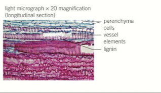
Xylem
- transports water and mineral from the roots
- no top and bottom walls for continuous hollow tubes
- cells are 'dead' so free movement of water
- outer walls thickened by lignin for support
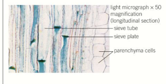
phloem
- transports dissolved sugars and amino acids
- made of living cells supported by companion cells
- cells joined end to end and contain holes in end cell walls to allow translocation
- very few sub-cellular structures to aid the flow of materials
What is an organ
a collection of tissues that are adapted to perform a particular function in an organism
what is an organ system +examples
ciliated epithelial cells structure and function
structure
- contains cilia which beat in a coordinated way to shift material
function
- moving substances across the surface of a tissue (in respiratory tract and fallopian)
squamous epithelial cells structure and function
structure
- thin and flat polygonal cells
- small round nuclei to shorten diffusion pathway
- held in place by basement membrane
function
-provides a surface covering on the alter layer of organs, allowing for rapid selective diffusion
cell division in yeast: budding
- a bud emerges as a lateral bulge in the cell
- nucleus, cytoplasm and organelles move into new bud
- nucleus divides by mitosis in 2
- 1 stays in the parent cell and the other moves to the bud
- once the bud is fully grown, it detaches or remains to form a colony
cell division in bacteria: binary fission
- circular DNA or plasmids are replicated and divided by meiosis (no nucleus so not mitosis)
- cell splits into 2
- each contains a single copy of DNA