Unit 2 Lab Practical - Summer Anatomy 1
1/43
There's no tags or description
Looks like no tags are added yet.
Name | Mastery | Learn | Test | Matching | Spaced |
|---|
No study sessions yet.
44 Terms
Simple squamous epithelium
- Single layer of flat, thin cells.
- Nucleus is disc-shaped and centrally located.
- Appears as a smooth, tile-like sheet under the microscope.
- Found in areas such as blood vessels and body cavities.
- Ideal for diffusion and filtration, and has a nearly transparent appearance.
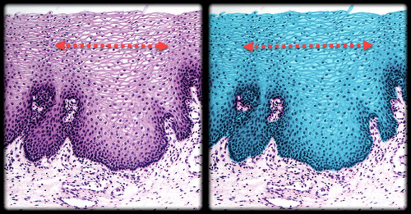
Stratified squamous epithelium
- Multiple layers of cells, with the outermost cells being flat.
- Deeper layers appear more cuboidal or columnar.
- Top layer looks thin and flat, while underlying cells become rounder toward the basement membrane.
- Found in the skin, mouth, and esophagus, offering protection.
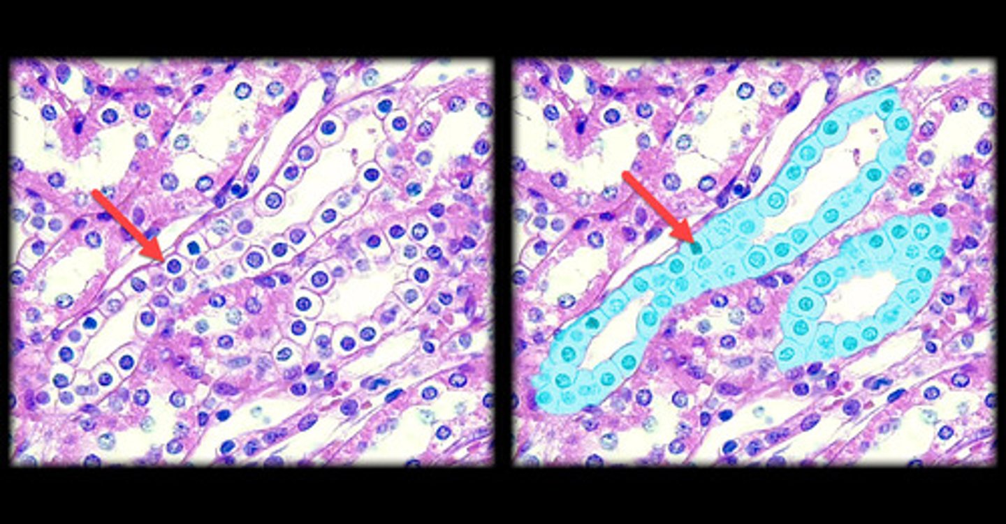
Simple cuboidal epithelium
- Single layer of cube-shaped cells.
- Nuclei are round and centrally located.
- Under the microscope, it appears as a neat row of box-like cells.
- Commonly lines glands and ducts, such as kidney tubules and the thyroid.
Simple columnar - ciliated epithelium
- Single layer of tall, column-like cells.
- Nuclei are oval and located near the basal end.
- Cilia on the surface create a fuzzy appearance under the microscope.
- Found in the respiratory tract and uterine tubes, where cilia help move substances.
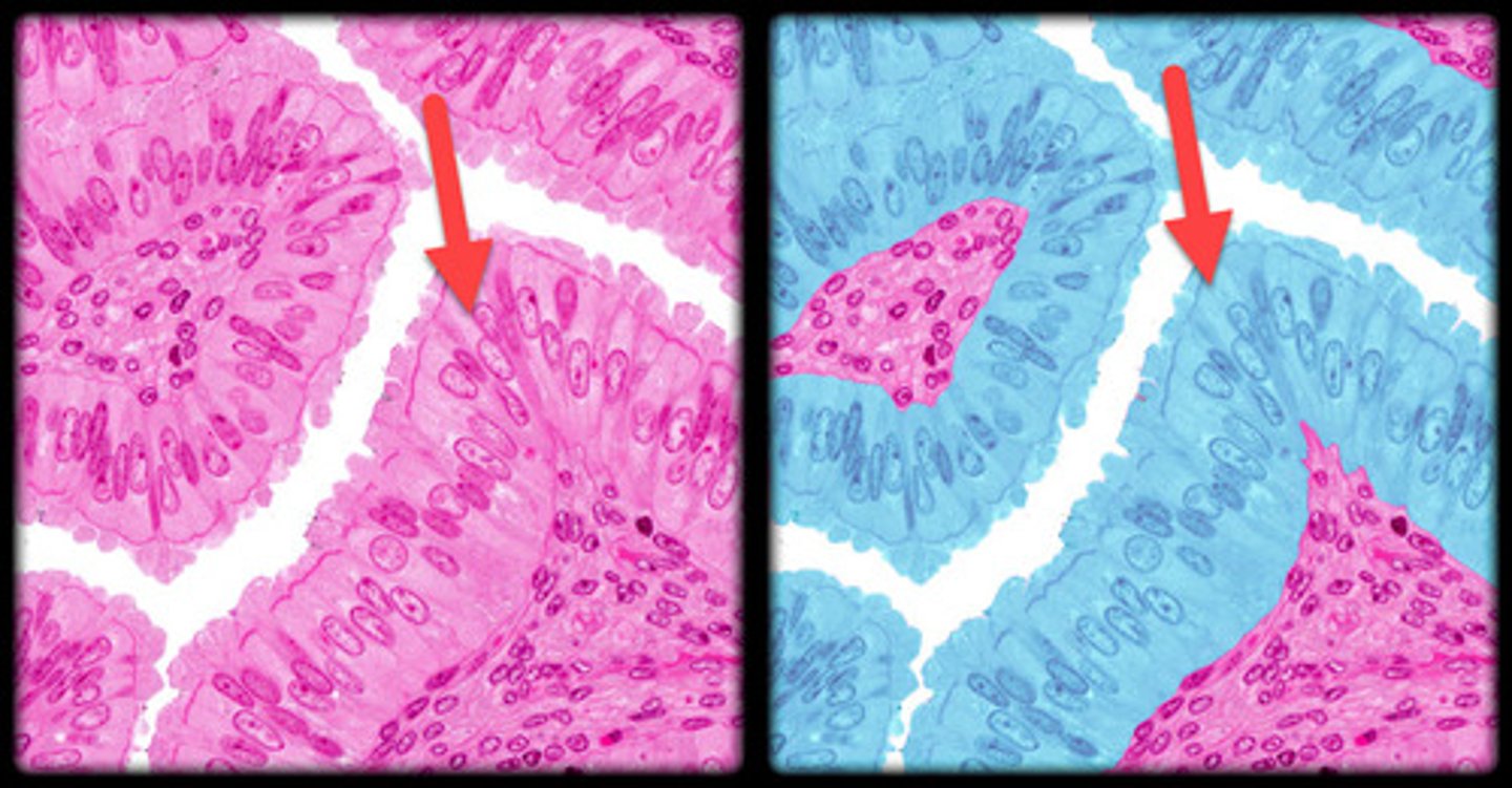
Simple columnar - nonciliated epithelium
- Single layer of tall, columnar cells without cilia.
- Nuclei are oval and aligned at the base of the cells.
- Absence of cilia gives it a smooth appearance under the microscope.
- Commonly found in the digestive tract, such as the stomach and intestines.
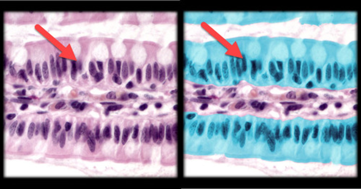
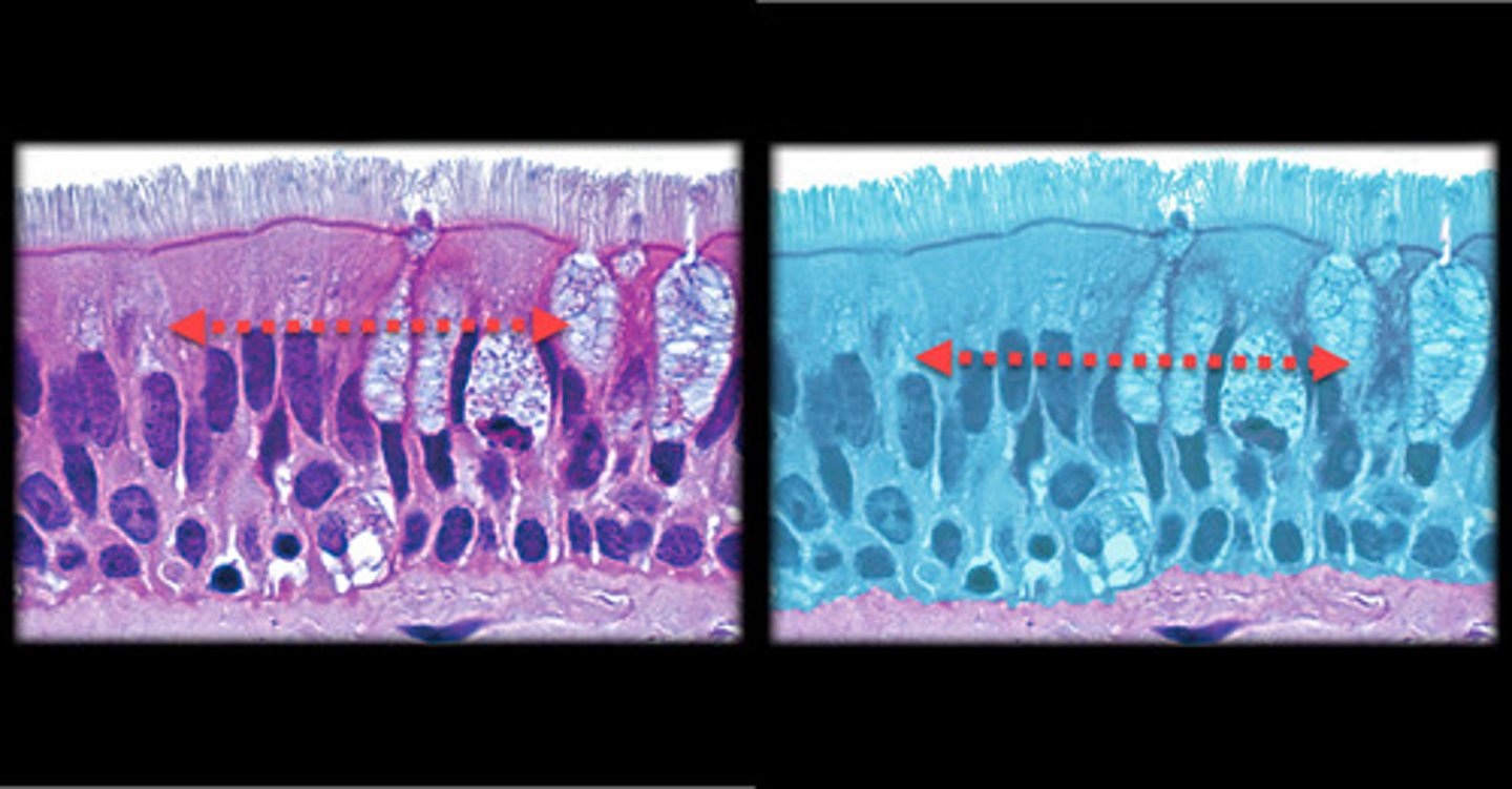
Pseudostratified columnar - ciliated epithelium
- Appears layered due to nuclei at different heights, it is actually a single layer.
- All cells contact the basement membrane.
- Cilia are present on the surface.
- Found in the trachea and upper respiratory tract, where the cilia help move mucus.
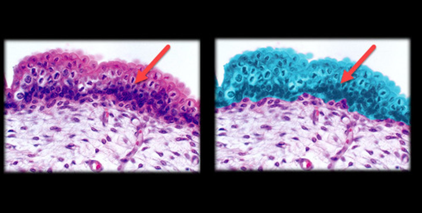
Transitional epithelium
- Cells change shape depending on whether the tissue is stretched or relaxed.
- In the relaxed state, the cells appear large and dome-shaped. When stretched, the cells flatten and resemble stratified squamous epithelium.
- Found in the bladder and urinary tract, specialized for stretching.
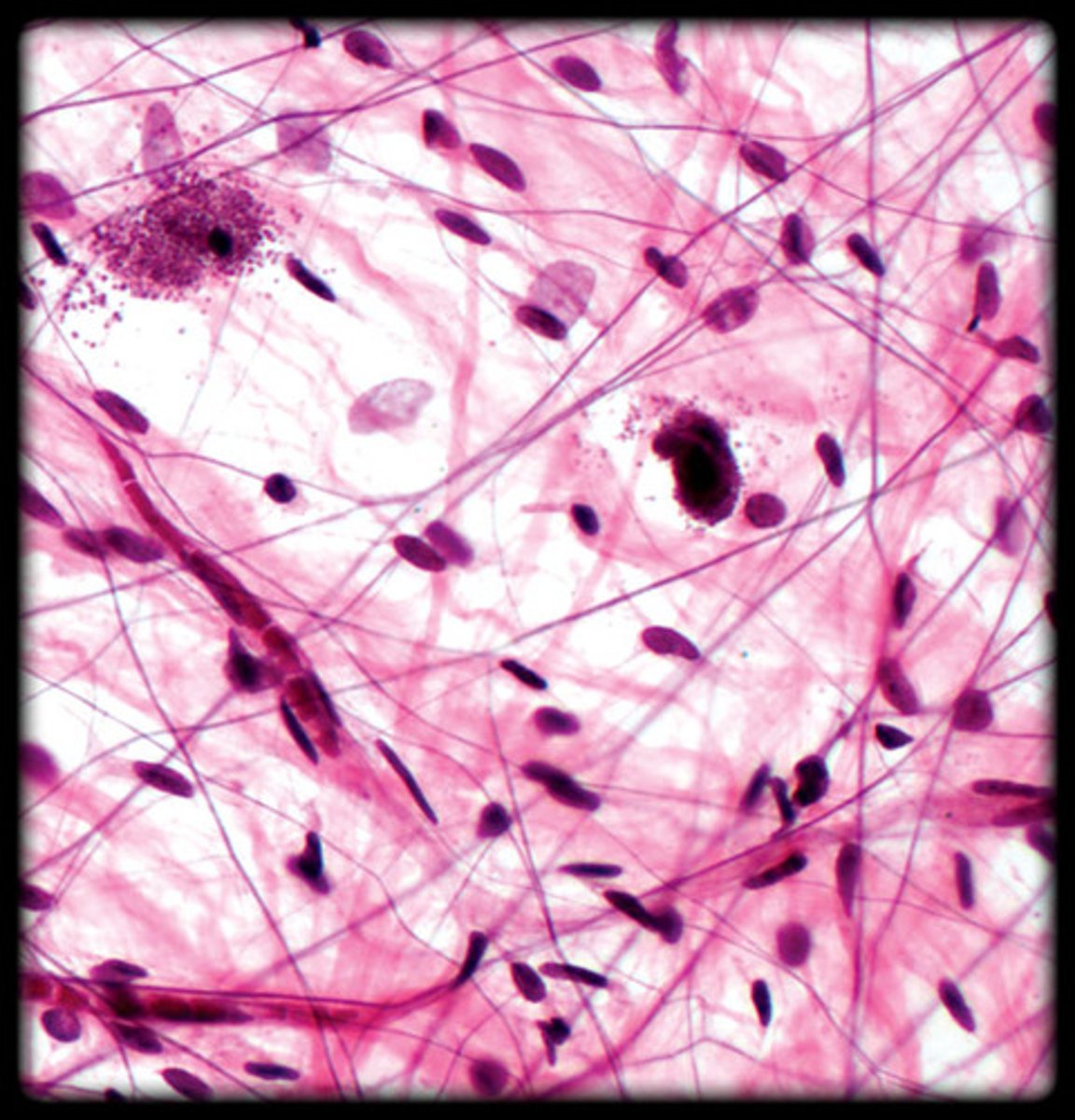
Areolar (connective tissue)
- Loose connective tissue with an open framework.
- Contains a mix of collagen, elastic, and reticular fibers.
- Visible under the microscope as loosely arranged fibers and numerous cells (fibroblasts).
- Found under the skin, around organs, and between muscles.
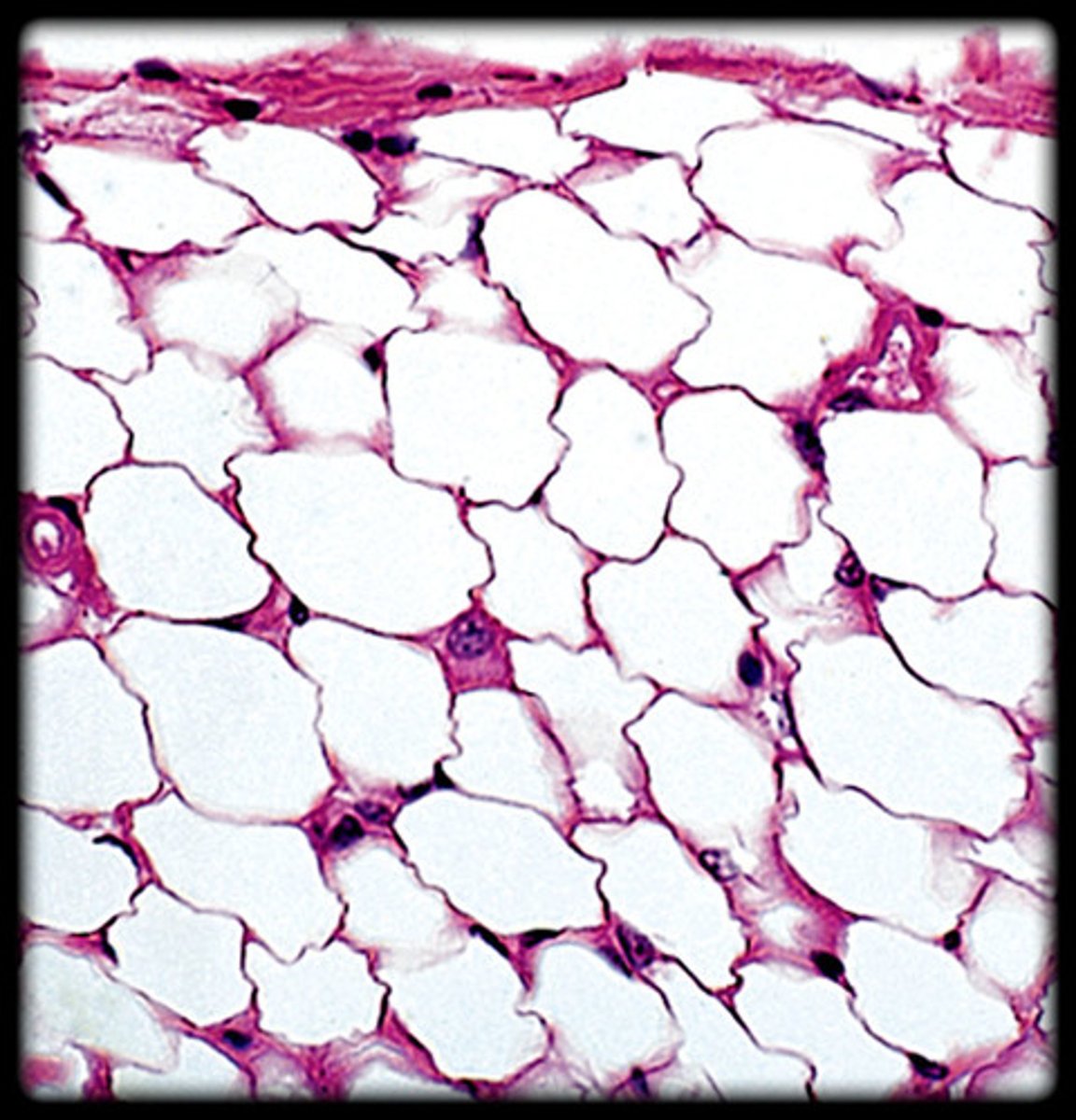
Adipose (connective tissue)
- Fat-storing tissue.
- Large, empty-looking cells with nuclei pushed to the edge. - - Large, circular cells with clear centers.
- Found under the skin, around organs, and in bone marrow.
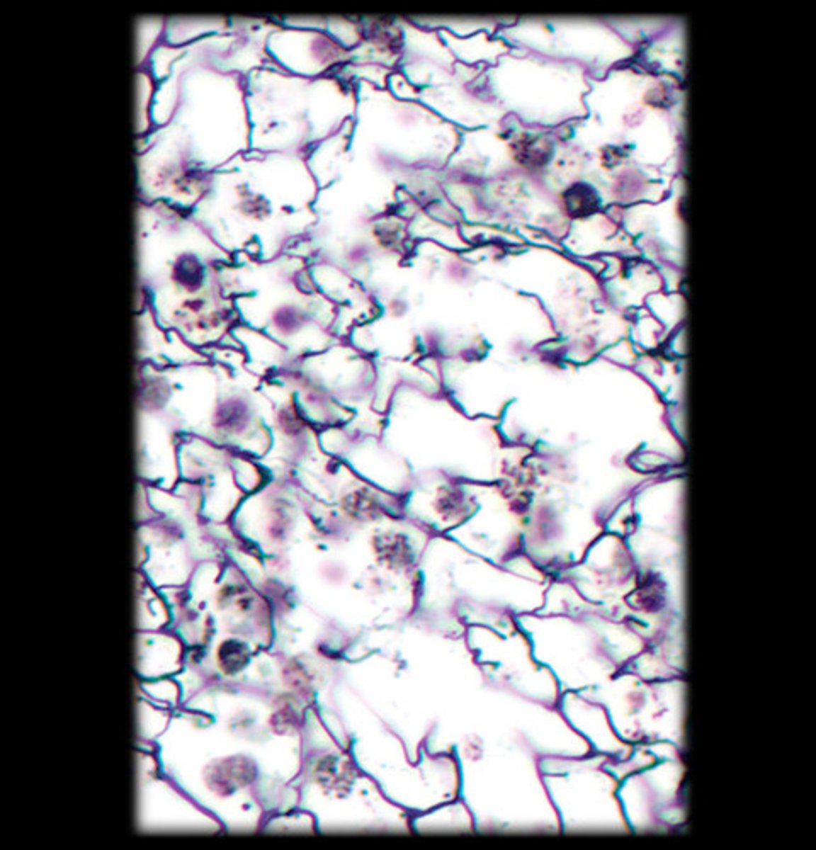
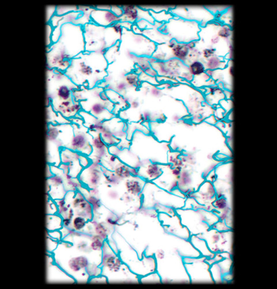
Reticular (connective tissue)
- Network of thin, branching reticular fibers.
- Appears as a dark, web-like network of fibers under the microscope.
- Forms the supporting framework of organs like the liver, spleen, and lymph nodes.
Collagen fibers (of reticular tissue)
- Thick, strong fibers that provide tensile strength.
- Appear as wavy, pink or reddish strands in stained tissue sections.
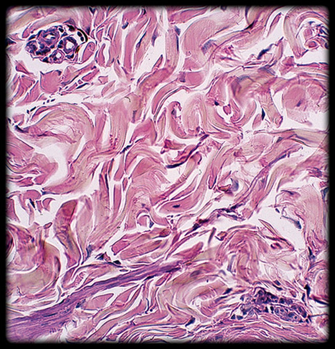
Dense irregular (connective tissue)
- Thick bundles of collagen fibers arranged in random directions.
- Provides strength in multiple directions.
- Appears as densely packed, wavy collagen fibers under the microscope.
- Found in the dermis of the skin and fibrous capsules of organs.
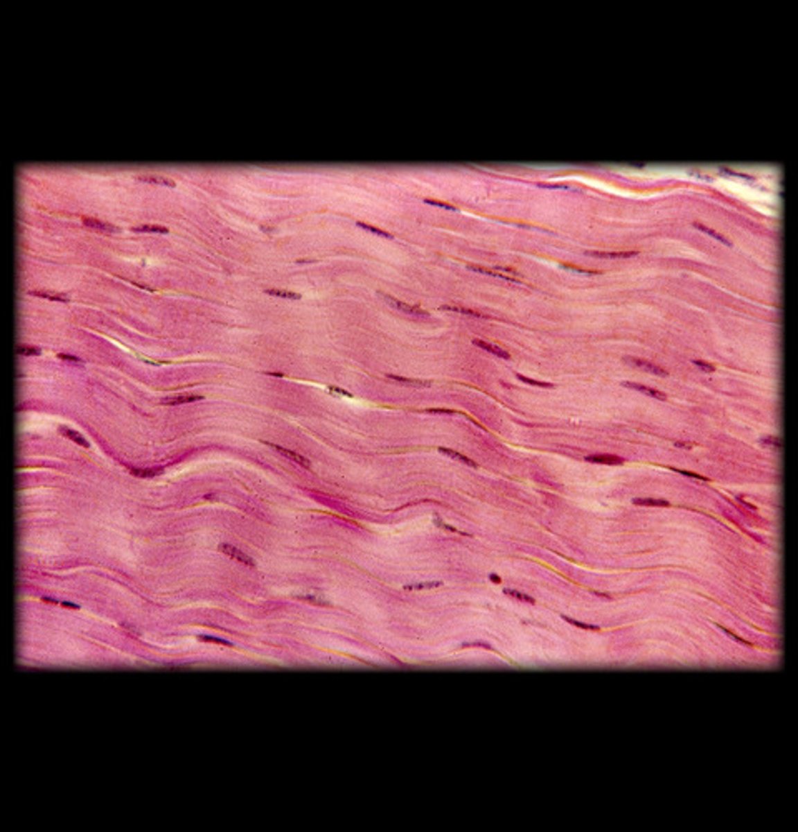
Dense regular (connective tissue)
- Parallel bundles of collagen fibers.
- Provides tensile strength in one direction.
- Appears as neat, parallel waves of collagen fibers.
- Found in tendons and ligaments.
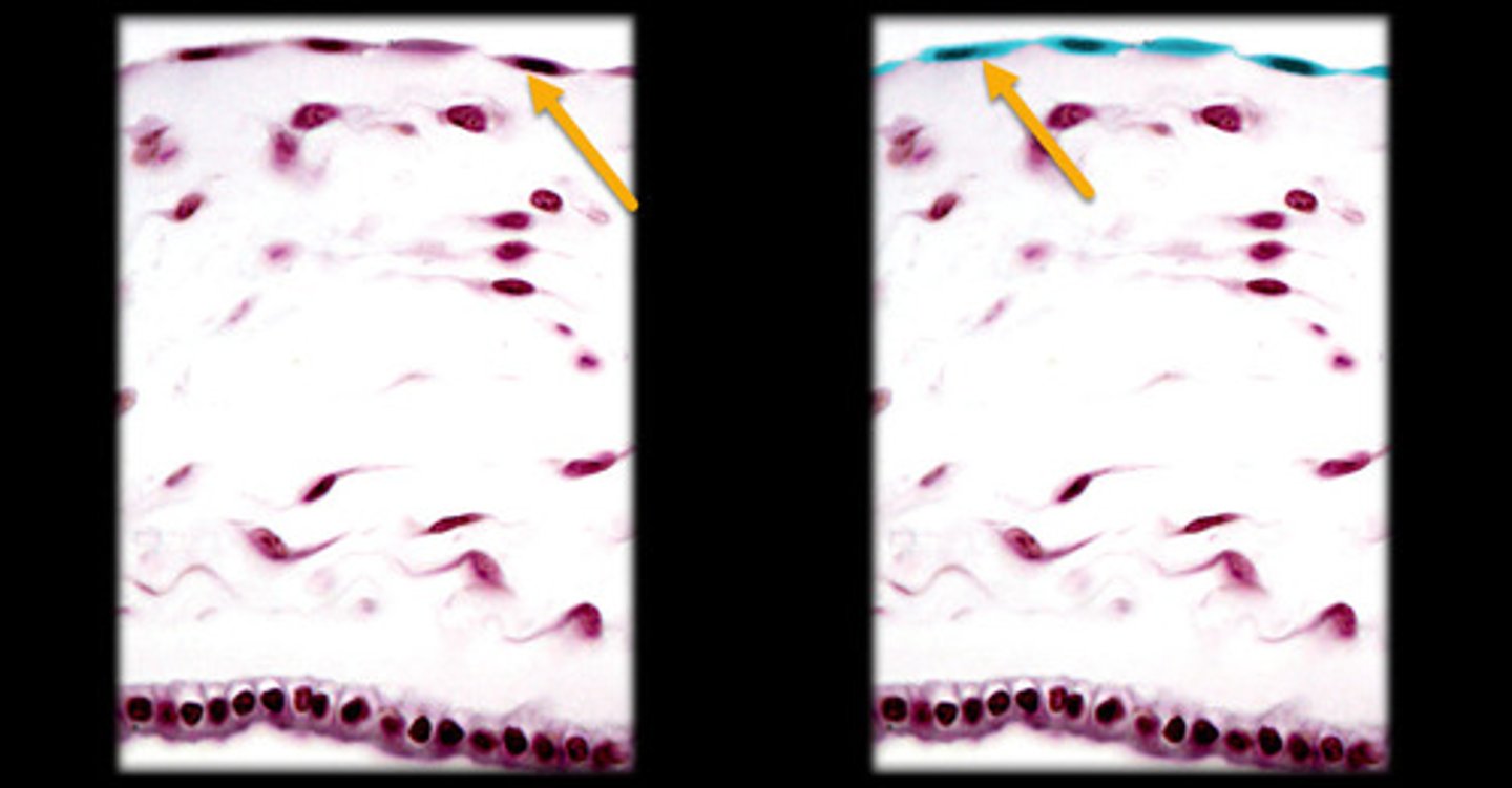
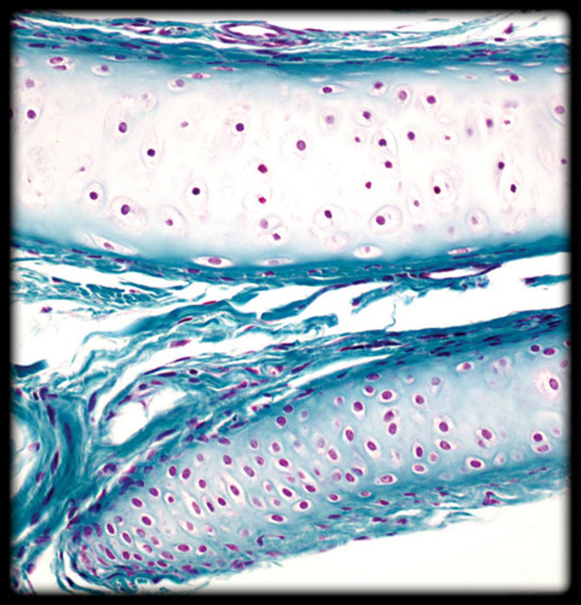
Hyaline cartilage
- Glassy, smooth appearance due to the fine collagen fibers. - - Chondrocytes (cartilage cells) are visible in lacunae (small spaces).
- Appears as a smooth, clear matrix with scattered cells under the microscope.
- Found in the nose, trachea, and at the ends of long bones.
Elastic cartilage
- Dense network of elastic fibers.
- Chondrocytes in lacunae are visible, but the matrix is more flexible than hyaline cartilage.
- Appears as dark, thread-like elastic fibers surrounding the chondrocytes.
- Found in the ear and epiglottis.
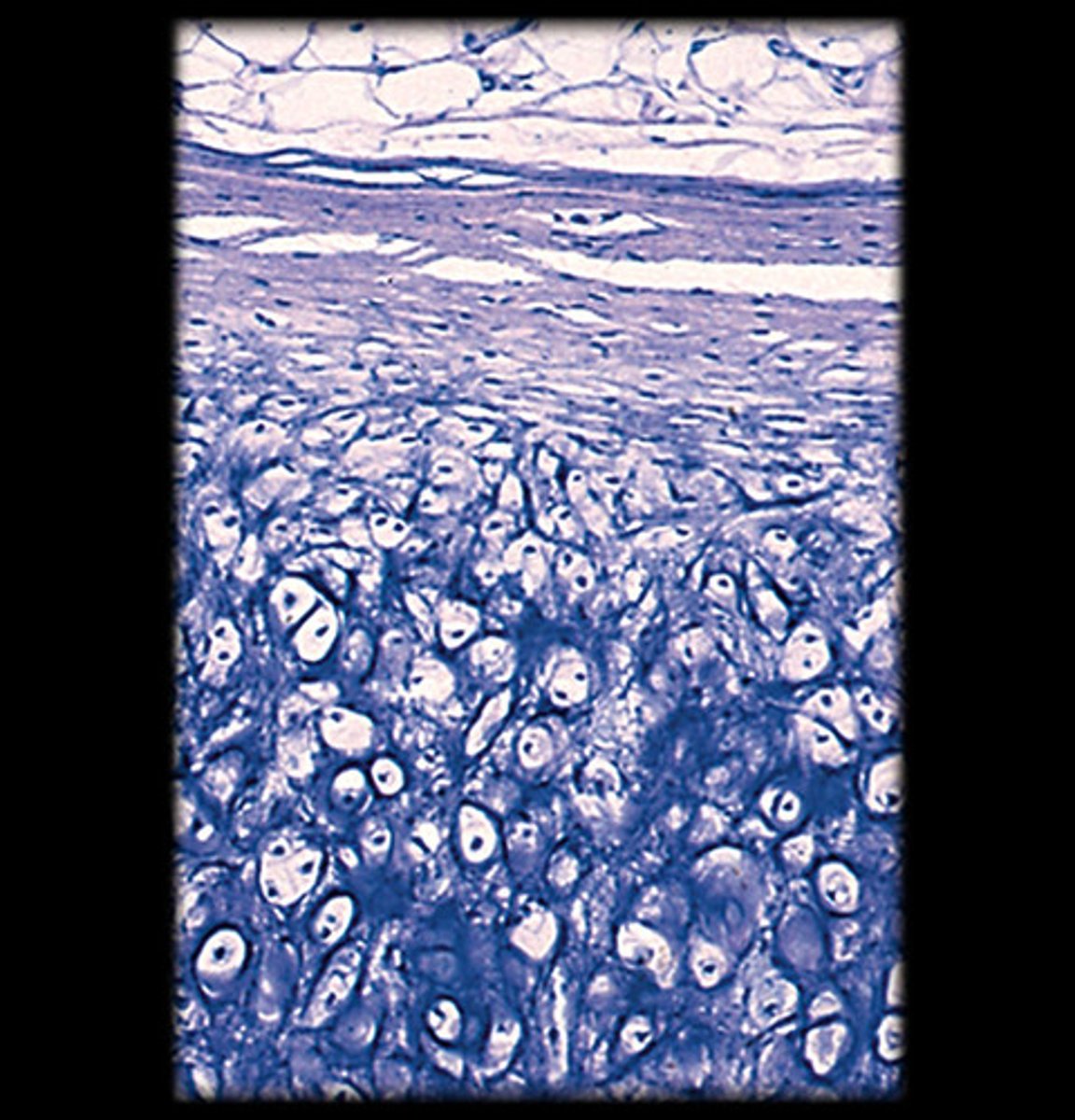
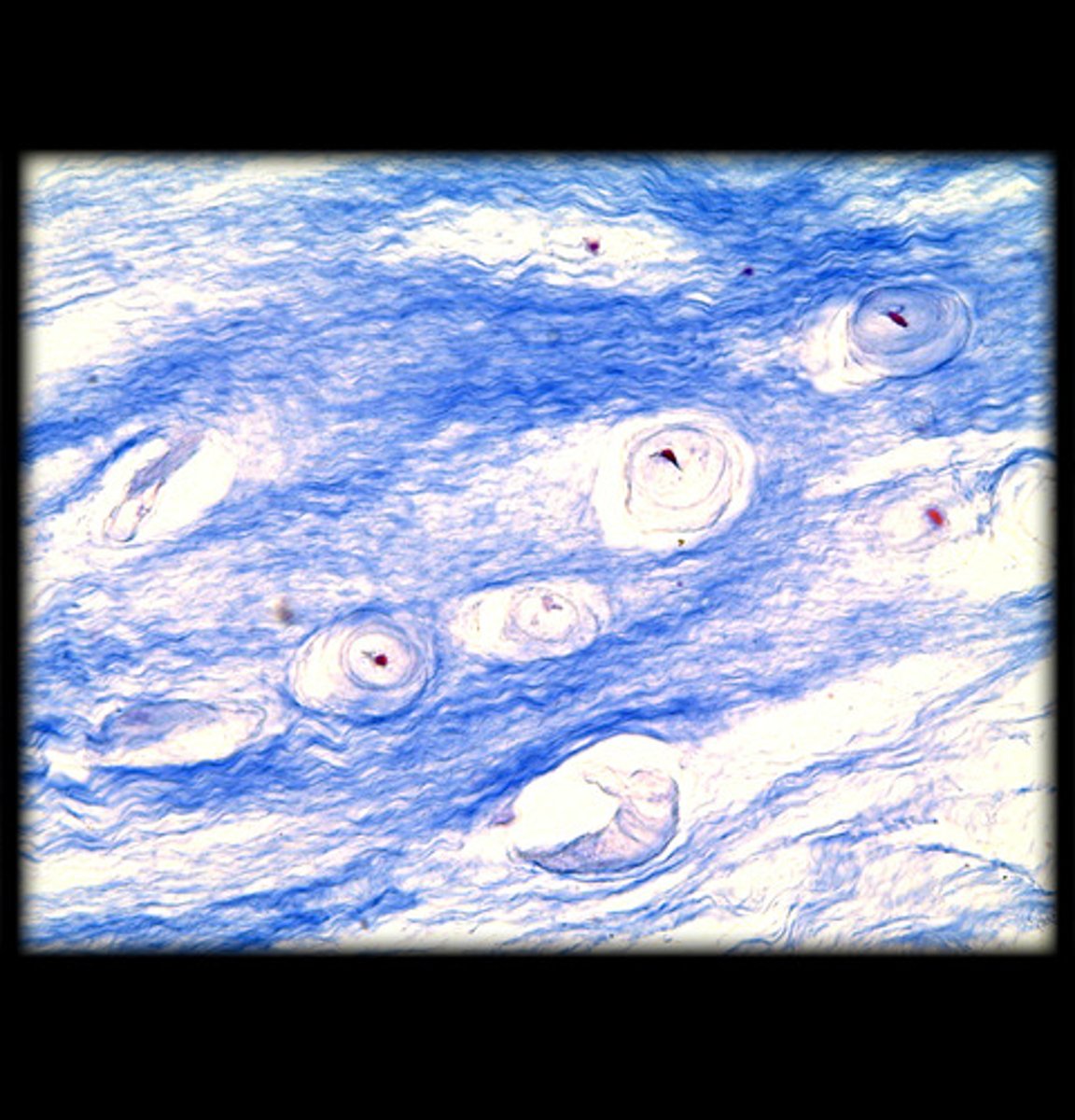
Fibrocartilage
- Thick, dense bundles of collagen fibers in the matrix. Chondrocytes are visible but surrounded by a more fibrous matrix.
- Appears as wavy, densely packed collagen fibers, often arranged in rows.
- Found in intervertebral discs and pubic symphysis.
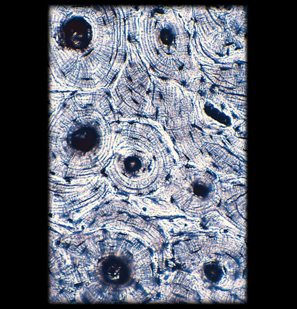
Compact bone
- Dense, solid matrix with a layered (lamellar) structure.
- Osteons (cylindrical structures) are visible with a central canal surrounded by concentric lamellae.
- Appears as tree-ring-like circular structures (osteons) under the microscope with central canals and radiating lines (canaliculi).
- Found in the outer layer of bones.
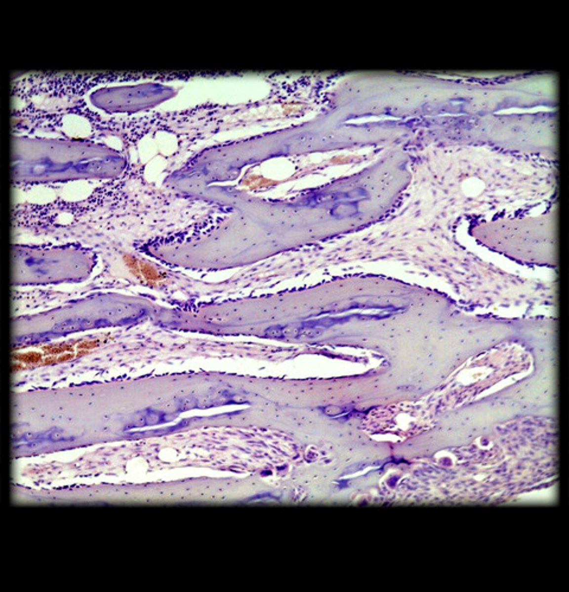
Spongy bone
- Thin, branching plates of bone forming a lattice-like structure.
- Bone marrow is often visible between the trabeculae.
- Appears as a network of thin, bony plates under the microscope.
- Found in the ends of long bones and inside flat bones.
Blood
- Fluid connective tissue with various cell types suspended in plasma.
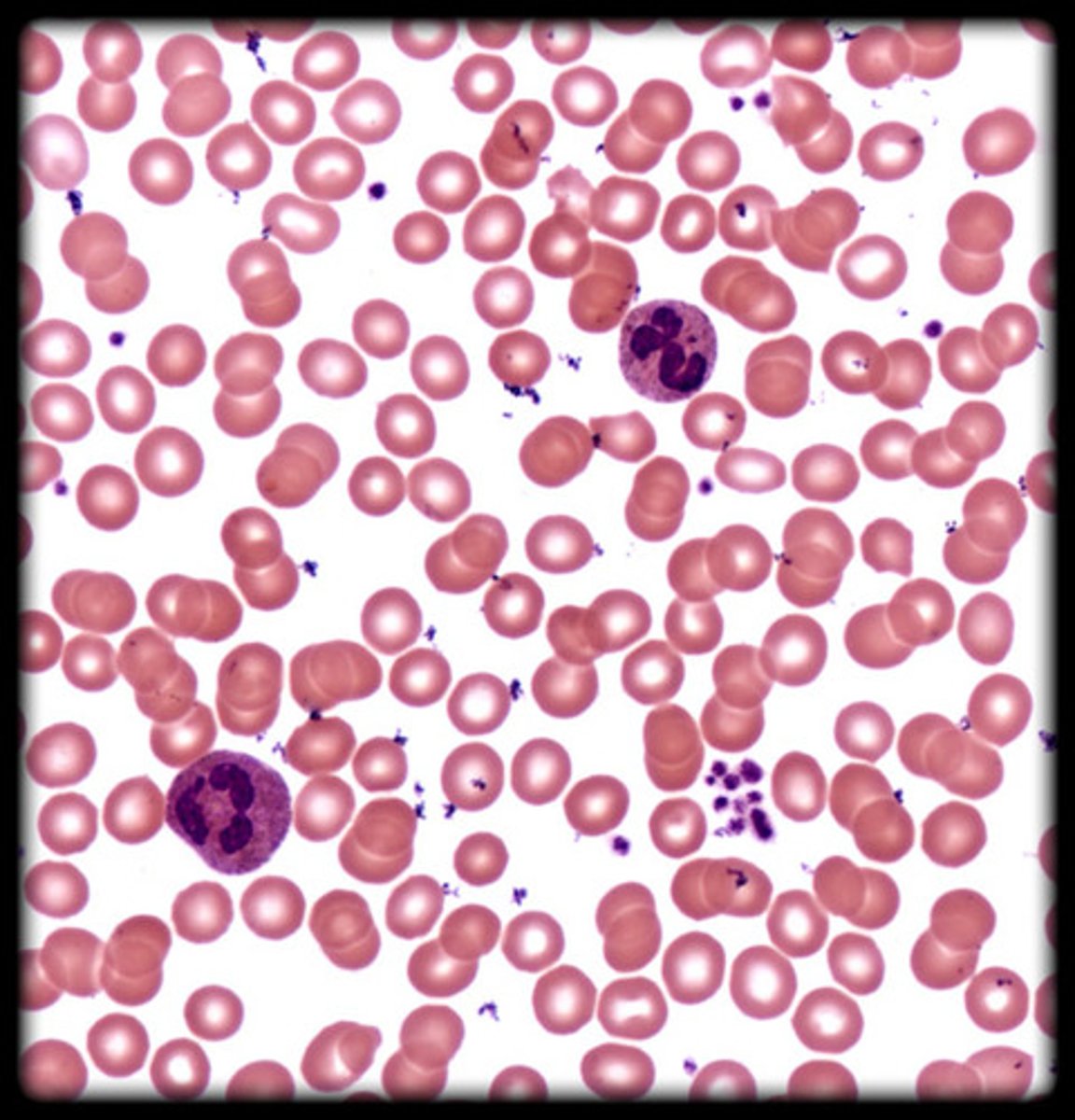
Erythrocyte (red blood cell)
- Small, biconcave discs with no nucleus.
- Appear as round, pale pink cells under the microscope.
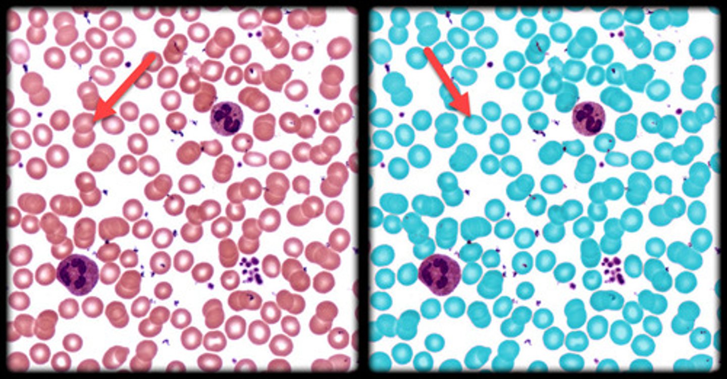
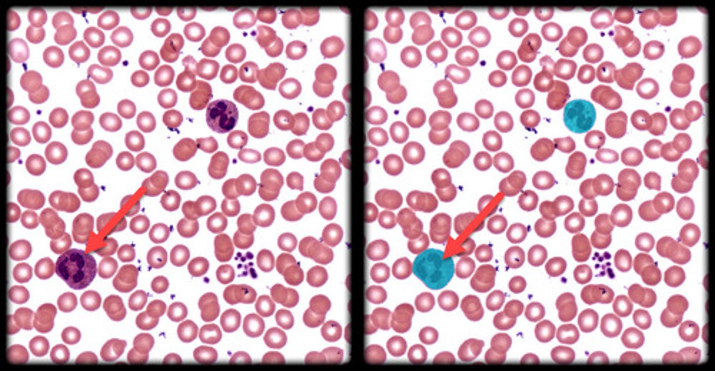
Leukocyte (white blood cell)
- Larger, irregularly shaped cells with a visible nucleus.
- Appear darker and larger than erythrocytes, often stained purple or blue.
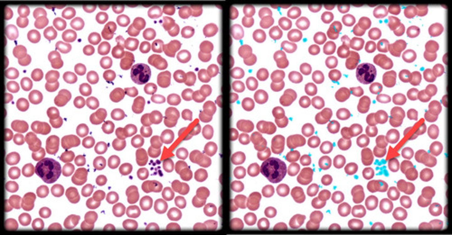
Platelet
- Tiny, cell fragments involved in blood clotting.
- Appear as small, dark dots scattered among red and white blood cells.
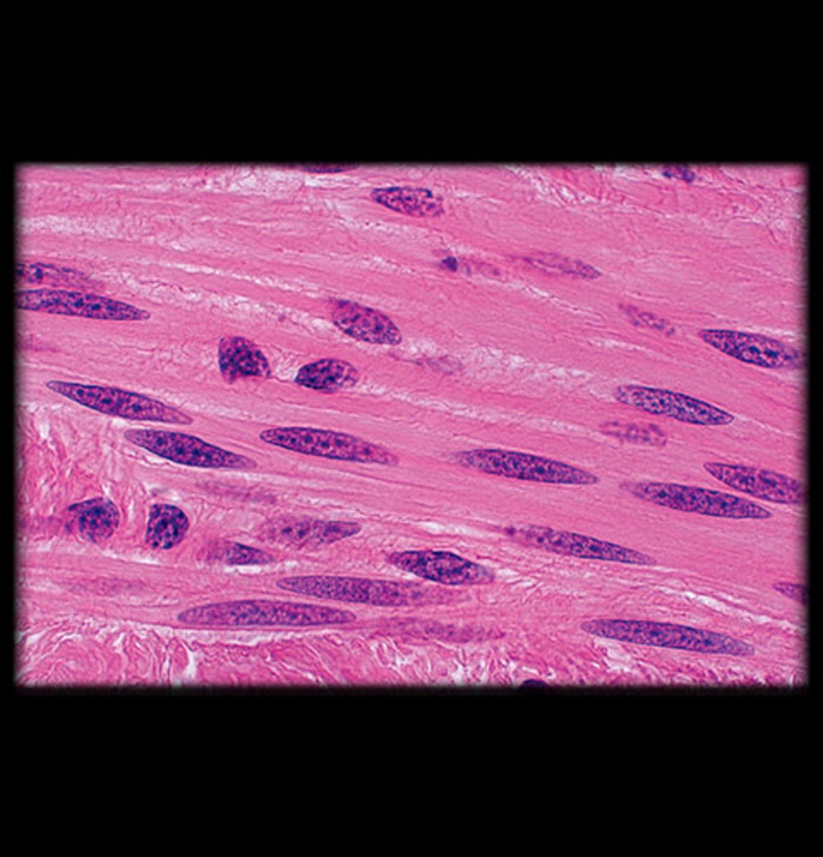
Smooth muscle
- Non-striated, involuntary muscle.
- Cells are spindle-shaped (long and tapered) with a single, centrally located nucleus.
- Appears as smooth, uniform layers of elongated cells under the microscope with no visible striations.
- Found in the walls of hollow organs like the intestines, blood vessels, and uterus.
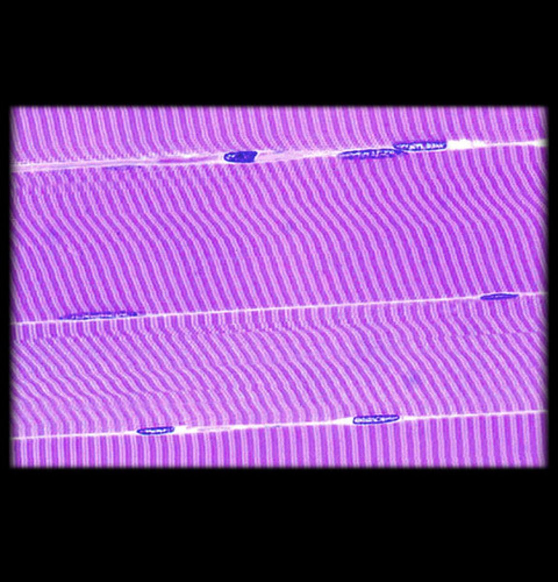
Skeletal muscle
- Striated, voluntary muscle.
- Long, cylindrical cells with multiple peripheral nuclei.
- Visible striations (alternating light and dark bands) due to the arrangement of actin and myosin filaments.
- Appears as parallel bundles of long fibers with clear striations under the microscope.
- Found attached to bones for voluntary movement.
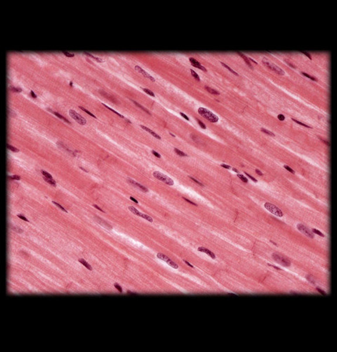
Cardiac muscle
- Striated, involuntary muscle with branching fibers.
- Cells have a single, centrally located nucleus and are connected by intercalated discs (visible as dark lines).
- Appears with striations, but fibers are branched and connected at intercalated discs, giving a "net-like" appearance under the microscope.
- Found in the heart.
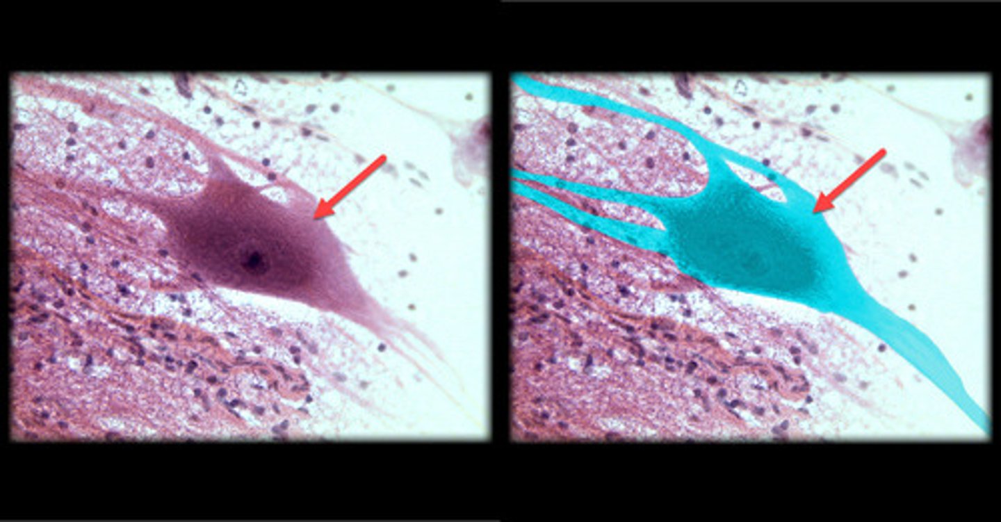
Neuron
- Large, star-shaped cells with multiple processes (dendrites) radiating from the cell body and a single, long axon.
- The cell body (soma) contains a prominent nucleus and a nucleolus.
- Appear with a clearly defined soma, multiple branching dendrites, and a single axon extending away from the cell body.
- Found in the brain, spinal cord, and peripheral nerves, these are the most common type of neurons in the nervous system.
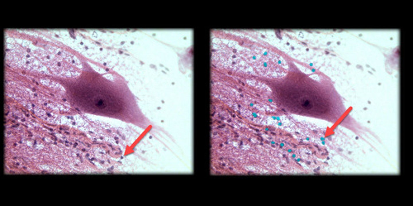
Neuroglial cells
- Smaller, more numerous cells that provide support, protection, and nourishment for neurons.
- Appear as small, round or oval-shaped cells scattered around the larger neurons, often with a more uniform or darkly stained nucleus.
- Can vary in shape depending on the specific type (e.g., astrocytes, oligodendrocytes, microglia), but they are typically much smaller than neurons.
- Found throughout the central and peripheral nervous system, often near or surrounding neurons.
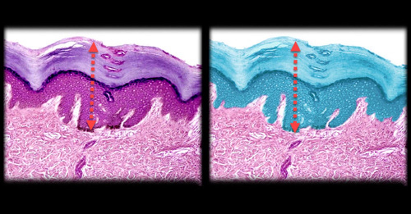
Epidermis
- Outermost layer of skin composed of tightly packed epithelial cells.
- Appears as a thin, multi-layered structure with the outermost cells being flat and dead, gradually becoming more cuboidal or columnar toward the deeper layers.
- Often looks like a clear boundary separating the outer environment from the underlying dermis.
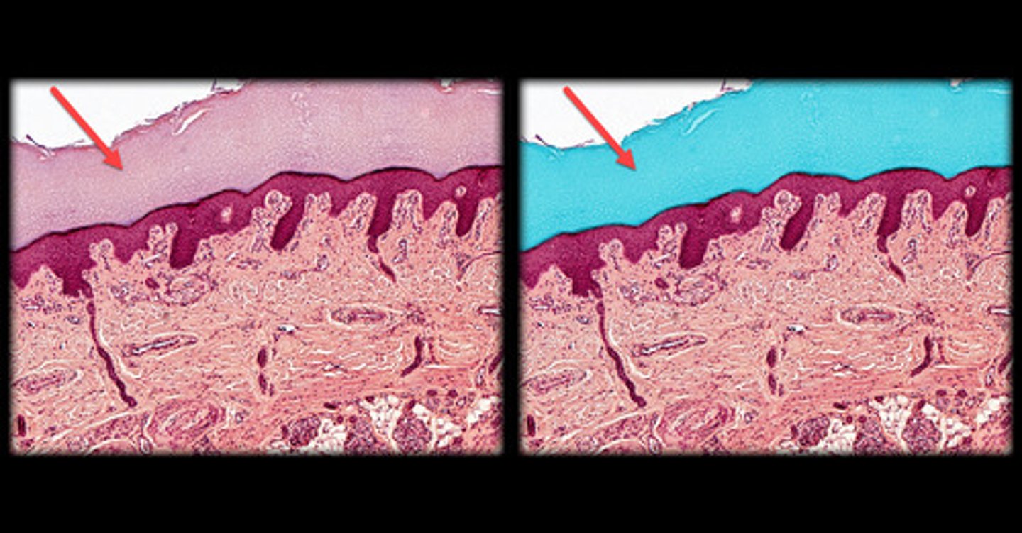
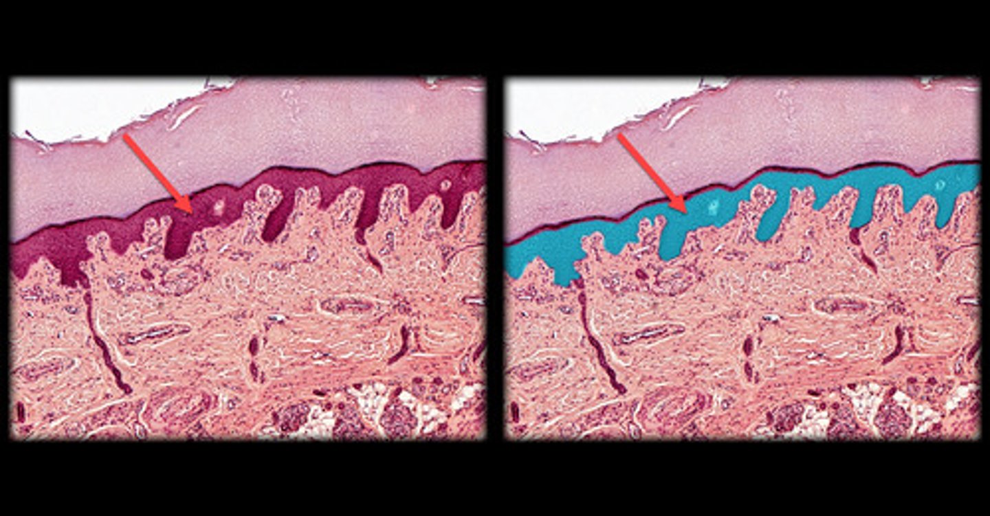
Stratum corneum (Epidermis)
- Outermost layer of the epidermis.
- Multiple layers of flat, dead keratinized cells.
- Appears as a thick, dense layer of flattened, anucleate cells (no visible nuclei) under the microscope, often peeling off or sloughing at the surface.
- Found in both thick and thin skin, but much thicker in areas like the palms and soles.
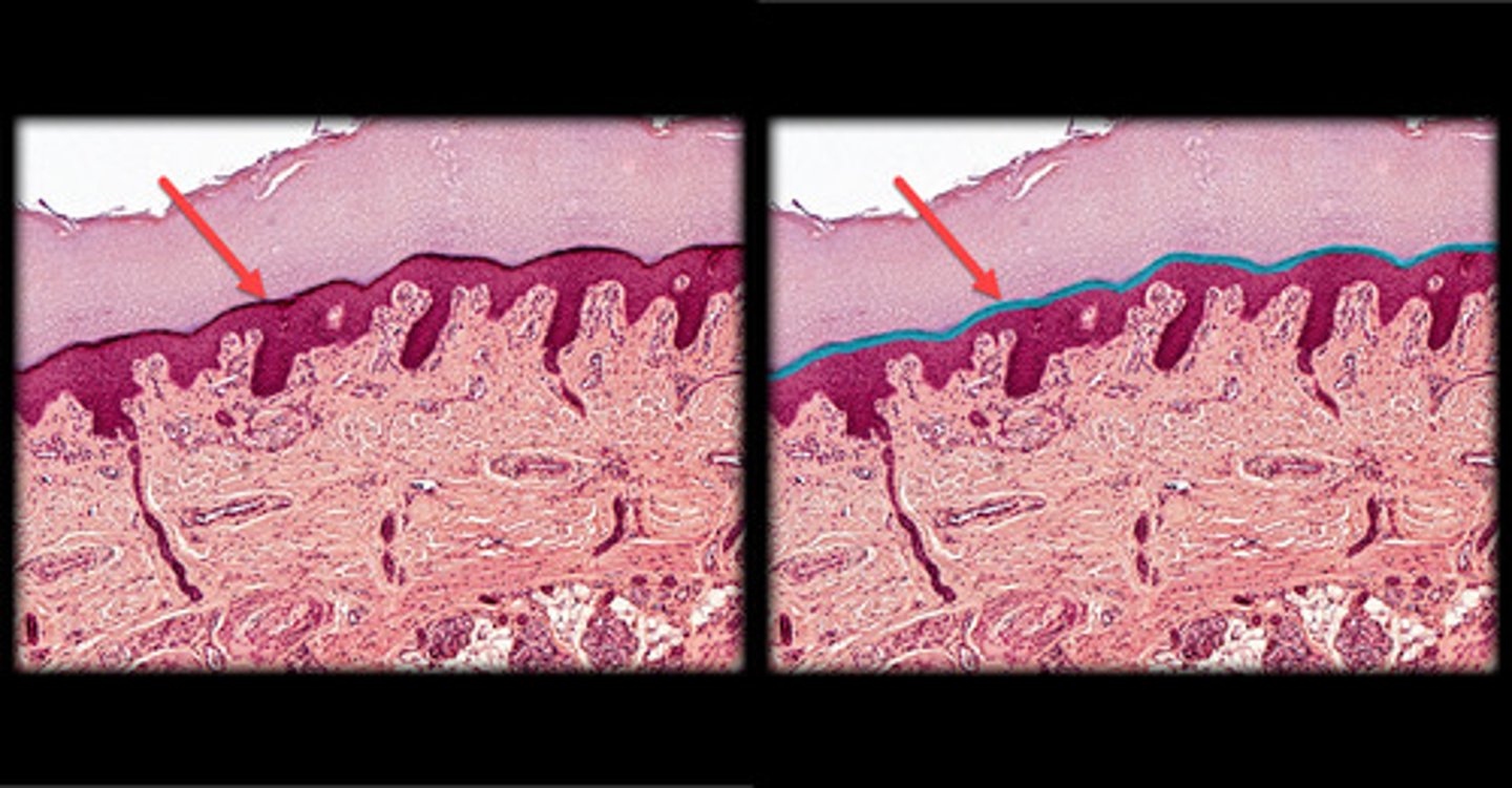
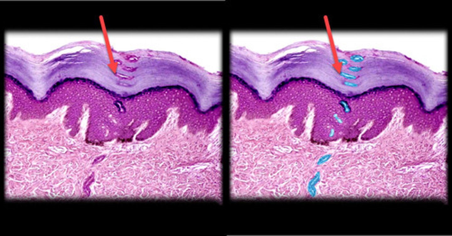
Stratum lucidum (Epidermis)
- Thin, clear layer found only in thick skin (e.g., palms of the hands, soles of the feet).
- Appears as a thin, translucent layer just beneath the stratum corneum.
- Cells are dead, flattened, and lack visible organelles or nuclei, giving the layer a clear or glassy appearance.
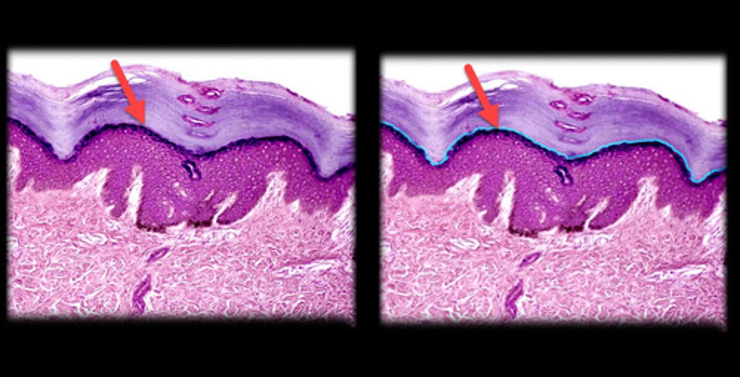
Stratum granulosum (Epidermis)
- Middle layer, typically 3-5 cell layers thick.
- Cells appear more flattened with dark-staining keratohyalin granules in the cytoplasm, giving a grainy appearance under the microscope.
- Nuclei start to break down as cells prepare to become part of the stratum corneum.
- Forms a thin, dark band under the microscope, especially visible in thick skin.
Stratum spinosum (Epidermis)
- Directly below the stratum granulosum.
- Cells are polygonal with a "spiny" appearance due to desmosome connections between cells, which are more visible in this layer.
- Appears as a thicker layer with cells that are more rounded and less flattened than in the layers above.
- Nuclei are still present and prominent in these cells.
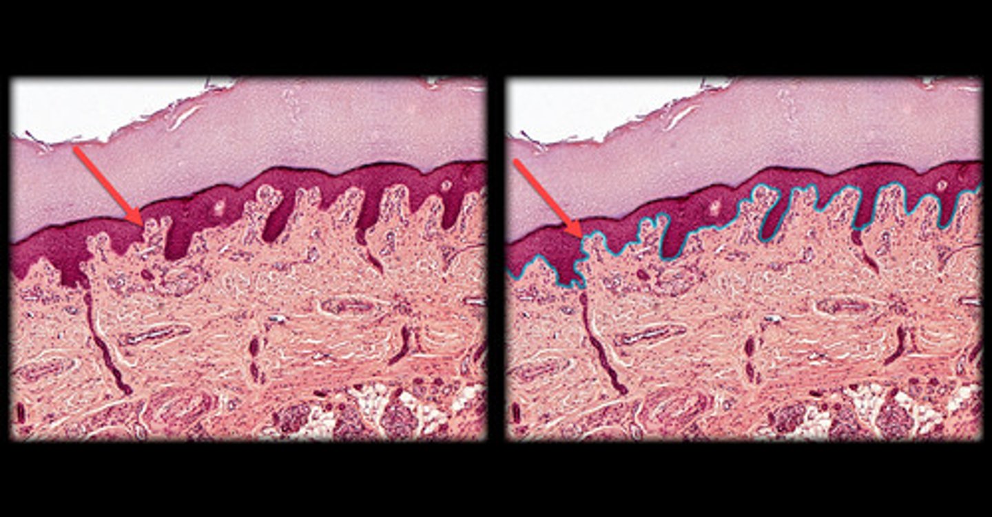
Stratum basale (Epidermis)
- Deepest layer of the epidermis, resting directly on the basement membrane.
- Single layer of columnar or cuboidal cells, often with visible mitotic activity (cells dividing).
- Cells appear larger and darker-stained, with a large nucleus. - - Responsible for generating new cells that migrate upwards to form the outer layers of the skin.
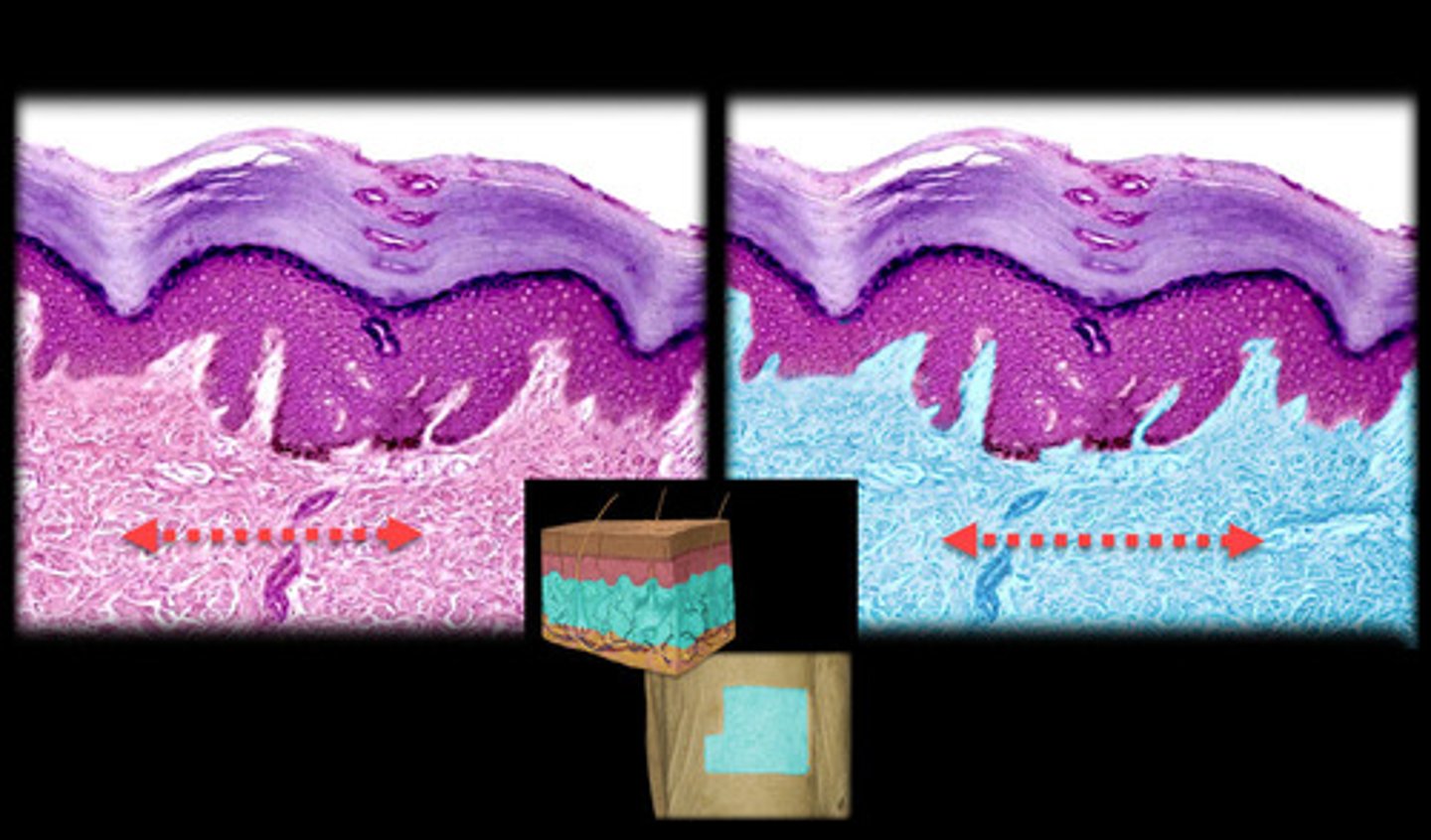
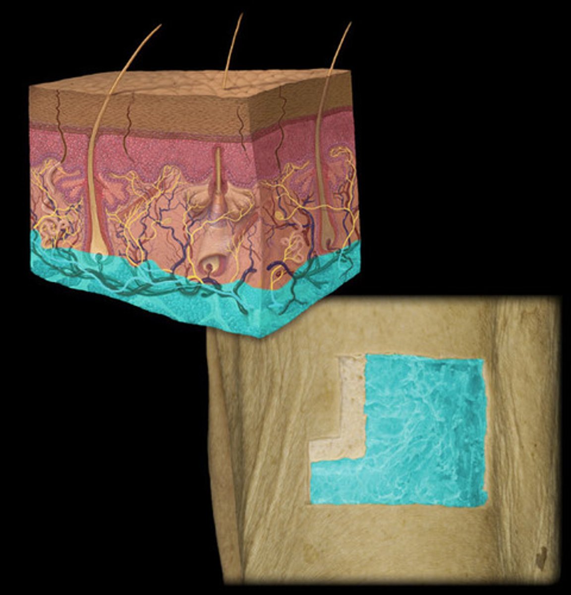
Dermis
- Located directly below the epidermis.
- Composed of two layers, the papillary layer (loose connective tissue) and the reticular layer (dense irregular connective tissue).
- Appears as a thicker, more fibrous layer compared to the epidermis, containing collagen and elastic fibers.
- Includes blood vessels, hair follicles, and glands embedded in the tissue.
- Papillary layer has finger-like projections (dermal papillae) that interlock with the epidermis, while the reticular layer appears denser with fewer cells.
Hypodermis (subcutaneous layer)
- Lies beneath the dermis.
- Composed mainly of adipose tissue (fat cells), providing insulation and cushioning.
- Appears as large, round, empty-looking cells (adipocytes) due to fat being dissolved during slide preparation.
- Can also contain larger blood vessels and some connective tissue.
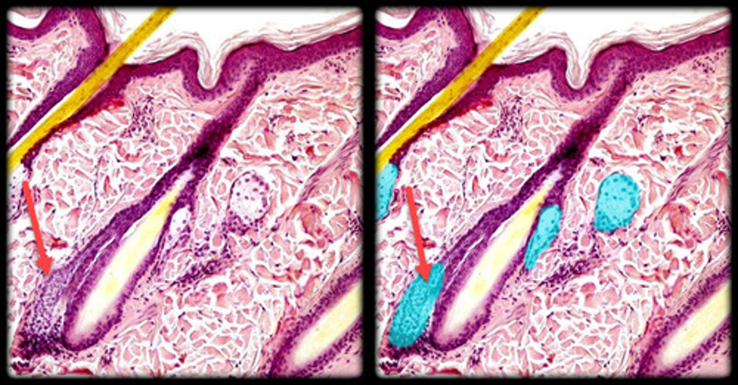
Sebaceous gland
- Associated with hair follicles, producing oily sebum.
- Appears as clusters of rounded, lobular cells located near hair follicles.
- Cells may have a bubbly or foamy appearance due to lipid content.
- Found in the dermis, usually connected to hair follicles, and absent in thick skin (e.g., palms, soles).
Merocrine (eccrine) sweat glands
- Simple, coiled tubular glands.
- Appear as tightly coiled ducts with small, dark-stained cells and a visible lumen in cross-section.
- Found throughout the skin, especially in areas like the palms, soles, and forehead.
- Primarily involved in thermoregulation and producing watery sweat.
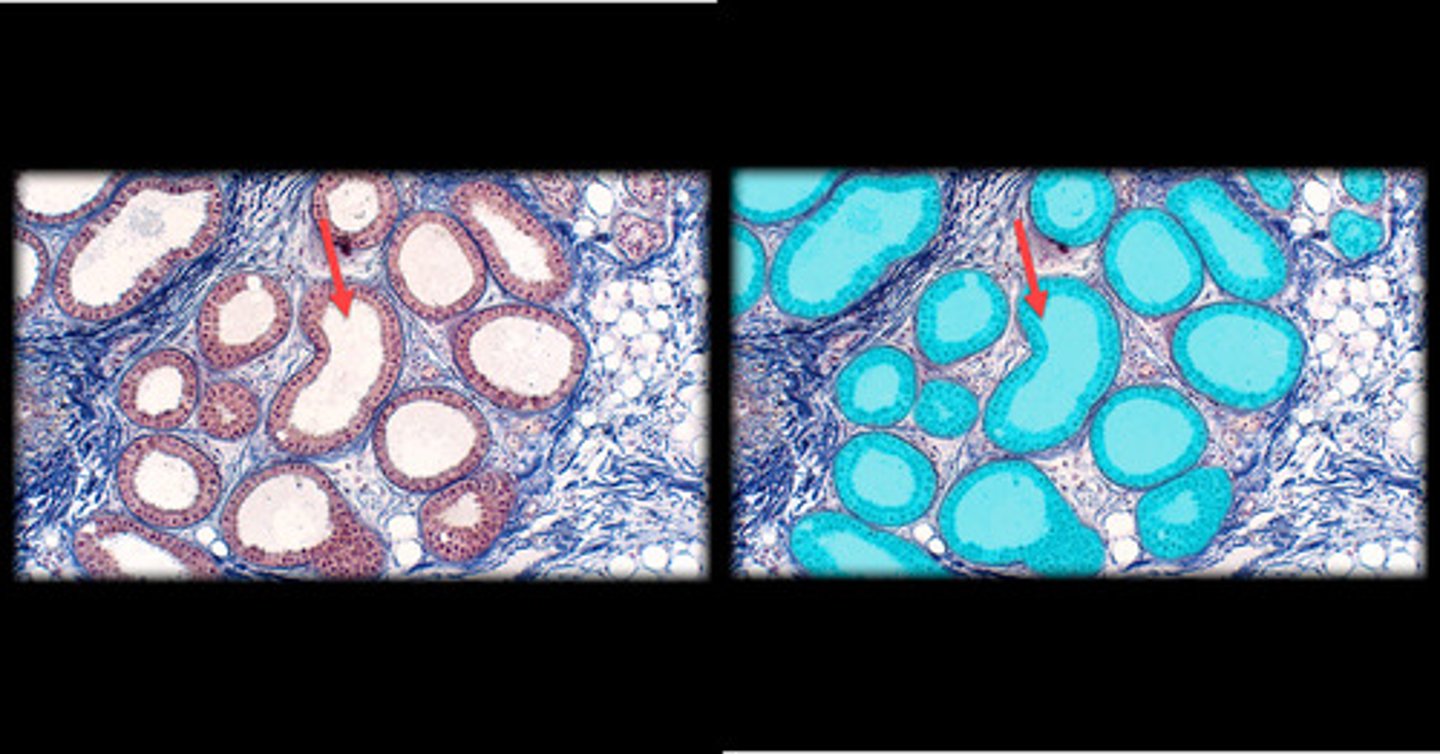
Apocrine sweat glands
- Larger and located deeper in the dermis or hypodermis. - - - - Appear as larger coiled glands with a wider lumen than merocrine glands.
- Found in specific areas like the axilla (armpits), groin, and around the nipples.
- Produce thicker, lipid-rich sweat and are associated with hair follicles, becoming active during puberty.
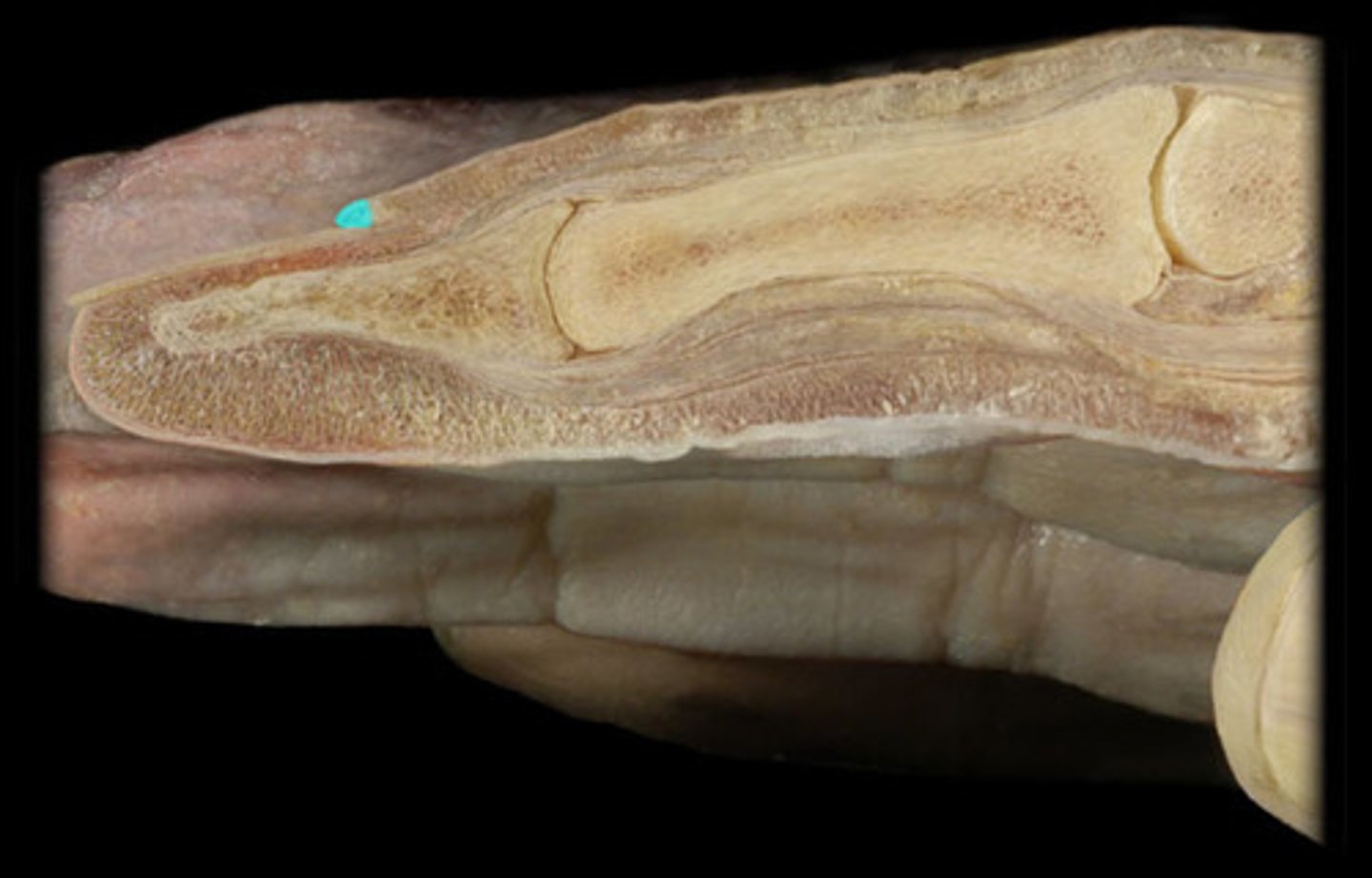
Nail body
- Visible part of the nail, composed of hard keratin.
- Found on the surface, extending from the nail root to the free edge.
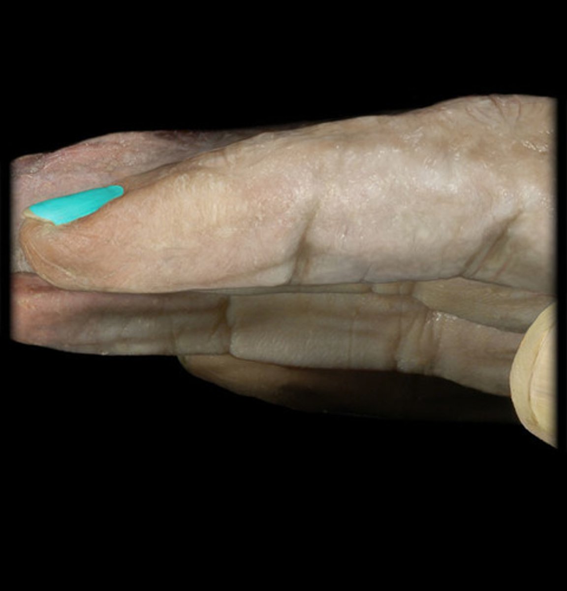
Nail bed
- Skin beneath the nail body, supplying it with nutrients.
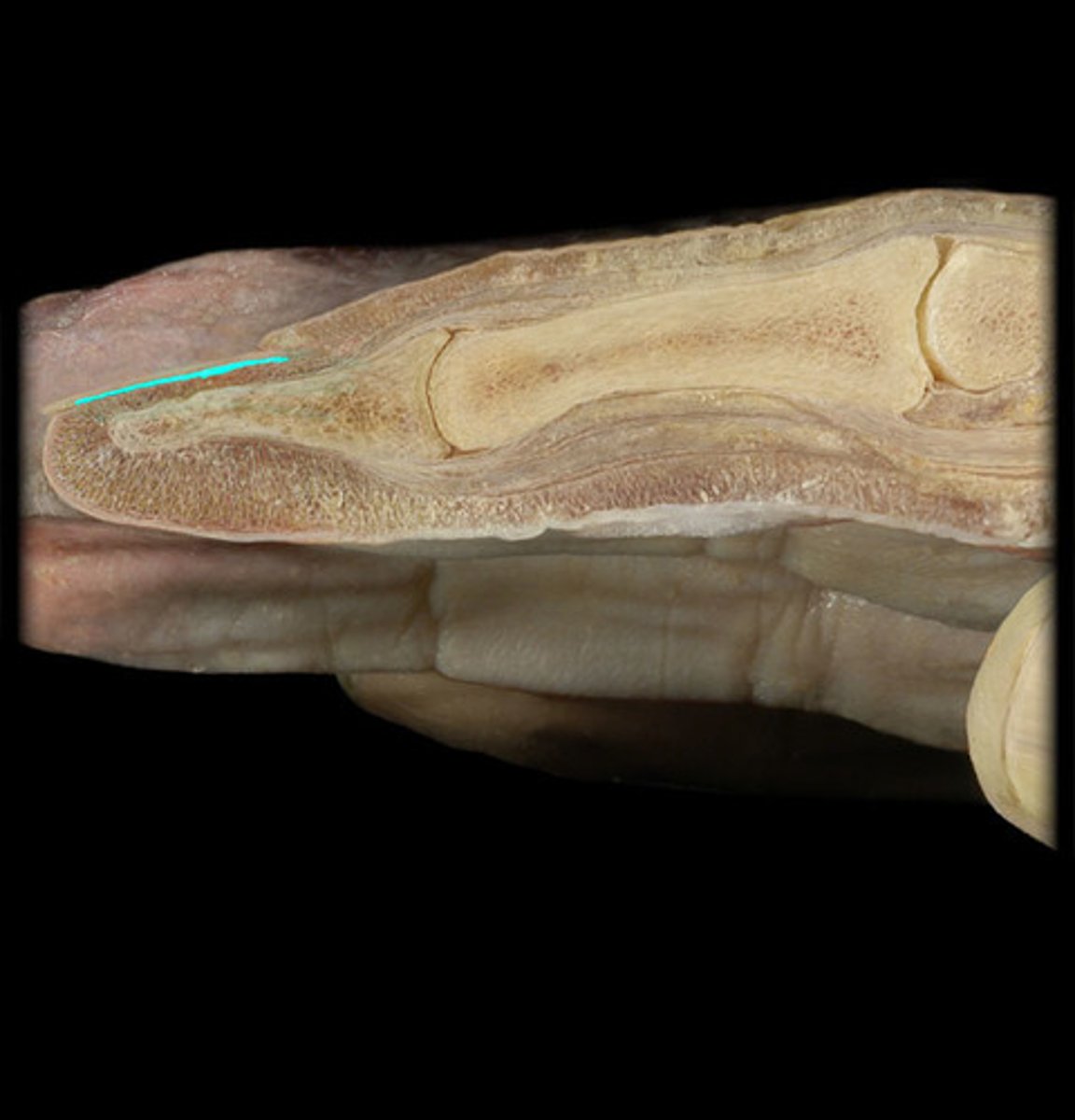
Cuticle (eponychium)
- Thin layer of skin at the base of the nail, where the skin meets the nail body.
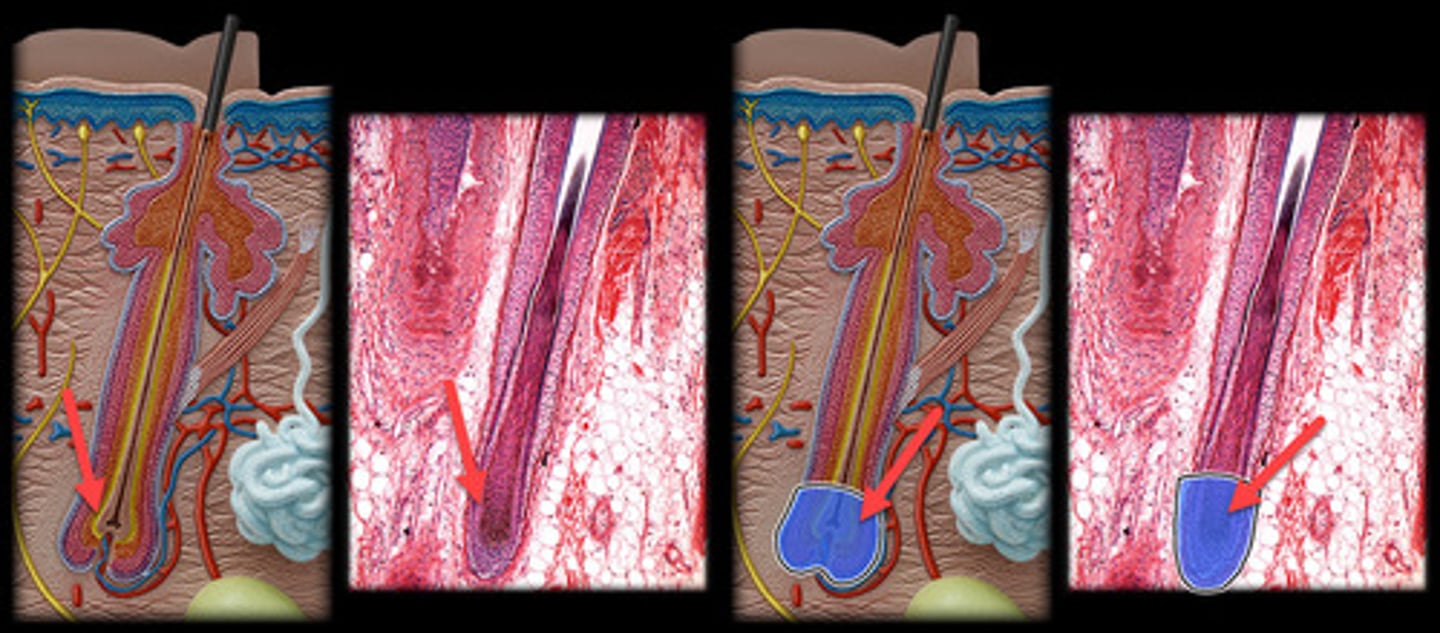
Hair bulb
- Portion of the hair located below the skin's surface.
- Appears as a bulbous, swollen end of the hair follicle located deep in the dermis or hypodermis.
- Contains the hair matrix, where actively dividing cells give rise to the hair shaft.
- Typically stained darker due to the high cellular activity and concentration of melanocytes responsible for hair pigment.
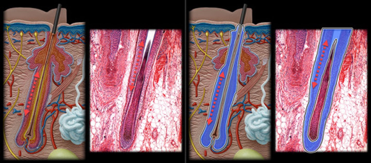
Hair follicle
- Tubular structure in the dermis or hypodermis that surrounds the hair root.
- Appears as a sheath of epithelial cells encasing the hair root.
- Visible in histological slides as a distinct, circular or oval structure around the hair, containing several layers of cells.
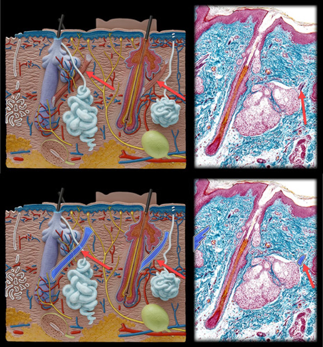
Arrector pili
- Small, smooth muscle fibers attached to the base of the hair follicle.
- Appear as thin, elongated muscle fibers extending from the base of the hair follicle to the dermal layer.
- Responsible for causing hair to stand up ("goosebumps"), found adjacent to hair follicles in the dermis.