CH. 4 Amino Acids and Proteins
 Call Kai
 Learn
Learn Practice Test
 Spaced Repetition
 Match
Match Flashcards
 Knowt Play
1/51

There's no tags or description
Looks like no tags are added yet.
Name | Mastery | Learn | Test | Matching | Spaced | Call with Kai | Chat |
|---|
No analytics yet
Send a link to your students to track their progress
52 Terms
Amino Acids
Proteins are biological macromolecules that act as an enzyme, hormones, receptors, channels, transporters, antibodies, and support structures inside and outside cells.
Proteins are composed of 20 different amino acids linked together in polymers.
The composition and sequence of amino acids in the polypeptide chain is what makes each protein unique and able to fulfill its special role in the cell.
Amino acids are the building blocks of proteins1
Amino acid structure and nomenclature
Understanding the structure of amino acids is key to understanding both their chemistry and the chemistry of proteins.
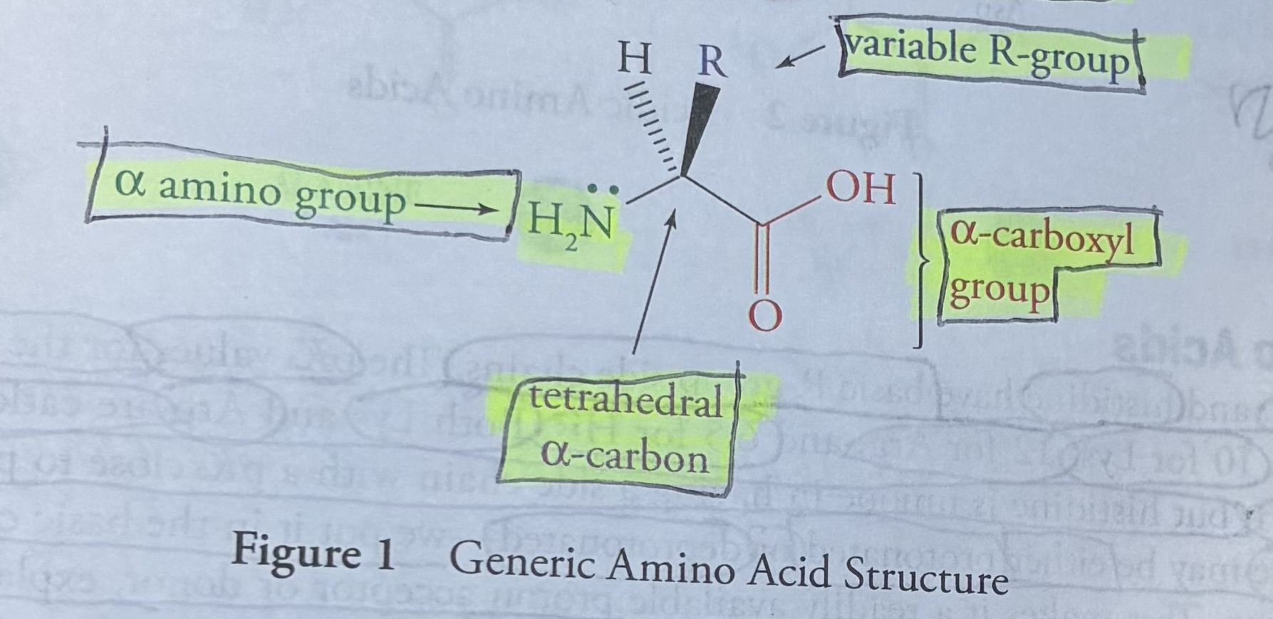
In this flashcard is shown the generic formula for all 20 Amino Acids
Generic Formula For All 20 Amino Acids
Alpha carboxyl group
Tetrahedral alpha carbon
Variable R-group
Alpha amino group
The amino and carboxyl groups attached to the α-carbon atom are termed the α-amino and α-carboxyl groups, to distinguish them from similar groups which may be present as part of the side chain.
The unique feature of each amino acid is the side chain (variable R group), which it the physical and chemical properties that distinguish it from the other nineteen.
Classification of Amino Acids
You should categorize the 20 amino acids in four broad categories:
nonpolar
polar
acidic
basic
Acidic Amino Acids
Aspartic acid (D) and glutamic acid (E) are the only amino acids with carboxylic “acid” functional groups (pKa=4) in their side chains, therefore making their side chains acidic. The “acid” part tells you it’s an acid and has a proton.
There are three functional groups in these amino acids that may act as acids-the two backbone groups and the R group.
Aspartate and Glutamate simply refer to an anionic (deprotonated) form of each molecule, which is how these amino acids are observed at physiological pH. (pH>pKa, so the molecule is deprotonated).
Aspartic acid
Three letter code for Aspartic Acid: Asp
One letter code for Aspartic Acid: D
Glutamic acid
Three letter code for glutamic acid: Glu
One letter code for glutamic acid: E
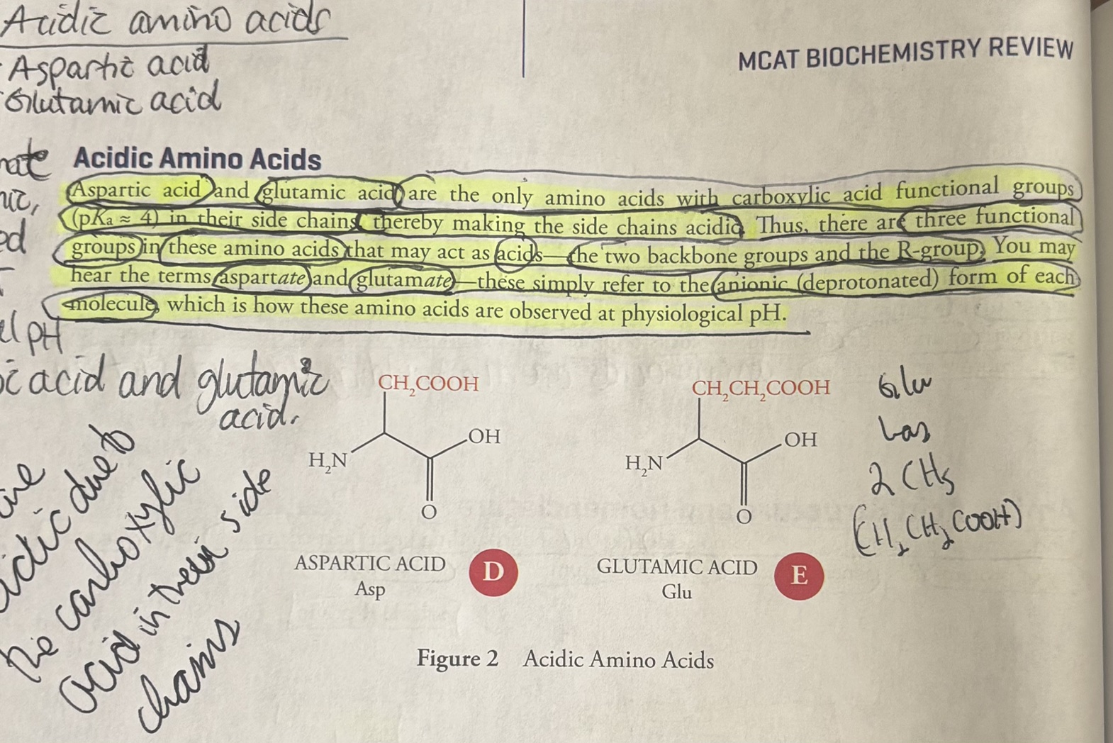
what is the pKa of aspartic acid and glutamic acid? 4.
![<p><u>Basic Amino Acids</u></p><ol><li><p>Lysine (Lys, K)</p></li><li><p>Arginine (Arg, R)</p></li><li><p>Histidine (His, H)</p></li></ol><p>The <strong>pka </strong>values for the basic amino acids are 10 for Lys, 12 for Arg, and 6.5 for His.</p><p>Therefore, both Lysine and Arginine are protonated at phsiological pH (pH=7.4).</p><p>Look at the figure. Notice the basic amino acids have <strong>nitrogen</strong> in their side chains.</p><p><strong>Basic amino acids have a high</strong> <strong>pKa</strong> <strong>because their side chains contain nitrogen atoms (like amines or guanidino groups) that act as strong proton acceptors</strong>. <strong>When they accept a proton, the resulting protonated form is</strong> <strong>remarkably stable</strong>, meaning they hold onto protons tightly and require a very high pH (low proton concentration) to give them up.</p><p>Strong affinity for protons: The unshared electron pair on the nitrogen of groups like the aliphatic amine in lysine or the guanidine in arginine readily attacks and binds free H+ ions.</p><p>Resonance stabilization (Arginine): Arginine’s side chain (guanidino group) can delocalize its positive charge across three equivalent nitrogen atoms once protonated. pKa of Arginine is 12.5.</p><p>Inductive and electrostatic effects (Lysine): Lysine features a primary alkyl amino group with an alkane chain that helps stabilize the localized positive charge on the ammonium ion, resulting in a high pKa (~10.5)</p><p><strong>Histidine </strong>is unique in having a side chain with a pKa close to physiological pH. Therefore, at physiologial pH (7.4), Histidine may be either protonated or deprotonated (meaning histidine can act as both an acid and base). This means histidine is a readily available proton donor and acceptor. “His goes both ways”.</p><p>[By the way hist<em>amine</em> is a small molecule that has to do with allergic responses, itching, inflammation, and other processes. You have heard of antihistamine drugs, for example). Do not confuse <em>histidine</em> with the amino acid <em>histidine</em>.]</p>](https://assets.knowt.com/user-attachments/d1a6fbf6-161c-42b6-a895-101318905c50.jpg)
Basic Amino Acids
Lysine (Lys, K)
Arginine (Arg, R)
Histidine (His, H)
The pka values for the basic amino acids are 10 for Lys, 12 for Arg, and 6.5 for His.
Therefore, both Lysine and Arginine are protonated at phsiological pH (pH=7.4).
Look at the figure. Notice the basic amino acids have nitrogen in their side chains.
Basic amino acids have a high pKa because their side chains contain nitrogen atoms (like amines or guanidino groups) that act as strong proton acceptors. When they accept a proton, the resulting protonated form is remarkably stable, meaning they hold onto protons tightly and require a very high pH (low proton concentration) to give them up.
Strong affinity for protons: The unshared electron pair on the nitrogen of groups like the aliphatic amine in lysine or the guanidine in arginine readily attacks and binds free H+ ions.
Resonance stabilization (Arginine): Arginine’s side chain (guanidino group) can delocalize its positive charge across three equivalent nitrogen atoms once protonated. pKa of Arginine is 12.5.
Inductive and electrostatic effects (Lysine): Lysine features a primary alkyl amino group with an alkane chain that helps stabilize the localized positive charge on the ammonium ion, resulting in a high pKa (~10.5)
Histidine is unique in having a side chain with a pKa close to physiological pH. Therefore, at physiologial pH (7.4), Histidine may be either protonated or deprotonated (meaning histidine can act as both an acid and base). This means histidine is a readily available proton donor and acceptor. “His goes both ways”.
[By the way histamine is a small molecule that has to do with allergic responses, itching, inflammation, and other processes. You have heard of antihistamine drugs, for example). Do not confuse histidine with the amino acid histidine.]
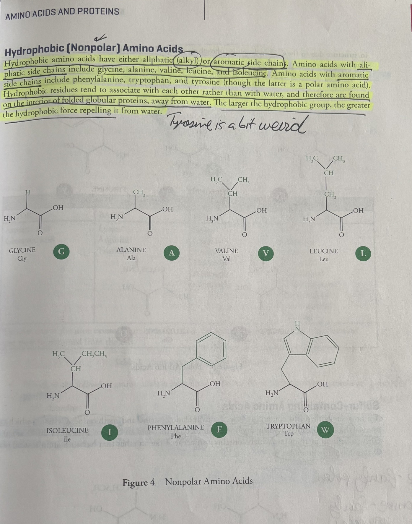
Hydrophobic (Nonpolar) Amino Acids
Hydrophobic amion acids have either alipathic (alkyl) or aromatic side chains.
Aliphatic refers to organic chemical compounds in which carbon atoms link together in open chains or non-aromatic rings, meaning they lack a benzene ring or aromatic stability.
Hydrophobic Amino Acids With Alipathic (Alkyl) Side Chains
Glycine (Gly, G), not glutamine
alanine (Ala, A)
valine (Val, V)
leucine (Leu, L)
isoleucine (Ile, I)
Hydrophobic Amino Acids With Aromatic Side Chains
Phenylalanine (Phe, F)
Tryptophan (Trp, W)
Hydrophobic Amino Acid Characteristics
Hydrophobic residues tend to associate with other hydrophobic residues and are found in the inside of globular folded proteins, away from water (because water is the solvent and its on the outside).
The larger the hydrophobic group, the greater the hydrophobic force repelling it from water.
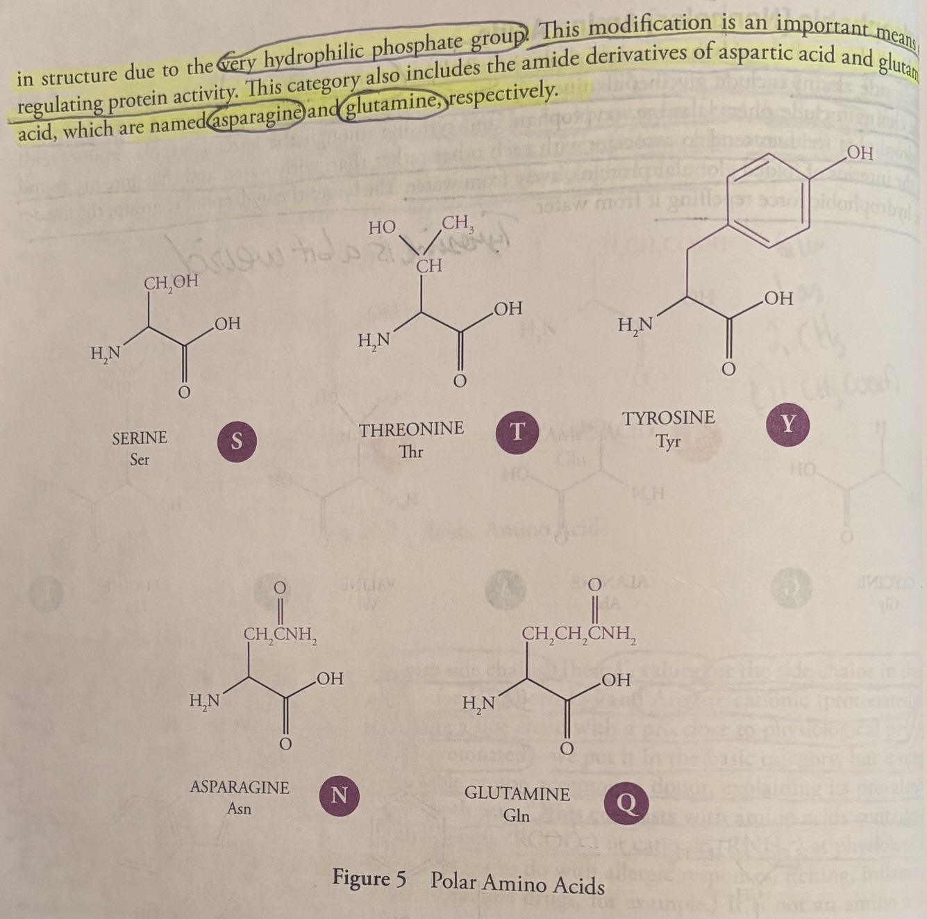
Polar Amino Acids
Serine (Ser, S)
Threonine (Thr, T)
Tyrosine (Tyr, Y)
Asparagine (Asn, N)
Glutamine (Gln, Q)
Polar amino acids are characterized by an R-group that is polar enough to form hydrogen bonds with water, but the polar amino acids do not act as an acid or base.
This means they are hydrophilic and will interact with water whenever possible.
The hydroxyl groups of serine, threonine, and tyrosine residue (tyrosine is an aromatic polar amino acid) are often modified by the attachment of a phosphate group by a kinase. The result is a change in structure due to the very hydrophilic phosphate group. This phosphate add on is an important means of regulating protein activity.
Polar amino acids also include the amide derivatives of aspartic acid and glutamic acid, which are named aparagnine and glutamine.
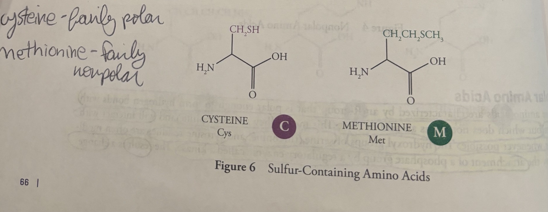
Sulfur-containing Amino Acids
Amino Acids with sulfur-containing side chains include
cysteine (Cys, C)
methionine (Met, M)
Cysteine, which contains a thiol (also called sulfhydryl) like an alcohol that has an S atom instead of an O atom), is fairly polar, and methionine, which contains a thioether (like an ether that has an S atom instead of an O atom), is fairly nonpolar.
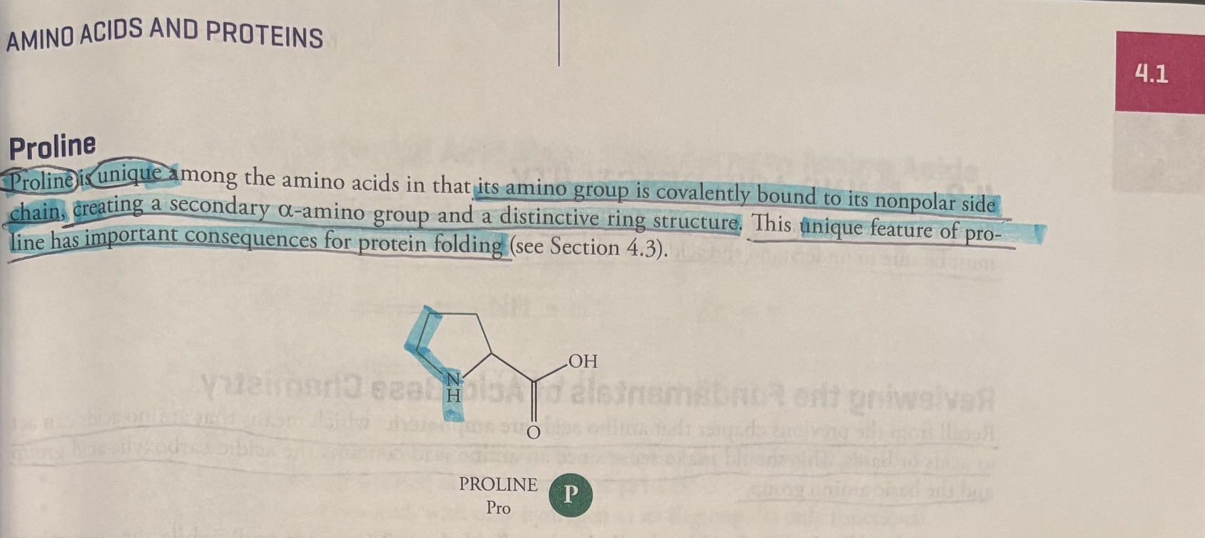
Proline
Poline is unique among the amino acids in that its amino group is covalently bound to its nonpolar side chain, creating a secondary a-amino group and a distinctive ring structure. This unique feature of proline has important consequences for protein folding.
Nine essential amino acids (those that cannot be synthesized by the human diet)
Lysine
Histidine
Threonine
Valine
Isoleucine
Phenylalanine
Tryptophan
Methionine
Through evolution, our animal ancestors lost these costly pathways because they easily obtained these nutrients from their diet.
Which of the following amino acids is most likely to be found on the exterior of a protein at pH 7.0?
A) Leucine
B) Alanine
C) Serine
D) Isoleucine
Answer: The exterior of a protein is surrounded by water, and the only polar amino acid here is Serine.
Amino Acid Reactivity
Since amino acids are composed of an acidic group (the carboxylic acid) and a basic group (the amine), we must be sure to understand the acid/base chemistry of amino acid.
Reviewing the fundamentals of acid/base chemistry
-amino acids are amphoteric (act as both acids and bases), this makes sense because amino acids contain carboxylic acid and amine.
-carboxyl groups of amino acids generally have a pKa of 2 (strong acid), while the ammonia has a pKa of 9 or 10 (weaker acid).
Henderson-Hasselbach Equation
- Henderson-Hasselbach equation: the mathamatical formula that describes the relationship between pH, pka, and the position of equilibrium in acid-base reaction.
pH= pKa + log [base form]/[acid form]
Given the pH and pKa, we can calculate the ratio of the acid and base forms in equilibrium.
-when the pH of a solution is less than the pKa of an acidic group, the acidic group will be protonated
-when the pH of a solution is greater than the pKa of an acidic group, the acidic group will be deprotonated
-if the pH=pKa, There is a perfect 50:50 balance of the protonated and deprotonated forms.
![<p><u>Reviewing the fundamentals of acid/base chemistry</u></p><p>-<strong>amino acids</strong> <strong>are amphoteric </strong>(act as both acids and bases), this makes sense because amino acids contain <strong>carboxylic acid</strong> and <strong>amine</strong>. </p><p>-carboxyl groups of amino acids generally have a pKa of 2 (strong acid), while the ammonia has a pKa of 9 or 10 (weaker acid). </p><p><strong><u>Henderson-Hasselbach Equation</u></strong></p><p>- <strong>Henderson-Hasselbach equation: </strong>the mathamatical formula that describes the relationship between pH, pka, and the position of equilibrium in acid-base reaction. </p><p><strong>pH= pKa + log [base form]/[acid form] </strong></p><p>Given the pH and pKa, we can calculate the <strong>ratio</strong> of the <strong>acid</strong> and <strong>base forms</strong> in <strong>equilibrium</strong>. </p><p>-when the pH of a solution is less than the pKa of an acidic group, the acidic group will be protonated</p><p>-when the pH of a solution is greater than the pKa of an acidic group, the acidic group will be deprotonated</p><p>-if the pH=pKa, There is a perfect 50:50 balance of the protonated and deprotonated forms.</p>](https://assets.knowt.com/user-attachments/dc83c8e4-ce50-4a1c-8d19-5ef3a45f0f5d.jpg)
Question: Which functional group of amino acids has a stronger tendency to donate protons: carboxyl groups (pKa=2.0) or ammonium groups (pKa=9)? carboxyl groups
Question: Which group will donate protons at the lowest pH? carboxyl groups
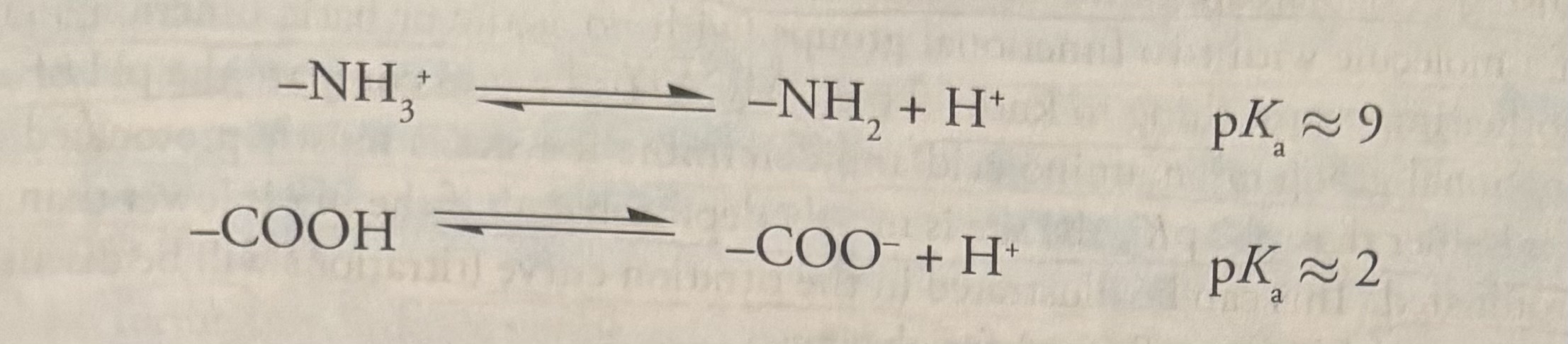
Application of Fundamental Acid/Base Chemistry to Amino Acids
All amino acids contain an amino group that acts as a base and carboxyl group (pKa=2) that acts as an acid.
-so, amino acids are put into classifications of groups (basic, acidic, nonpolar, polar) based off their R groups, but all amino acids have an amine group (base) and a carboxyl group (acid) due to their natural structure.
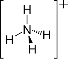
In its protonated, or acidic form, the amine is called an ammonium group, and it has a pKa between 9-10. So, at physiological pH (7.4), the ammonium group is protonated.
this is the ammonium group, the acidic, protonated form of ammonia.
Question: assuming a pKa of 2, will a carboxylate group be protonated or deprotonated at pH 1.0?
answer: pH<pKa, so the carboxylate group will be protonated.
Question: will the amino group be protonated or deprotonated at pH 1.0?
answer: pH <pKa, so the amino group will be protonated (and be ammonium ion).
Question: Glycine is the simplest amino acid, with only hydrogen as its R-group. Its only functional charge are the backbone groups discussed above (amino and carboxyl). What will be the net charge on the glycine molecule at pH 12?
answer: pH 12 is above the pKa of both the amino and carboxyl.
pH>pka= deprotonated
-the amino group (pKa of 9), will be deprotonated (neutral charge)
-the carboxyl group (pKa of 2), will be deprotonated (negative charge)
Therefore, the Glycine will have a negative charge.
Question: At pH 6.0, between the pKas of the ammonium and carboxyl groups, what will be the net charge on a molecule of glycine?
answer: pH 6.0 is above the pKa of carboxyl group (the carboxyl group will be deprotonated and have a negative charge), and pH 6.0 is below the pKa of the ammonium group (the ammonium group will be protonated and have a positive charge).
-therefore, negative + positive= neutral.
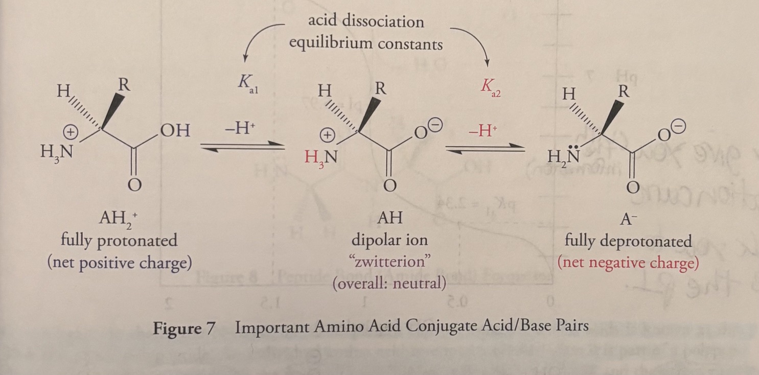
The Isoelectric Point of Amino Acids
Isoelectric point (pI): a pH at which amino acids are zwitterionic and have a neutral charge.
A molecule with positive and negative charges that balance is referred to as a dipolar ion or zwitterion.
“Zwitter” German for “hybrid”, implying that an amino acid has both (+) and (-) charges.
The Isoelectric Point of Amino Acids (cont)
-For a molecule with two functional groups, such as glycine, average the pKas of the two functional groups.
Titration Curves
-picture attached
-titration curves compare the pH of a solution to the pKa of a functional group on the amino acid. You need to be able to determine if a site is mostly protonated or deprotonated.
-if the pH<pKa, the site is protonated
-if the pH>pKa, the site is deprotonated.
The MCAT may give you a titration curve
Then ask you to calculate the pI.
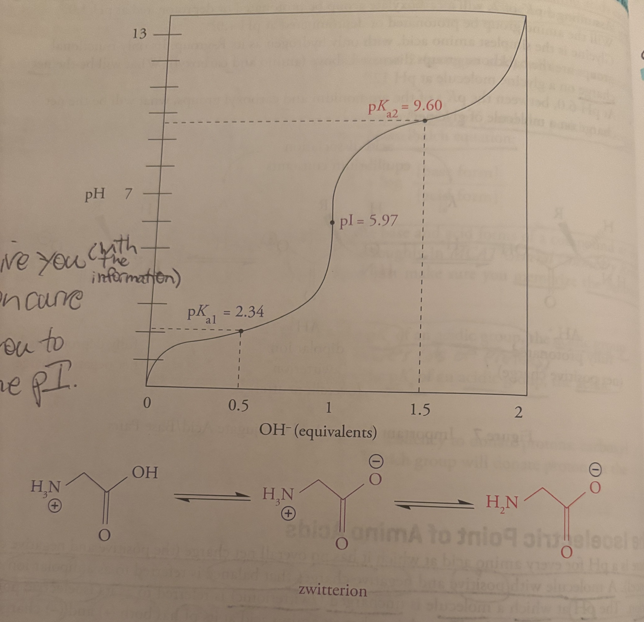
Question: what is the pI of glycine?
Answer:
Glycine is nonpolar
the pka of the carboxyl group is 2.34
th pka of the amino group is 9.60
-the way to calculate the pI for a nonpolar amino acid is to average out the pKas of the functional groups
2.34+9.60/2= 11.94/2= 5.97
Protein Structure
-the two common types of covalent bonds between amino acids in proteins:
peptide bonds
disulfide bridges
peptide bonds link amino acids together in a polypeptide chain
disulfide bridges are bridges between cystine R-groups that stabilize tertiary structure.
The Peptide Bond
-A peptide bond is formed between the carboxyl group of one amino acid and the a-amino group of another amino acid with the loss of water.
The image shows the formation of a dipeptide the amino acids glycine and alanine.
A dipeptide is an organic molecule formed by two amino acids joined by a single peptide bond.
-a peptide bond is really an amide bond.
A peptide bond is called an amide bond because it is chemically an amide functional group.
An amide is a type of chemical compound and functional group containing a carbonyl group linked to a nitrogen atom.
In a polypeptide chain, the N-C-C-N-C-C pattern formed from the amino acids is known as the backbone of the polypeptide.
an individual amino acid is termed a residue when it is part of a polypeptide chain.
The amino terminus is the first end made during polypeptide synthesis, and the carboxy terminus is made last. Hence, by convention, the amino-terminal residue is always written first.
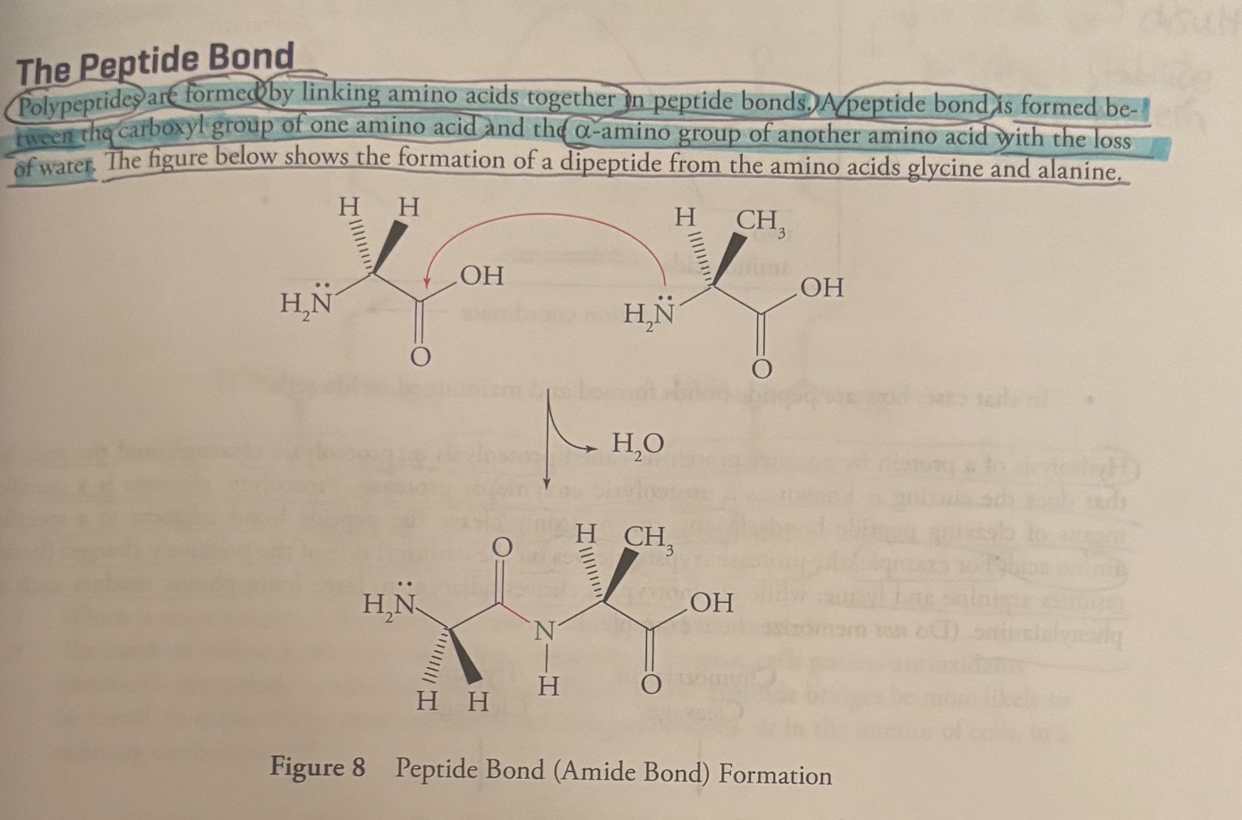
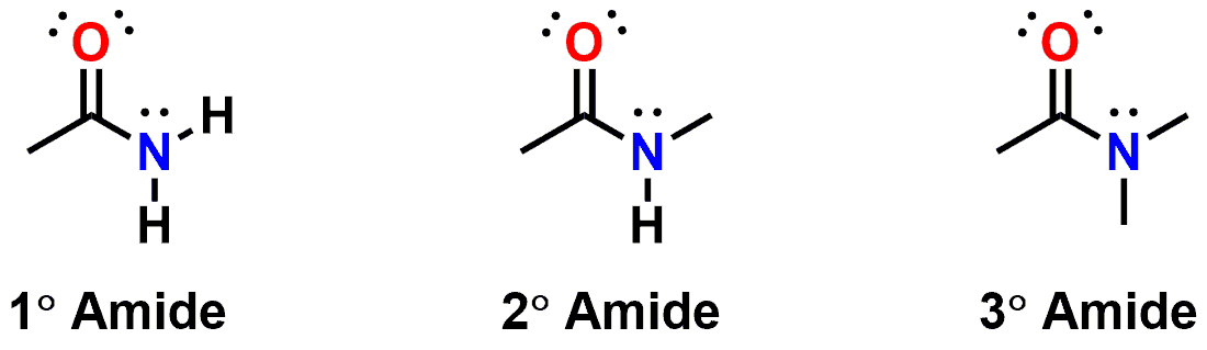
This is a picture of an amide bond.
Question: In the oligopeptide Phe-Glu-Gly-Ser-Ala, state the number of acid and base functional groups, which residue has a free a-amino group, and which residue has a free a-carboxyl group.
Answer:
Number of acid and base functional groups: Phe (nonpolar): 2, Glu (glutamic acid, acidic): 3, Gly (nonpolar): 2, Ser (polar); 2, Ala (nonpolar):2. 2+3+2+2+2=11
WRONG! acid or base functional groups as in acid/base side chains: Glu has one acid side chain, then there are two terminal backbone groups (one free carboxyl group, one free amino group from the ends), giving a total of 3 acid/base functional groups.
Which residue has a free a-amino group: The a-amino group is written first, therefore, the residue with the free a-amino group is Phe.
Which residue has a free a-carboxyl group: the a-carboxyl group is written last, therefore, the residue with the free a-carboxyl group is Ala.
Question: How many unique dipeptides (made from linking two amino acids) can be synthesized using only alanine and glycine residues?
Answer: Four (Gly-Gly, Ala-Ala, Gly-Ala, Ala, Gly). Note that Ala-Gly and Gly-Ala are not identical peptides. In Ala-Gly, the N-terminus is Ala and the C-terminus is Gly. In Gly-Ala, the N-terminus is Gly and the C terminus is Ala.
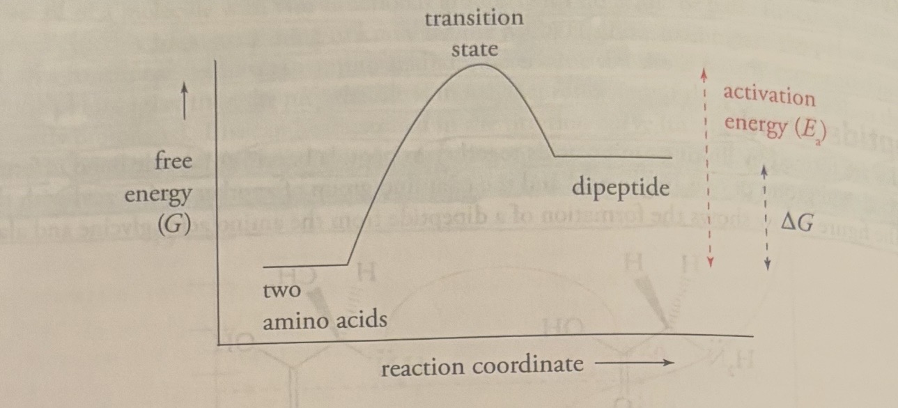
Question: Thermodynamics states that free energy must decrease for a reaction to proceed spontaneously and that such a reaction will spontaneously move toward equilibrium. The diagram below shows the free energy changes during peptide bond formation. At equilibrium, which is the thermodynamicaly favored: the dipeptide or the individual amino acids? (diagram attached to flashcard).
answer: the individual amino acids
Question: if the answer to the last question is amino acids, then how are peptide bonds formed and maintained inside cells?
Answer: during protein synthesis, stored energy is used to force peptide bonds to form. Once the peptide bond is formed, even though its destruction is thermodynamically favorable, it remains stable because the activation energy for the hydrolysis reaction is so high.
Proteolysis
Hydrolysis of a protein by another protein is called proteolysis or proteolytic cleavage, and the protein that does the cutting is known as a proteolytic enzyme or protease.
Proteolytic cleavage is a specific means of cleaving peptide bonds.
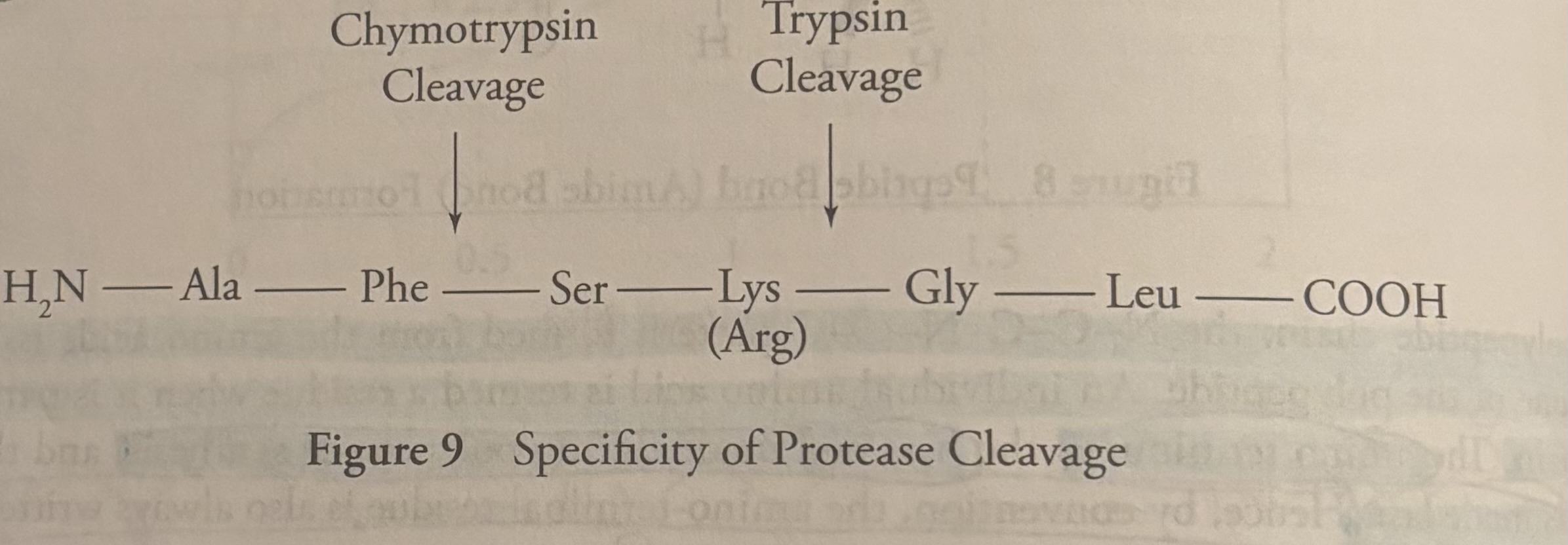
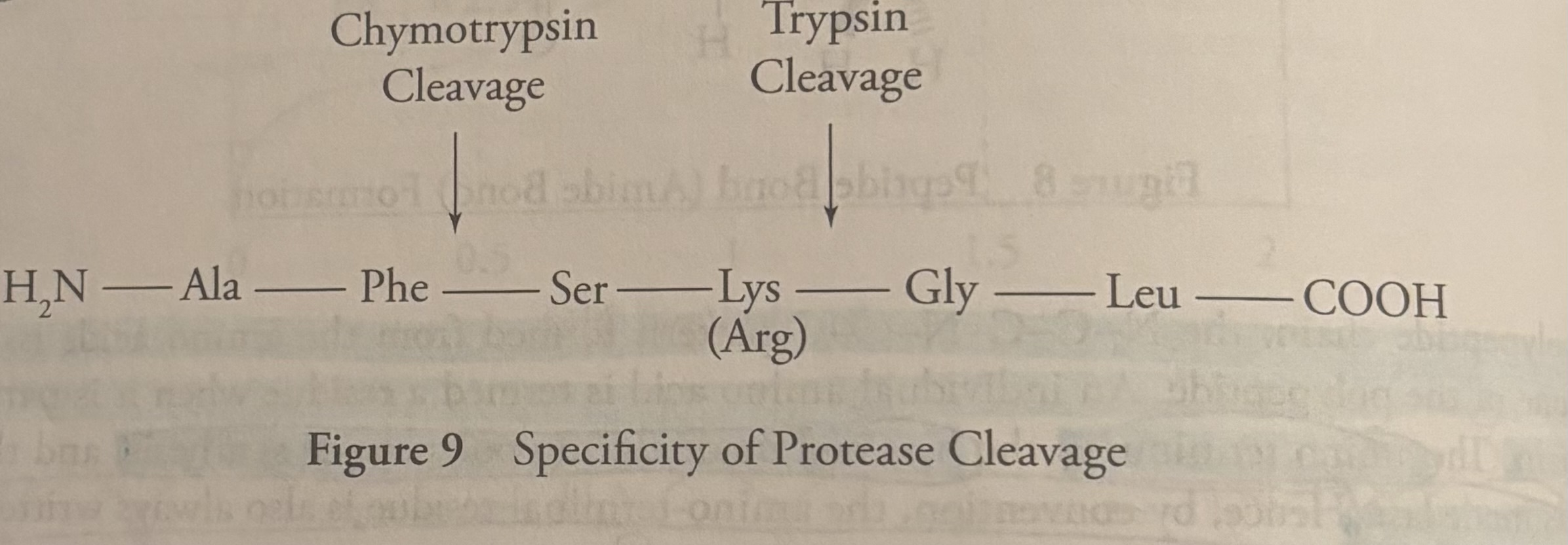
Many enzymes only cleave the peptide adjacent to a specific amino acid. For example, the protease trypsin cleaves the carboxyl side of the positively charged residues arginine and lysine, while chymotrypsin cleaves adjacent to large hydrophobic residues such as phenylalanine.
Question: Based on the image in this flashcard, if the following peptide is cleaved by Trypsin, what amino acid will be on the new N-terminus and how many fragments will result: Ala-Gly-Glu-Lys-Phe-Phe-Lys?
Answer: Trypsin cleaves at Lys (Arg). There will be two fragments: 1. Ala-Gly-Glu-Lys and 2. Phe-Phe-Lys. The amino acid that will be on the new N-terminus (the terminus that comes first) is Phe
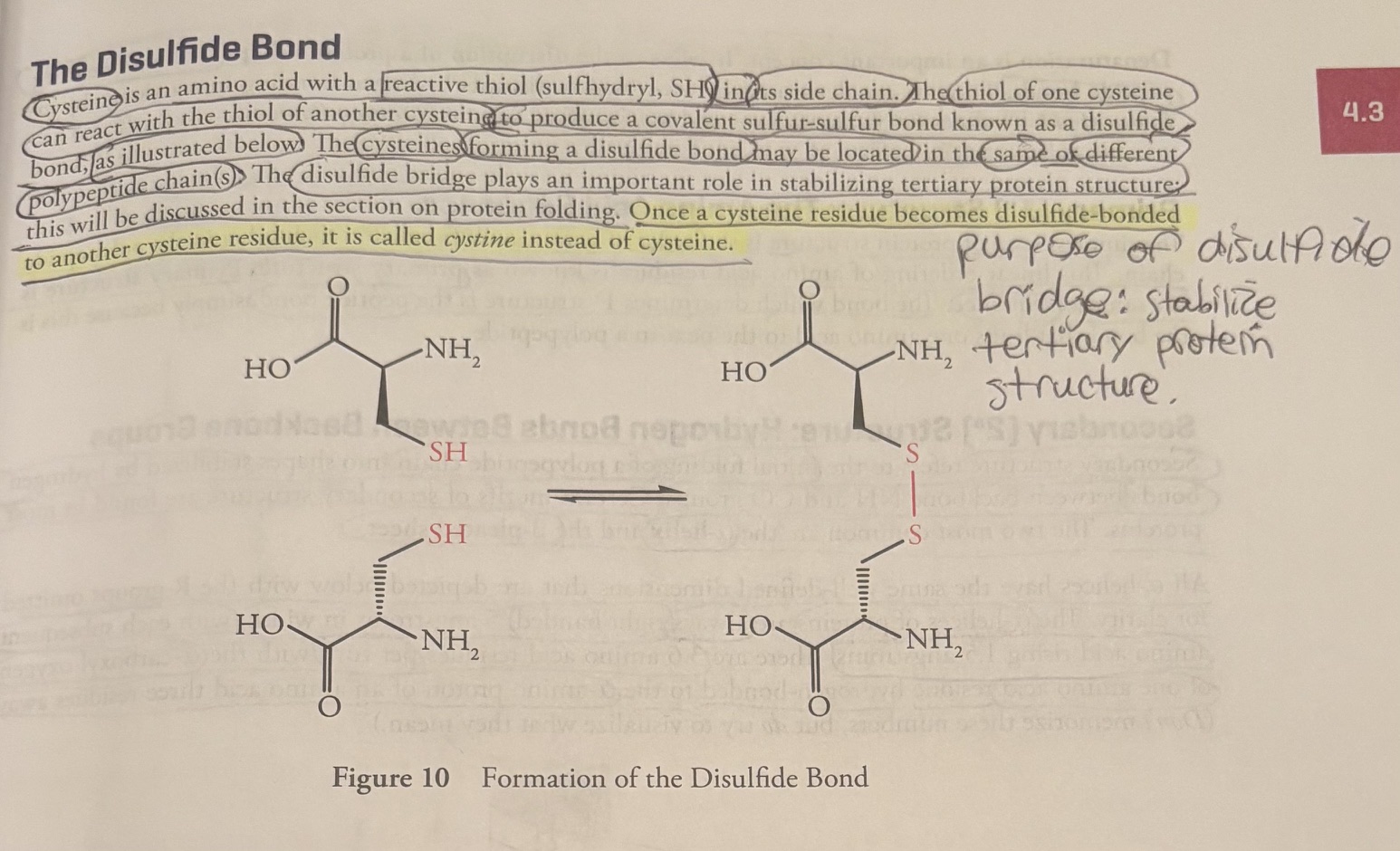
The Disulfide Bond
Cystine is an amino acid with a reactive thiol (sulfyhydryl, SH) in its side chain.
The thiol of one cysteine can react with the cysteine of another cysteine to produce a covalent sulfur-sulfur bond known as a disulfide bond, as illustrated below.
The cysteins forming a disulfide bond can be located at the same or different polypeptide chains (s).
The disulfide bond stabilizes tertiary protein structure.
Once a cysteine residue becomes disulfide bonded to another cysteine residue, it is called cystine instead of cysteine (no e in the middle).
Question: which is more oxidized: the sulfur cysteine or the sulfur in cystine?
answer: the sulfur in cysteine has an hydrogen, the sulfur in cystine does not have a hydrogen. Therefore, the sulfur in cystine is more oxidized.
Question: the inside of cells is known as a reducing environment because cells possess antioxidants (chemicals that prevent “anti” oxidation reactions). Where would disulfide bridges be more likely to be found, in extracellular proteins, under oxidizing conditions, or in the interior of cells, in a reducing environment?
answer: disulfide bridges is an oxidization of sulfur, so disulfide bridges would be more likely found in extracellular proteins, under oxidizing conditions.
Denaturation
Denaturation is the disruption of a protein’s shape without breaking the peptide bonds.
Proteins are denatured by:
urea (breaks the hydrogen bonds)
extremes of pH
extremes of temperature
changes in salt concentration (tonicity)
Primary (1o) Structure: The Amino Acid Sequence
-the simplest level of protein structure is the order of amino acids, bonded to each other by the polypeptide chain.
This linear ordering of amino acid residues is known as primary structure. Primary structure is the same as sequence.
-the bond that determines primary structure is the peptide bond, because peptide bonds are the bonds that link one amino acid to the next in a polypeptide.
Secondary (2o) Structure: Hydrogen Bonds Between Backbone Groups
Secondary Structure refers to the initial folding of a polypeptide chain into shapes stabilized by hydrogen bonds between backbone NH and CO groups.
The two most common secondary secondary structures are the a-helix and B-pleated sheet.
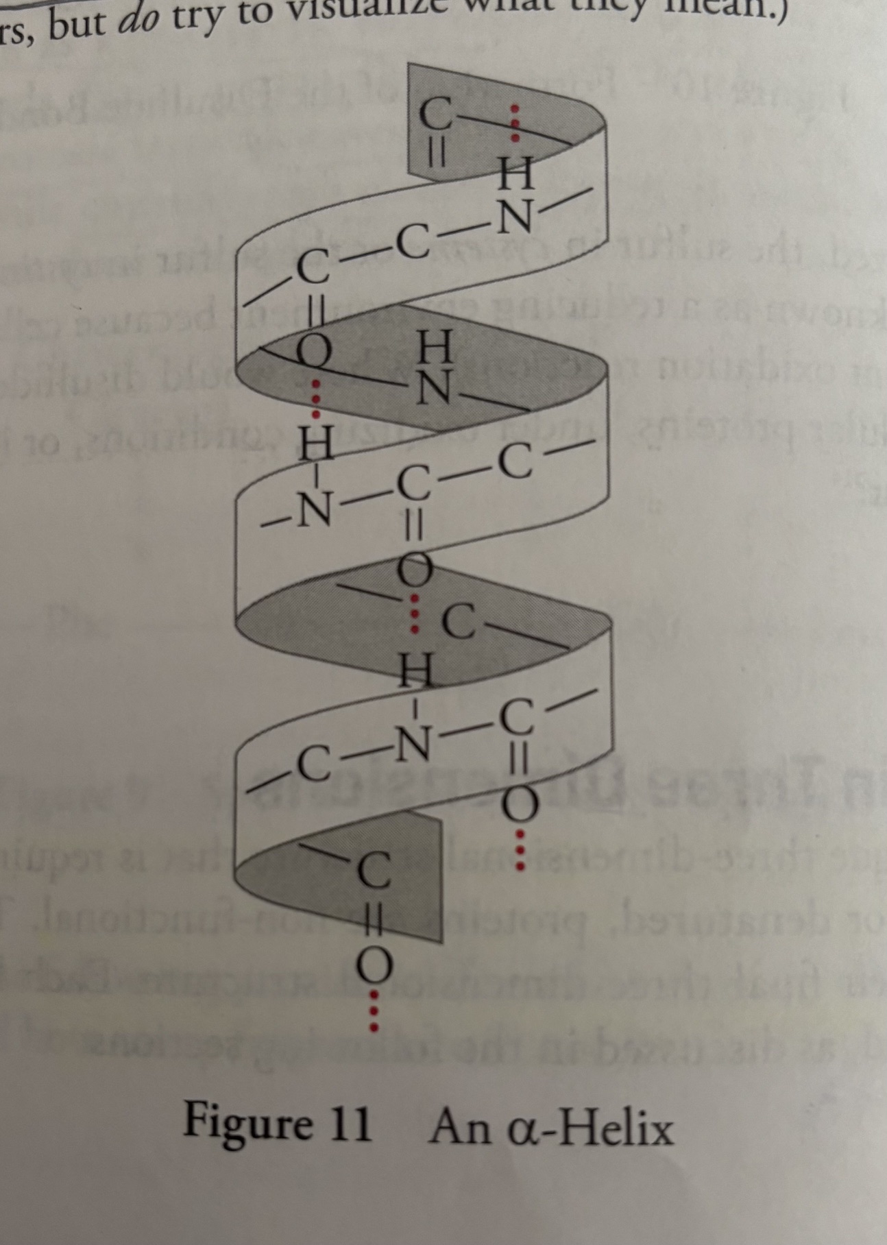
All a-helices have the same well defined dimensions that are depicted below.
The Picture of the Secondary Structure
-All a-helices have the same well-defined dimensions that are depicted below with the R-groups omitted for clarity.
-The a-helices of proteins are always right-handed, 5 angstoms in width, with each subsequent amino acid rising 1.5 angstroms.
-There are 3.6 amino acid residues per turn with the a-carboxyl oxygen of one amino acid residue hydrogen-bonded to the a-amino proton of an amino acid three residues away.
-the oxygen is the hydrogen bond acceptor, the nitrogen is the hydrogen bond donor.
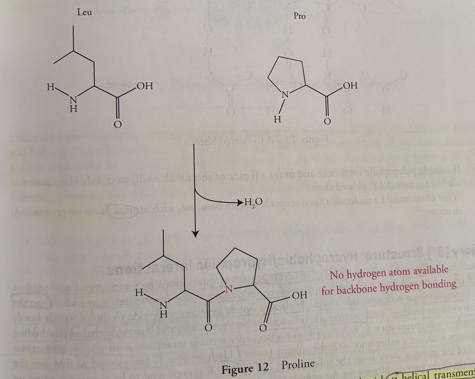
The unique side chain of proline causes two problems in side chains
The formation of a peptide bond with proline (shown in the picture attached to this flashcard) eliminates the only hydrogen atom on the nitrogen atom of proline. The absence of the N-H bond disrupts the backbone hydrogen bonding in the polypeptide chain.
The unique structure of proline forces it to kink the polypeptide chain (because the ring stays).
Hydrophobicity and secondary structure a-helix
Proteins such as hormone receptors and ion channels are often found with a-helical transmembrane regions integrate into the hydrophobic membranes of cells.
The a-helix is a favorable structure for a hydrophobic transmembrane region because all polar NH and CO groups in the backbone are hydrogen-bonded to each other on the inside of the helix, and thus don’t interact with the hydrophobic membrane interior.
Inside vs outside
The core of an alpha-helix features hydrogen bonds between the polar NH and CO groups of the backbone. For transmembrane helices, the outer surface is hydrophobic to interact with the cell membrane's lipid core. However, it spans the hydrophobic core of the membrane, not just "the hydrophobic transmembrane."
Inside of the Helix
Polar peptide groups form hydrogen bonds.
Every backbone C=O group bonds to the N-H group four residues down.
This stabilizes the rod-like shape.
Outside of the Helix
Side chains (R groups) point outward.
Transmembrane helices have hydrophobic side chains.
These amino acids face the lipid bilayer.
A-helical regions that span membranes also have hydrophobic R-groups, which radiate out form the helix, interacting with the hydrophobic interior of the membrane.
B-pleated sheets
B-pleated sheets, because they are secondary structures, are also stabilized by hydrogen bonding between NH and CO groups in the polypeptide backbone.
In B-pleated sheets, however, hydrogen bonding occurs between residues distant from each other in the chain or even on separate polypeptide chains.
also, the backbone of a B-pleated sheet is extended rather than coiled, with side groups above and below the plane of the B-sheet.
two types of B-pleated sheets
There are two types of B-sheets, one with adjacent polypeptide strands running in the same direction (parallel B-pleated sheet) and another in which the polypeptide strands run in opposite directions (antiparallel B-pleated sheet).
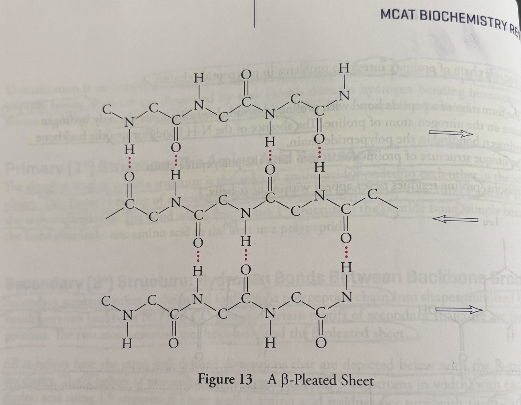
Question: if a single polypeptide folds once and forms a B-pleated sheet with itself, would this be a parallel or antiparallel B-pleated sheet?
Answer: Antiparallel
Question: What effect would a molecule that disrupts hydrogen bonding, such as urea, have on protein structure?
Answer: Urea disrupts hydrogen bonding. In protein structure, secondary, tertiary, and quaternary foldings have hydrogen bonding, therefore, urea would disrupt secondary, tertiary, and quaternary structures. Urea would not disrupt primary structure, because primary structure is held by peptide bonds.
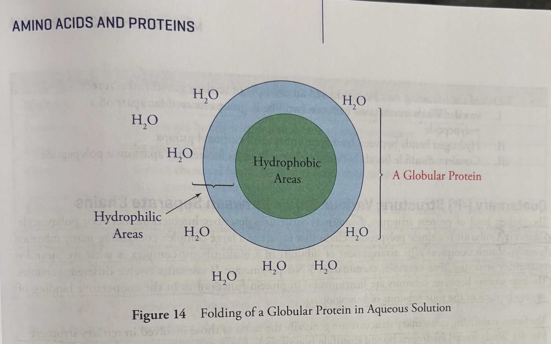
Tertiary (3o) Structure: Hydrophobic/Hydrophilic Interactions
The next level of protein folding: tertiary structure
-interactions between amino acid residues located more distant from each other in the polypeptide chain. These interactions include van der Waals forces between non-polar side chains, hydrogen bonds between polar side chains, disulfide bonds between cysteine residues, and electrostatic interactions between acidic and basic side chains.
Van der Waals forces are weak electric attractions between neutral atoms or molecules, driven by London dispersion forces, dipole-dipole interactions, and temporary charge shifts.
Hydrophobic effect: The folding of secondary structures into tertiary structures is driven by interactions of R-groups with each other and with the solvent (water). Hydrophobic R-groups tend to fold into the interior of the protein, away from from the solvent, and hydrophilic R-groups tend to be exposed to water on the surface of the protein (shown for globular protein).
Under the right conditions, the forces driving hydrophobic avoidance of water and hydrogen bonding will fold a polypeptide spontaneously into the correct conformation, the lowest energy conformation.
In a classic experiment by Christian Anfinsen and coworkers, the effect of a denaturing agent (urea) and a reducing agent (B-mercaptoethanol) on the folding of a protein called ribonucleas were examined. In the following questions, you will re-enact their through processes.
Question: Ribonuclease has eight cysteines that form four disulfide bonds (2 cysteines are needed for 1 disulfide bond). What effect would a reducing agent have on its tertiary structure?
answer: the formation of a disulfide bridge is oxidation (reducing hydrogen), a reducing agent adds hydrogens. Therefore, a reducing agent,, adding hydrogen, would destroy disulfide bonds, disrupting tertiary structure.
Question: If the disulfides serve only to lock into place a tertiary protein structure that forms first on its own, then what effect would the reducing agent have on correct protein folding?
answer: You know that a tertiary protein structure has more than disulfide bonds holding it together. A reducing agent would disrupt the disulfide bonds, making the tertiary structure less stable, but the tertiary structure would still be held together by the hydrophobic R groups on inside and hydrophilic R groups outside, hydrogen bonds, van der waals interactions.
Question: Would a protein end up folded normally if you (1) first put it in a reducing environment, (2) then denatured it by adding urea, (3) next removed the reducing agent, allowing disulfide bridges to reform, and (4) finally removed the denaturing agent?
answer: NO! this was tricky. Notice how you have to wait for the 1,2,3,4 structures to form. The answer is no because if you allow the disulfide bridges to form while the protein is still denatured, the protein will become locked into abnormal shape.
Question: what if you did the same experiment but in this order: 1,2,4,3?
answer: you removed the denaturing agent (4), allowing hydrogen bonds to form, then you removed the reducing agent, allowing disulfide bridges to reform, then YES, you should end up with the correct protein structure.
Question: which of the following may be considered an example of tertiary protein structures?
I. van der Waals interactions between two Phe R-groups located far apart on a polypeptide
II. Hydrogen bonds between backbone amino and carboxyl groups
III. Covalent disulfide bonds between cysteine residues located far apart on a polypeptide?
answer: Tertiary protein structures have van der Waals interactions, the two Phe R-groups are able to communicate with each other because they are non-polar, and located far on polypeptide is normal for tertiary structure. I is correct.
II: wrong, that’s more secondary structure
III: covalent disulfide bonds between cysteine residues located far apart on a polypeptide, true. The disulfide bonds stabilize the tertiary protein structure.
I and III are correct.
Quaternary (4o) Structure: Various Bonds Between Separate Chains
The highest level of protein structure is quaternary structure.
Quaternary structure describes interactions between polypeptide subunits. A subunit is a single polypeptide chain that is part of a large complex containing many subunits (multisubunit complex).
The arrangement of subunits in a multisubunit complex is what we mean by quaternary structure.
For example, mammalian RNA polyermerase II contains twelve different subunits. The interactions between subunits are instrumental in protein function, as in cooperative binding of oxygen by each of the four subunits of hemoglobin.
The forces stabilizing quaternary structure are the same forces stabilizing tertiary structure- van der Waals forces, hydrogen bonds, disulfide bonds, and electrostatic interactions. It is key to understand, however, that there is one bond that may be not be involved in quaternary structure- the peptide bond- because the peptide bond defines sequence (1o structure).
Question: what is the difference between a disulfide bridge involved in quaternary structure and one involved in tertiary structure?
Answer: The disulfide bridge involved in quaternary structure is formed between chains that are not linked by peptide bonds. The disulfide bridge formed in tertiary structure is formed between chains that are linked by peptide bonds.
Proteins As Enzymes
Enzymes are biological catalysts
Enzymes increase the rate of a spontaneous reaction by lowering the activation energy by stabilizing the transition state, but enzymes do not affect the deltaG of a reaction. This is because enzymes play a kinetic role, not a thermodynamic one.
Enzymes are able to alter the rate of a reaction enormously: a reaction that would take one hundred years to reach equilibrium without an enzyme may occur in just seconds with an enzyme.
Enzyme Table
-Enzymes are classified based on reaction type.
Enzyme Class
Hydrolase: hydrolyzes chemical bonds (includes ATPases, proteases, and others)
Isomerase: rerranges bonds within a molecule to form an isomer
Ligase: forms a chemical bond (ex: DNA ligase)
Lyase: breaks chemical bonds by means other than oxidation or hydrolysis (ex: pyruvate decarboxylase)
Kinase: transfers a phosphate group to a molecule from a high energy carrier, such as ATP (ex: phosphofructokinase [PFK])
Oxidoreductase: runs redox reactions (includes oxidases, reductases, dehydrogenases, and others)
Polymerases: polymerization (ex: addition of nucleotides to the leading strand of DNA by DNA polymerase III)
Phosphatase: removes phosphate group from a molecule
Phosphorylase: transfers a phosphate group to a molecule from inorganic phosphate (ex: glycogen phosphorylase)
Protease: hydrolyzes peptide bonds (ex: trypsin, chymotrypsin, pepsin, etc.)
ATP As An Energy Source: Reaction Coupling
Enzymes increase the rate of spontaneous reactions. The spontaneous reactions would occur on their own (without an enzyme), but the spontaneous reaction would occur far more slowly than with an enzyme.
-However, there are many reactions in the body that will occur with a positive G. The biosynthesis of macromolecules such as DNA and protein is not spontaneous (G>0), but clearly these reactions do take place (or else we would not be here). This is because thermodynamically unfavorable reactions in the cell can be driven forward by reaction coupling.
Reaction coupling: one very favorable reaction is used to drive an unfavorable one. Reaction coupling is possible because free energy changes are additive.
In the lab, deltaGo’ for the hydrolysis of one phosphate group from ATP is -7.3kcal/mol, therefore, the hydrolysis of of one phosphate group from ATP is a very favorable reaction.
In the cell, deltaG (yes, just deltaG) is about -12kcal/mol, so in the cell, the hydrolysis of ATP is an even more favorable reaction.
The value of delta G depends on the concentrations of reactants and products, which are able to be variable in the body.
delta G: Gibb’s free energy
deltaGo’: Gibb’s free energy with all solutes at 1 M concentration for all solutes except H+ and a pH of 7.
How does ATP hydrolysis drive unfavorable reactions? There are many ways. One example is ATP hydrolysis can cause a conformational change in a protein, in this way, ATP hydrolysis can be used to power energy-costly events like transmembrane transport.
Another example is by transfer of a phosphate group from ATP to a substrate. Take the unfavorable reaction A+B→ C. Let’s say that Reactant A must proceed through an intermediate, APO42- in order to participate. Let’s say delta G of this reaction is +7 kcal/mol for the overall reaction. What if the two partial reactions have deltaG’s as follows (look at the picture attached to this flashcard):
A+ PO42- → APO42-, delta G= + 2 kcal/mol
APO42- + B → C + PO42-, delta G= +5 kcal/mol
The total delta G: + 7 kcal/mol
Thus, these reactions will not proceed because the overall delta G will be +7kcal/mol. What will be the overall deltaG if we couple the reaction to the hydrolysis of one ATP? All we have to do is add all the deltaG values, as follows (look at the picture on this flashcard):
ATP→ ADP + PO42-, delta G= -12 kcal/mol
A+ PO42- → APO42-, delta G= + 2 kcal/mol
APO42- + B → C + PO42-, delta G= +5 kcal/mol
total deltaG= -5 kcal/mol, through reaction coupling, the reaction is now thermodynamically favorable because we have coupled the unfavorable reaction A+B+ATP → C+ ADP + PO42-, deltaG= -5 kcal/mol.
Question: What is the favorable reaction that the cell can use to drive unfavorable reactions?
Answer: ATP hydrolysis!
Hydrolyzing ATP is the reaction that is used to drive unfavorable reactions. Now you see why ATP is so important.
Question: what’s the difference between the situation of in vitro (lab) under standard conditions and in vivo (cell) under nonstandard conditions?
Answer: Qcell does not equal Keq’. This means relative concentrations of ATP and ADP+Pi are not at equilibrium levels in the cell (because Qcell does not equal Keq). Actually, Q(cell)«Keq because the cell keeps a high concentration of ATP around.
If the reaction quotient (Q) is less than the equilibrium constant (K), the reaction has more reactants than products.
Enzyme Structure and Function
Most enzymes are proteins that fold into specific three-dimensional structures to act as catalysts.
(Some enzymes are RNA or contain RNA sequences with catalytic activity. Most catalyze their own splicing, and the rRNA in ribosomes helps in peptide-bond formation).
The reason for the importance of folding in enzyme function is the proper formation of the active site, the region of the enzyme that is directly involved in catalysis.
The reactants in an enzyme-catalyzed reaction are called substrates.
Products do not have a special name, they are just products.
What is the role of the active site/ how do enzymes work?
The active site model, commonly referred to as lock and key hypothesis, states that the substrate and active site are perfectly complementary.
This differs from the induced fit model, which asserts that the substrate and active site differ slightly in structure and that the binding of the substrate induces a conformation change in the enzyme.
The induced fit model has gained greater acceptance in recent years, but regardless of the model, enzymes accelerate the rate of a given reaction by helping to stabilize the transition state. Stabilizing the transition state lowers the activation energy barrier between reactants and products.
Question: An enzyme may consist of a single polypeptide chain or several polypeptide subunits help together in a ___25? (primary, secondary, tertiary, quaternary structure)?
answer: quaternary structure.
Question: what shapes are enzymes more likely to have: fibrous/elongated or globular/spherical?
Answer: globular/spherical. Structural proteins such as collagen tend to be fibrous, but proteins that act as catalysts (enzymes) tend to be roughly spherical to form an active site in a cleft in the sphere.
Question: if a transition state intermediate possess a transient negative charge, what amino acid residues might be found at the active sites to stabilize the transition state?
Answer: 1. transition state intermediate possesses a transient negative charge. The goal is to stabilize the transition state. 2. Therefore, the amino acid residues found at the active site will have positive charges to stabilize the transient negative charge. The amino acid residues are ones that hold a positive charge, such as His, Arg, or Lys (the basic amino acids). Alternatively, the hydrogen of the NH2 group in glutamine or asparagine could hydrogen bond with the negative charge.
Question: Is it possible that amino acids located far apart from each other in the primary protein sequence may play a role in the formation of the same active site?
Answer: Yes, the amino acids at the active site may be distant from each other in a polypeptide’s primary sequence but be near each other in the final folded protein. This is why protein folding is crucial for enzyme function.
Question: If, during an enzyme-catalyzed reaction, an intermediate forms in which the substrate is covalently linked to the enzyme via a serine residue, can this occur at any serine residue or must it occur at a specific serine residue?
Answer: it must occur at a specific serine residue that sticks out at the active site. (because the substrate passes through the active site).
-note, if you have an answer, know why the answer is correct.
Question: Compound A converts into Compound B in solution: A doublearrow B. The reaction has the following equilibrium constant: Keq’=[B]eq/[A]eq=1,000. If pure A is dissolved in water at 298K, will deltaG for the reaction A doublearrow B be positive or negative? Is it possible to answer this question without knowing deltaGo’?
Answer: A Keq’>1 means products are favored (there will be 1000x more B than A in solution at equilibrium), meaning it’s a spontanous reaction, meaning the delta G will be negative. It is possible to answer this question without knowing deltaGo’.
Question: Regarding the reaction described in the previous question (the question above on this flashcard), if pure B is put into solution in the presence of an enzyme that catalyzes the reaction between A and B, which one of the following will be true?
A) All the B will be converted into A, until there is 1,000 times more A than B
B) All of the B will remain as B, since B is favored at equilibrium
C) The enzyme will have no effect, since enzymes act on the transition state and there is no transition state present.
D) The reaction that produces A will predominate until deltaG=0.
Answer: D.
you put pure B into solution in the presence of an enzyme (the reaction is not at equilibrium yet)
Le Chatelier’s Principle will try to offset the balance of too much B by having the reaction produce A until the reaction reaches equilibrium (delta G=0).
Enzyme Active Site Specificity
The active site for enzymes is highly specific in its substrate recognition, including stereospecificity (the ability to distinguish between stereoisomers).
For example, enzymes which catalyze reactions involving amino acids for D or L amino acids, and enzymes catalyzing reactions involving monosacharides may distinguish stereoisomers as well.
Many proteases (protein-cleaving enzymes) have an active site with a serine residue whose OH group can act as a nucleophile attacking the carbonyl carbon of amino acid residue in a polypeptide chain. (the carbonyl carbon is electrophilic due to the high electronegativity of the Oxygen). Examples are trypsin, chymotrypsin, and elastase. These enzymes also usually have a recognition pocket near the active site, which is a pocket that attracts certain residues on the substrate polypeptides.
The enzyme always cuts polypeptides at the same site, just to one side of the recognition residue. For example, chymotrypsin always cuts on the carboxyl side of one large hydrophobic or aromatic residue Tyr, Trp, Phe, and Met. Enzymes that act on hydrophobic substrates have hydrophobic amino acids in their active sites, while hydrophilic/polar amino acids will comprise the active site of enzymes with hydrophilic substrates.
Effect of temperature and pH on active site
Given the importance of the active site, it becomes clear that small alterations in its structure can drastically alter enzymatic activity. Therefore, both temperature and pH play a critical role in enzymatic function. As temperature increases, the thermal motion of the peptide and surrounding solution destabilize its structure. If the temperature rises sufficiently, the protein denatures and loses its orderly structure.
The pH of a surrounding medium also impacts protein stability; several amino acid possesses ionizable-R groups that change charge depending on pH. This can decrease the affinity of a substrate for the active site and, if the pH deviates sufficiently, the protein can denature.
Question: Which configurations are found in animals?
Answer: L amino acids and D sugars. Remember the L in aLanine.
Question: the transition state for a reaction possesses a transient negative charge. The active site for an enzyme catalyzing this reaction contains a His residue to stabilize the intermediate. If the His residue at the active site is replaced by a glutamate that is negatively charged at pH=7.0. What effect will this have on the reaction, assuming that the reactants are present in excess compared to the enzyme?
A) The repulsion caused by the negative charge in the glutamate at the altered activate state will increase the activation energy and make the reaction proceed more slowly than it would in a solution without an enzyme.
B) The rate of catalysis will be unaffected, but the equilibrium ratio of products and reactants will change, favoring reactants.
C) The transition state intermediate will not be stabilized as effectively by the altered enzyme, lowering the rate relative to the rate with catalysis by the normal enzyme
D) The rate of catalysis will decrease, and the equilibrium constant will change.
Answer:
transition state has a negative charge
active site normally has a His residue to stabilize the negative charge
Glutamate has a negative charge and replaces His.
transition state is no longer stabilized as effectively.
The correct answer is C. Even mutated or altered enzymes usually speed up chemical reactions much better than no catalyst at all, though their efficiency often drops compared to the original, normal enzyme.
Enzymatic function can also depend on the association of additional molecules. Cofactors, which are metal ions or small molecules (not themselves a protein), are required for activity for activity in many enzymes. In fact, the majority of vitamins in our diet serve as precursors for cofactors (ex: niacin [B3]) is ultimately transformed into NAD+).
When a cofactor is an organic molecule, it is referred to as coenzymes; these often bind to the substrate during the catalyzed reaction. One prime example of a coenzyme is coenzyme A (CoA
A cofactor is a general term for any non-protein helper molecule that assists an enzyme. When that helper molecule is specifically an organic (carbon-based) molecule, it is called a coenzyme to separate it from inorganic helpers like metal ions.
Regulation of Enzyme Activity
Metabolic pathways in the cell are not all continually on, but must be tightly regulated to maintain health.
For example, if glycogen synthesis and breakdown occur in the same cell as the same time, a great deal of energy will be wasted without accomplishing anything.
Therefore, the activity of key enzymes in metabolic pathways is usually regulated in one or more of the following ways:
Covalent modification: Proteins can have several different groups covalently attached to them, and this can regulate their activity, lifespan in the cell, and/or cellular location. The addition of a phosphorylation group from a molecule of ATP by a protein kinase to the hydroxyl of serine, threonine, or tyrosine residues is the most common example. Phosphorylation of these different sites on an enzyme can either activate or inactivate enzyme. Protein phosphorylases also phosphorylate proteins, but use free-floating inorganic phosphate (Pi) in the cell instead of ATP. Protein phosphorylation can be reversed by protein phosphatases
Proteolytic cleavage: Many enzymes (and other proteins) are synthesized in inactive forms (zymogens) that are activated by cleavage of a protease.
Association with other peptides: some enzymes have a catalytic activity in one polypeptide unit that is regulated by association with a separate regulatory subunit. For example, there are some proteins that demonstrate continuous rapid catalysis if their regulatory subunit is removed, this is known as constitutive activity (constitutive means continuous or unregulated). There are other proteins that require association with another peptide in order to function. Still other proteins can bind many regulatory subunits. There are numerous examples of this in the cell, and many of them have diverse and complex regulatory mechanisms that all revolve around the theme of “associations with other polypeptide can affect enzymes activity”.
Allosteric regulation: the modification of active-site activity through interactions of molecules with other specific sites on the enzyme (called allosteric sites). Let’s look at this in a little more detail.
Allosteric Regulation
-If the cell is to make use of the enzyme as a biochemical switch, it must be able to turn the enzyme on or off.
-one mechanism of regulation is the binding of small molecules to particular sites on an enzyme that are distinct from the active site; this is allosteric regulation.
-this name comes from the fact that the particular spot on the enzyme which can bind the small molecule is not the active site; allo means “other”, and steric refers to a location in space (as in steric hinderance). So, allosteric means “other space”.
-The binding of the allosteric regulator to the allosteric site is generally noncovalent and reversible. When bound, the allosteric regulator can alter the conformation of the enzyme to increase or decrease catalysis even though it may be bound to the enzyme at a site distant from the active site or even on a polypeptide.
Feedback Inhibition
Enzymes usually act as part of pathways, not alone.
-in pathways, there are usually one or two key enzymes that are regulated, these enzymes typically catalyze irreversible steps in a pathway.
Negative feedback (feedback inhibition): a self-regulating control system where the end product of a metabolic pathway or hormonal cascade inhibits the very process that produced it.
Positive feedback (feedback stimulation): a process where the product of a system accelerates the original stimulus, pushing the system further away from baseline until a specific endpoint is reached
feedforward stimulation: a situation where a metabolic product from an early step in a pathway goes downstream to activate an enzyme that catalyzes a later step in that same pathway.
In addition to acting as switches, enzymes also act as valves, because they regulate the flow of substances into products.
![<p><u>Enzyme Kinetics</u></p><p><strong>Enzyme kinetics </strong>is the study of the <strong>rate of formation</strong> <strong>of products</strong> from <strong>substrates</strong> in the presence of an enzyme. </p><p>The <strong>reaction rate </strong>(<strong>V</strong>, for velocity) <strong>is the amount of product formed per unit time, in moles per second (mol/s)</strong>. <strong>The reaction rate depends on the concentration of substrate, [S], and enzyme</strong>. If there is only a little substrate, then the rate, V, is directly proportional to the amount of substrate added. Double the amount of substrate and the reaction rate doubles, triple the amount of substrate and the reaction rate triples, and so forth. But eventually, there is so much substrate that the active sites of the enzymes are occupied much of the time, and adding more substrate doesn’t increase the reaction rate as much, that is, the slope of the V vs [S] curve decreases. </p><p>Finally, there is so much substrate that every active site is continuously occupied, and adding more substrate does not increase the reaction rate at all. At this point, the enzyme is <strong>saturated</strong>. The reaction rate when the enzyme is saturated is denoted <strong>Vmax</strong>. This is a property of each enzyme at a particular concentration of the enzyme. </p><p><u>Km</u></p><p>Michaelis constant (<strong>Km</strong>, the “m” stands for the Michaelis constant). Km is the substrate concentration at which the enzyme is at ½ of Vmax. </p><p>Km is <strong>unique for each enzyme</strong> and <strong>determines the affinity the enzyme has for the substrate. </strong></p><p><strong>-</strong>A low Km means a low substrate concentration is needed for the enzyme to reach half Vmax, therefore, the enzyme has a high affinity for the substrate</p><p>-a high Km means a high substrate concentration is needed for the enzyme to reach half Vmax, therefore, the enzyme has a low affinity for the substrate. </p>](https://assets.knowt.com/user-attachments/715f5f9a-cdc5-4dce-b412-b414d26c1710.jpg)
Enzyme Kinetics
Enzyme kinetics is the study of the rate of formation of products from substrates in the presence of an enzyme.
The reaction rate (V, for velocity) is the amount of product formed per unit time, in moles per second (mol/s). The reaction rate depends on the concentration of substrate, [S], and enzyme. If there is only a little substrate, then the rate, V, is directly proportional to the amount of substrate added. Double the amount of substrate and the reaction rate doubles, triple the amount of substrate and the reaction rate triples, and so forth. But eventually, there is so much substrate that the active sites of the enzymes are occupied much of the time, and adding more substrate doesn’t increase the reaction rate as much, that is, the slope of the V vs [S] curve decreases.
Finally, there is so much substrate that every active site is continuously occupied, and adding more substrate does not increase the reaction rate at all. At this point, the enzyme is saturated. The reaction rate when the enzyme is saturated is denoted Vmax. This is a property of each enzyme at a particular concentration of the enzyme.
Km
Michaelis constant (Km, the “m” stands for the Michaelis constant). Km is the substrate concentration at which the enzyme is at ½ of Vmax.
Km is unique for each enzyme and determines the affinity the enzyme has for the substrate.
-A low Km means a low substrate concentration is needed for the enzyme to reach half Vmax, therefore, the enzyme has a high affinity for the substrate
-a high Km means a high substrate concentration is needed for the enzyme to reach half Vmax, therefore, the enzyme has a low affinity for the substrate.
Question: If a small amount of enzyme in a solution is acting at Vmax and the substrate concentration is doubled, what is the new reaction rate?
Answer: the reaction rate has not changed because the enzyme is at Vmax.
the enzyme is at Vmax
substrate concentration is doubled
the enzyme is still at Vmax, so the reaction rate has not increased.
Cooperativity
Many multisubunit enzymes do not behave in the simple kinetic manner described above.
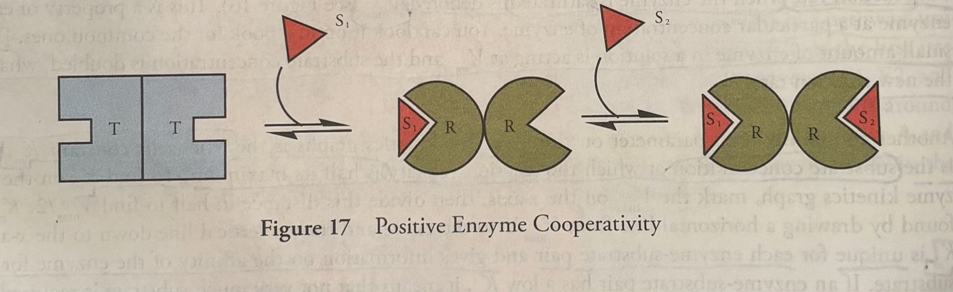
In multisubunit enzymes, the binding of substrate to one subunit modulates the affinity of other subunits for the substrate. Such enzymes are said to bind cooperatively. There are two types of cooperativity: 1. positive and 2. negative cooperativity.
In positive cooperativity, the binding of a substrate to one subunit increases the affinity of the other subunits for the substrate. The conformation of the enzyme prior to substrate binding with low substrate affinity is sometimes termed “tense”, and the conformation of enzyme with increased affinity is termed “relaxed”. (look at the figure attached to this photo).
-”tense” because the enzyme has tension. Adding a substrate breaks the tension.
In negative cooperativity, the binding of a substrate to one subunit decreases the affinity of other subunits for the substrate.
-Cooperative enzymes must have more than one active site. Cooperative enzymes are usually multisubunit complexes composed of more than one protein chain held together in a quaternary structure. They may also be a single-subunit enzyme with two or more active sites.
Positive cooperativity sigmoidal curve
A sigmoidal curve, or S-curve, is a mathematical graph shaped like the letter "S" that shows a slow start, a fast middle growth, and a flat finish.
A sigmoidal curve results from positive cooperative binding (figure attached to this flashcard).
Region 1 (bottom part) in the figure is explained by the notion that at low [S], the enzyme complex has a low affinity for substrate (the enzyme is in the tense state), and adding more substrate increases the rate little.
Region 2 (steep part) represents the range of substrate concentrations where adding substrate greatly increases the reaction rate, because the enzyme complex is in the relaxed state).
![<p><u>Positive cooperativity sigmoidal curve</u></p><p>A sigmoidal curve, or S-curve, is a mathematical graph shaped like the letter "S" that shows a slow start, a fast middle growth, and a flat finish.</p><p>A sigmoidal curve results from positive cooperative binding (figure attached to this flashcard).</p><p><strong>Region 1 </strong>(bottom part) in the figure is explained by the notion that at low [S], the enzyme complex has a low affinity for substrate (the enzyme is in the <strong>tense state</strong>), and adding more substrate increases the rate little.</p><p><strong>Region 2</strong> (steep part) represents the range of substrate concentrations where adding substrate greatly increases the reaction rate, because the enzyme complex is in the <strong>relaxed state</strong>).</p>](https://assets.knowt.com/user-attachments/5505d196-d874-4497-aab5-2d81f31383a5.jpg)
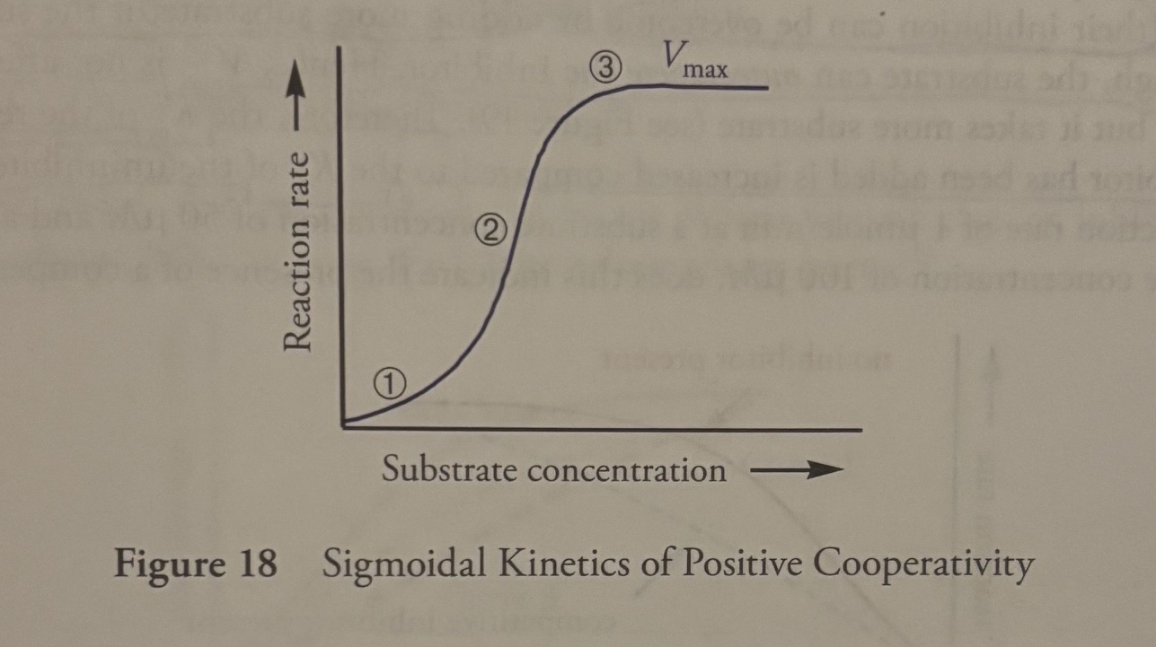
Question: the leveling off at the upper right part of the curve, Region 3, represents what?
Answer: the Vmax, the enzyme is saturated.
Cooperativity does not just apply to enzymes, it also applies to hemoglobin
Cooperativity does not apply to just catalytic enzymes. For example, hemoglobin (Hb) is a protein complex made of four polypeptide subunits, each of which contains a heme prosthetic group with a single O2-binding site. (So one Hemoglobin has four hemes and four binding sites). Hb is a carrier (of oxygen), not a catalyst of any reaction (not an enzyme). it exhibits positively cooperative O2 binding. This is why the Hb-O2 dissociation curve is sigmoidal.
Question: What is the relationship the two notions allosteric and cooperative?
Answer: Cooperativity is a special kind of allosteric interaction.
One active site acts like an allosteric regulatory site for the other active sites.
Cooperative enzyme complexes are often allosterically regulated also. Hb is an excellent example. Not only does O2 binding to one subunit increase the other subunit’s affinities but also several other molecules can bind to various sites to change the affinity of the complex. For example, Co2 stabilizes tense Hb, causing each of the four binding sites to have lower affinity for oxygen. As a result, in the presence of Co2, Hb tends to give up whatever O2 it has bound. The most important thing to remember though, is that the binding in cooperativity takes place at the active site, while the binding in allosteric regulation takes place at “other sites”
Inhibition of enzyme activity
Enzyme inhibitors can reduce enzyme activity by a few different mechanisms, including competitive inhibition, noncompetitive inhibition, uncompetitive inhibition, and mixed-type inhibition.
Pattern?
-if the inhibitor binds to the enzyme-substrate complex, then the enzyme’s affinity for substrate will increase, causing Km to decrease
-if the inhibitor binds to the free enzyme, then the enzyme’s affinity for substrate will decrease, causing Km to increase
Competitive inhibition
-molecules that compete with the substrate for binding at the active site.
-key thing to remember about competitive inhibition: competitive inhibition can be overcome by adding more substrate, because if the substrate concentration is high enough, the substrate can outcompete the inhibitor. Therefore, Km has increased
-Therefore, Vmax is not affected. you can get the same Vmax, but it takes more substrate
Km has increased
Vmax stays the same.
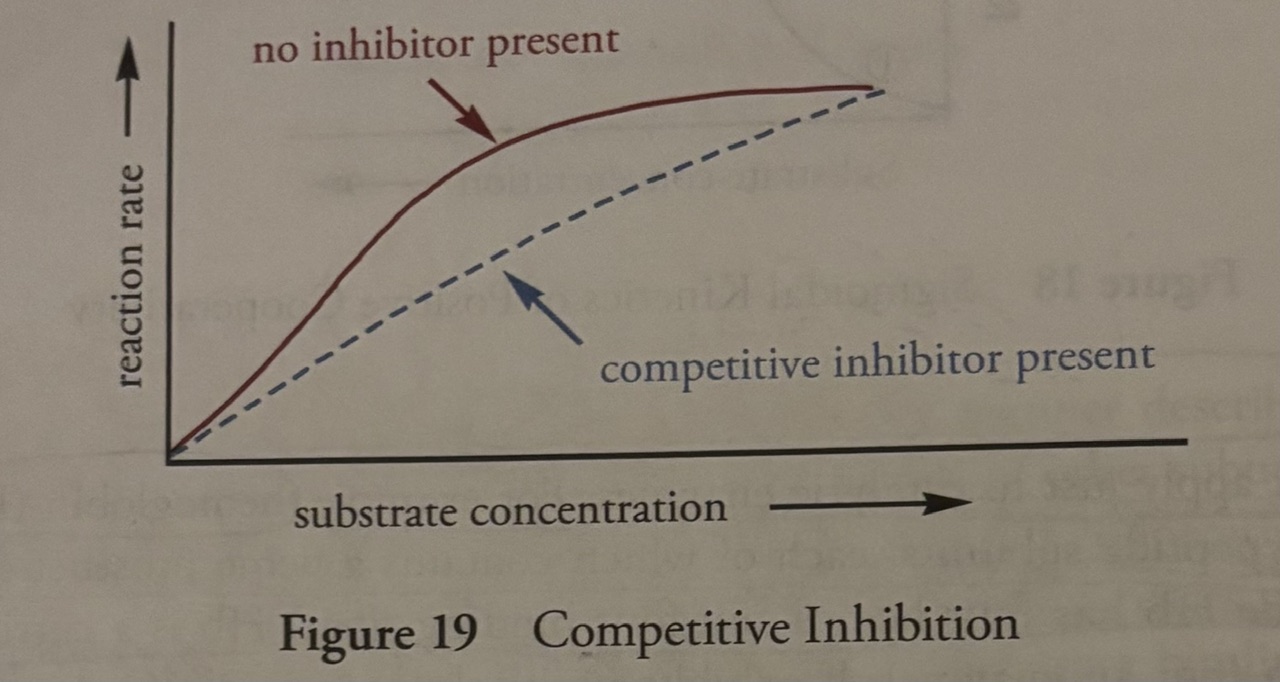
Question: you can predict that structurally, competitive inhibitors resemble what?
Answer: you can predict that structurally, competitive inhibitors resemble the substrate.
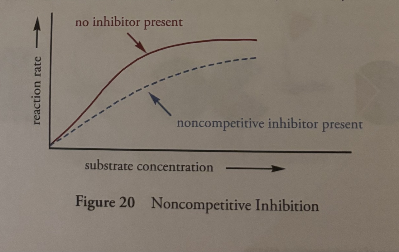
Non-competitive inhibition
-bind at the allosteric site, not at the active site. “non-competitive”. This means that no matter how much substrate you add, the inhibitor will not be displaced from its site of action.
-Km is not affected because the inhibitor binds to an allosteric site on both the free enzyme and the enzyme-substrate complex with equal affinity. Because the enzyme's affinity for the substrate does not change, Km remains unchanged.
-Vmax is diminished because fewer functional enzymes can turn substrate into product
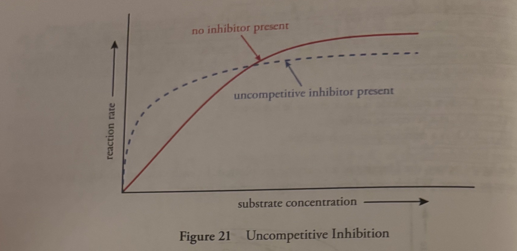
Uncompetitive Inhibition
-the inhibitor binds to the enzyme-substrate complex
-Km decreases because the inhibitor binds exclusively to the enzyme-substrate (ES) complex, the enzyme gains a higher affinity for the substrate and the Km decreases.
-Vmax decreases because the inhibitor binds exclusively to the enzyme-substrate complex (ES), rather than free enzyme, inhibiting the release of products
Mixed-type inhibition
-occurs when an inhibitor can bind to either the unoccupied enzyme by the enzyme-substrate complex.
-inhibitor binds to allosteric site
-if the inhibitor binds to the enzyme in free form, the enzyme’s affinity for substrate decreases, and Km increases
-if the inhibitor binds to the enzyme-substrate complex, the enzyme’s affinity for the substrate increases, and Km decreases.
-Vmax decreases
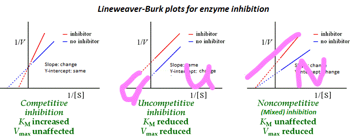
![<p><u>Line-weaver Burke Plot (Picture on Flashcard)</u></p><p>-a graphical representation of enzyme kinetics using the LineWeaver Burke Equation</p><p>(equation on the page)</p><p>-The equation is in the form of y=mx+b</p><p>-y-axis is the inverse of reaction rate (1/V). This means that as y goes up, the reaction rate decreases</p><p>-x-axis is the inverse of substrate concentration (1/[S]). This means that as x gets longer, the substrate concentration decreases.</p><ol><li><p>The slope of the graph is <strong>Km/Vmax</strong>. The purpose of this value—along with the graph's intercepts—is to linearize the Michaelis-Menten equation, allowing scientists to easily calculate the enzyme’s substrate affinity (Km) and maximum speed (Vmax) and to identify different types of enzyme inhibition.</p></li><li><p>The Y-intercept of the graph is 1/Vmax, allows you to easily find and calculate the maximum speed (Vmax)</p></li><li><p>The X-intercept of the graph is -1/Km, allows you to easily calculate and analyze the Michaelis constant Km of an enzyme.</p></li></ol><p></p>](https://assets.knowt.com/user-attachments/a3f09ef5-9da4-4259-afe2-74ce3ba4bba4.jpg)
Line-weaver Burke Plot (Picture on Flashcard)
-a graphical representation of enzyme kinetics using the LineWeaver Burke Equation
(equation on the page)
-The equation is in the form of y=mx+b
-y-axis is the inverse of reaction rate (1/V). This means that as y goes up, the reaction rate decreases
-x-axis is the inverse of substrate concentration (1/[S]). This means that as x gets longer, the substrate concentration decreases.
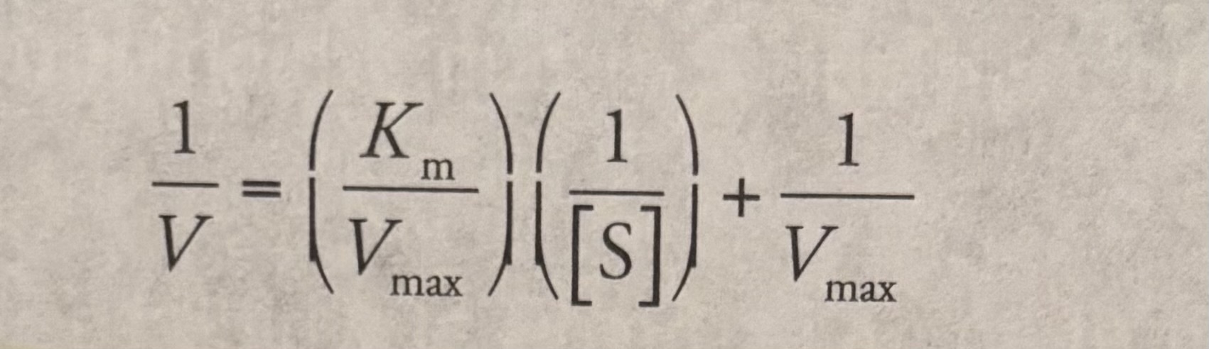
The slope of the graph is Km/Vmax. The purpose of this value—along with the graph's intercepts—is to linearize the Michaelis-Menten equation, allowing scientists to easily calculate the enzyme’s substrate affinity (Km) and maximum speed (Vmax) and to identify different types of enzyme inhibition.
The Y-intercept of the graph is 1/Vmax, allows you to easily find and calculate the maximum speed (Vmax)
The X-intercept of the graph is -1/Km, allows you to easily calculate and analyze the Michaelis constant Km of an enzyme.
Lineweaver Burke Plot Equation
Question: how would the lineweaver burke plot change when a noncompetitive inhibitor is added?
answer: a non-competitive inhibitor binds to the allosteric space, lowers Vmax, does not affect Km.
The y-axis would be increased on a lineweaver burke plot (the y-axis correlates to Vmax) AND the slope increases.
Nothing would happen to the x-axis (x-axis is the inverse of substrate concentration)
Competitive inhibition
-inhibitor competes with substrate for the active site
-Km increased (enzyme has a lower affinity for the substrate), the slope is rise/run, Km/Vmax, a bigger numerator and a small denominator means a bigger #, so the slope has increased.
-Vmax is unchanged
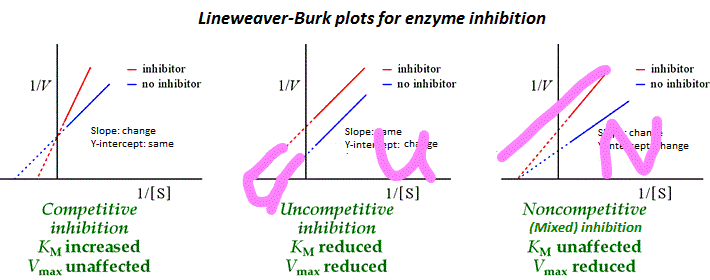
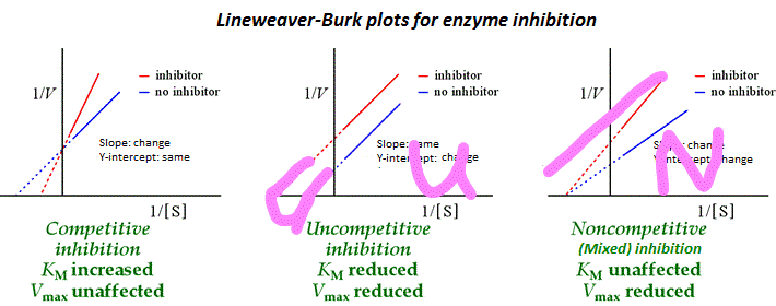
Non-competitive Inhibition
-inhibitor binds to allosteric site instead of active site
-Km does not change
-Vmax decreases
-the slope is rise/run, Km/Vmax, numerator does not change, denominator decreases, the slope will be bigger.
Uncompetitive Inhibition
-inhibitor binds to enzyme-substrate complex
-Enzyme affinity for substrate is increased because the inhibitor binds to enzyme substrate complex, therefore, Km is decreased
-Vmax decreases
-the slope is rise/run, Km/Vmax, numerator has decreased, denominator has decreased, slope does not change (because both values have decreased).