Bones and Bone Markings
 Call Kai
 Learn
Learn Practice Test
 Spaced Repetition
 Match
Match Flashcards
 Knowt Play
1/167

There's no tags or description
Looks like no tags are added yet.
Name | Mastery | Learn | Test | Matching | Spaced | Call with Kai | Chat |
|---|
No analytics yet
Send a link to your students to track their progress
168 Terms
skull (cranium)
composed of two sets of bones: the cranial bones and facial bones
frontal bone
bone that forms the forehead
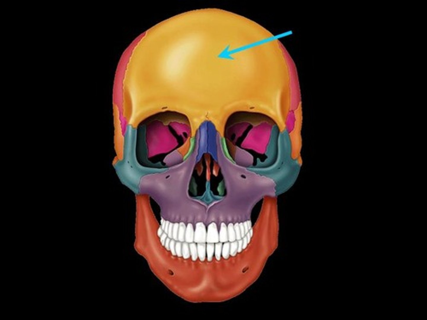
parietal bone
either of two skull bones between the frontal and occipital bones and forming the top and sides of the cranium
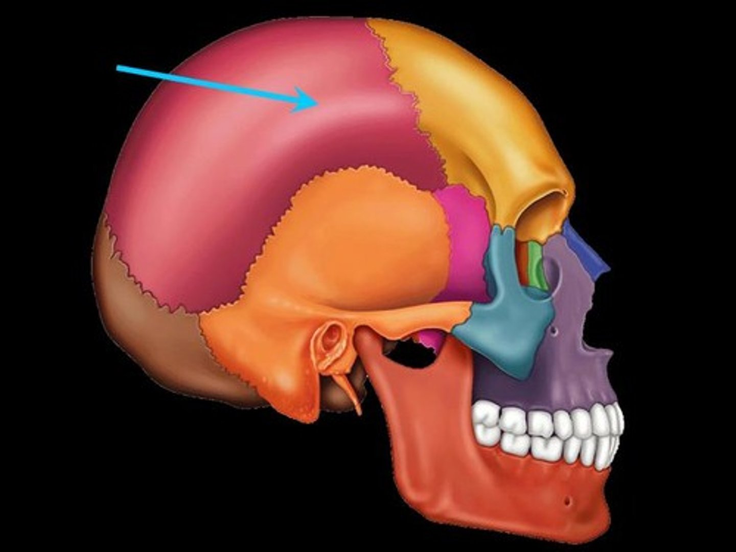
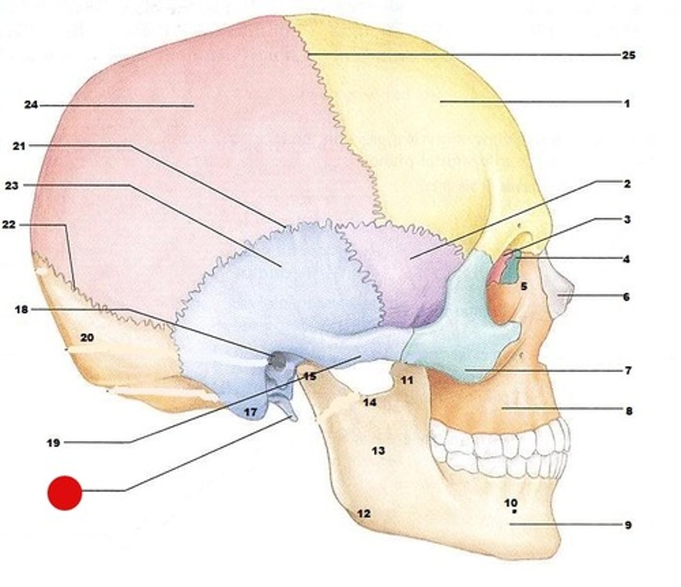
temporal bone
bone that forms parts of the side of the skull and floor of the cranial activity. There is a right and left temporal bone.
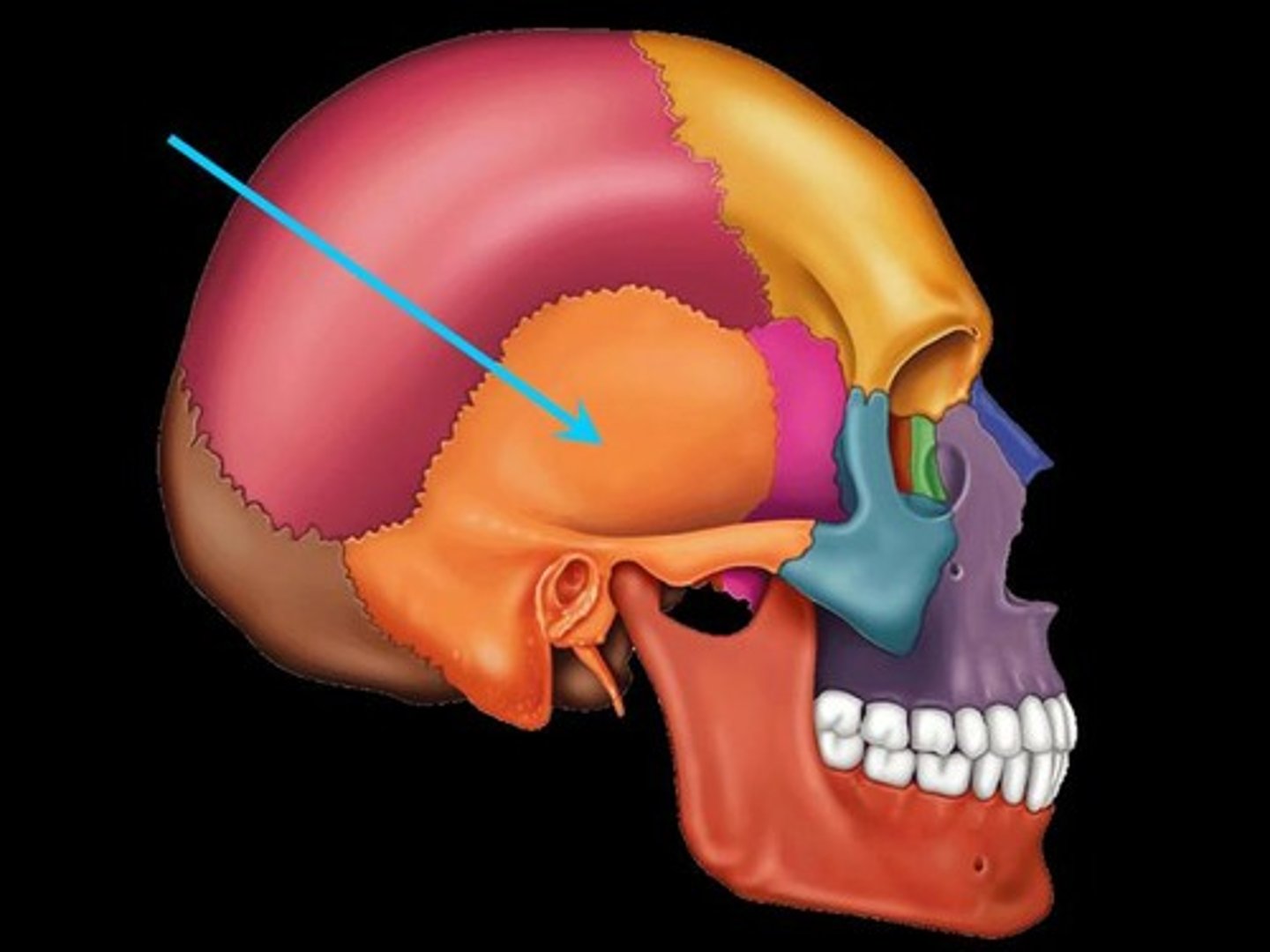
styolid process
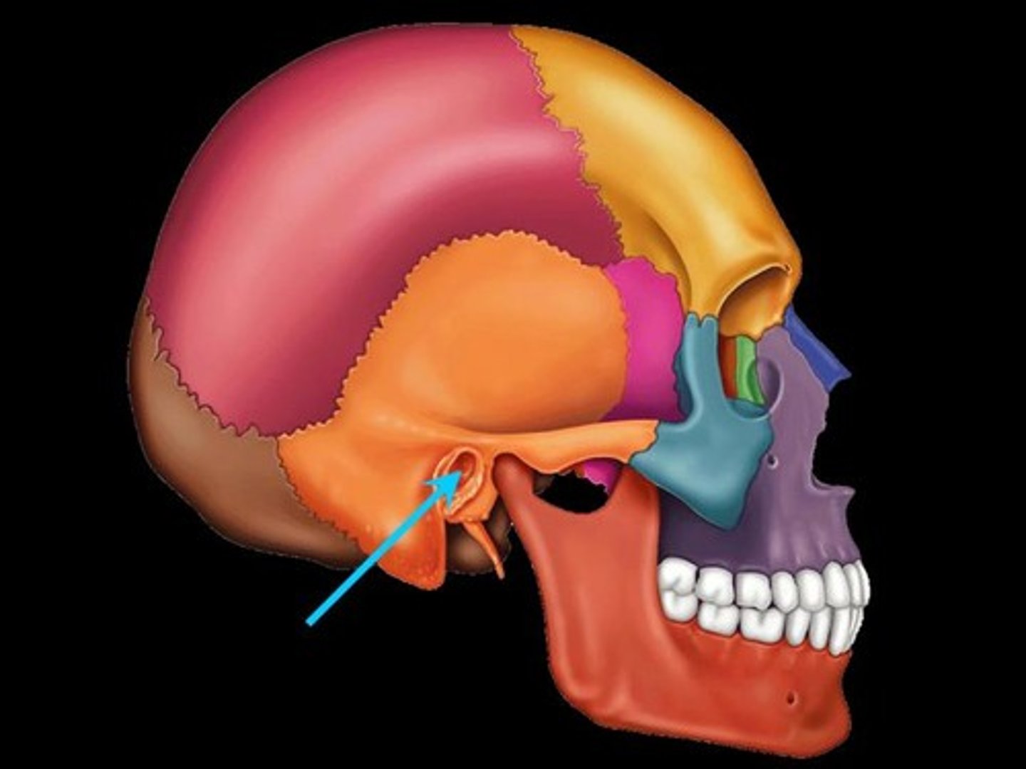
external auditory meatus
makes up the ear canal
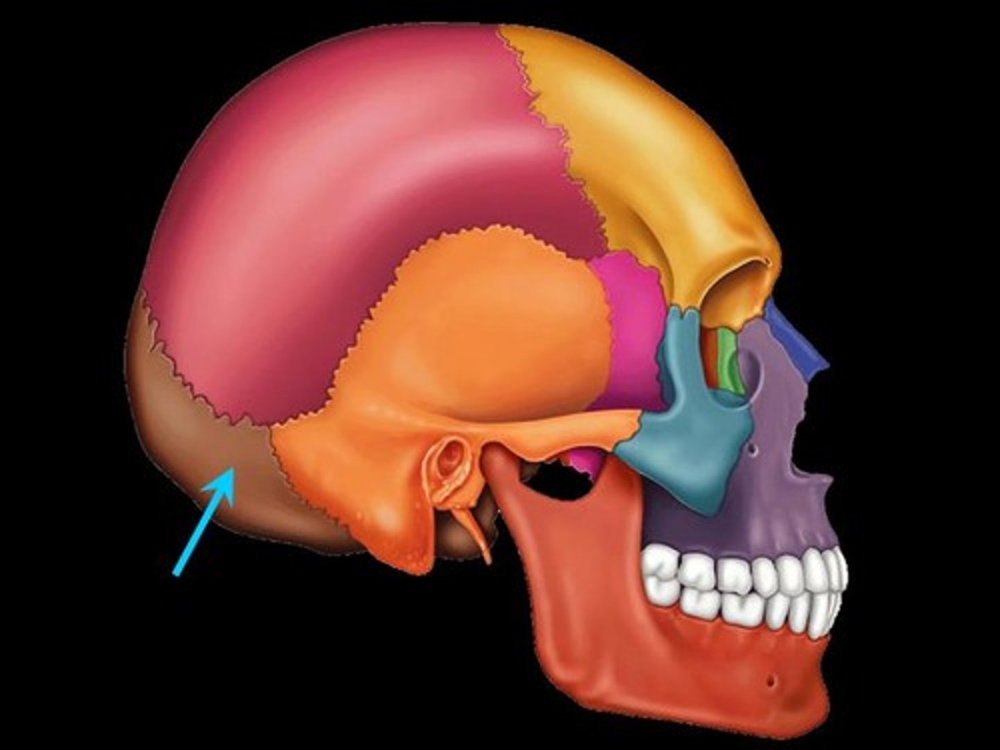
occipital bone
back of head
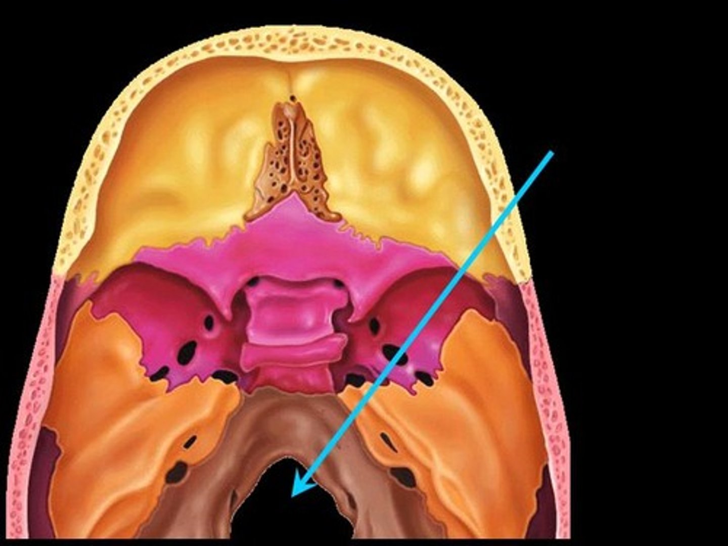
foramen magnum
A large opening at the base of the skull through which the brain connects to the spinal cord.
occipital condoyle
each of two rounded knobs on the occipital bone that form a joint with the first cervical vertebra.
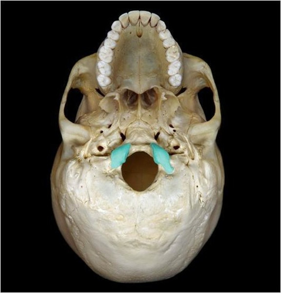
sphenoid bone
forms part of the base of the skull and parts of the floor and sides of the orbit
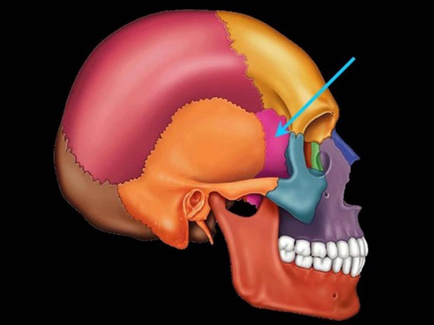
zygomatic process
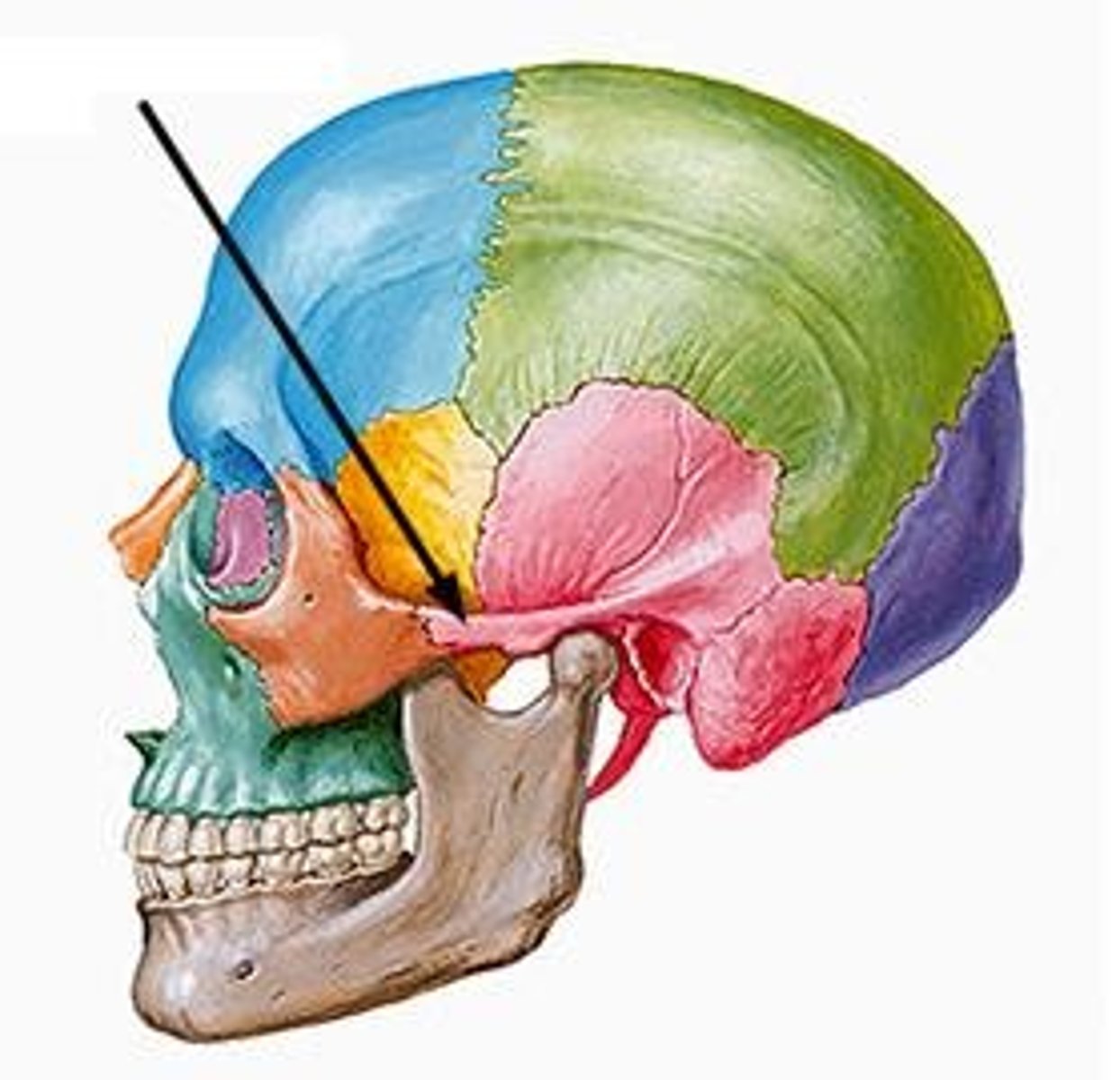
maxilla
upper jaw bone
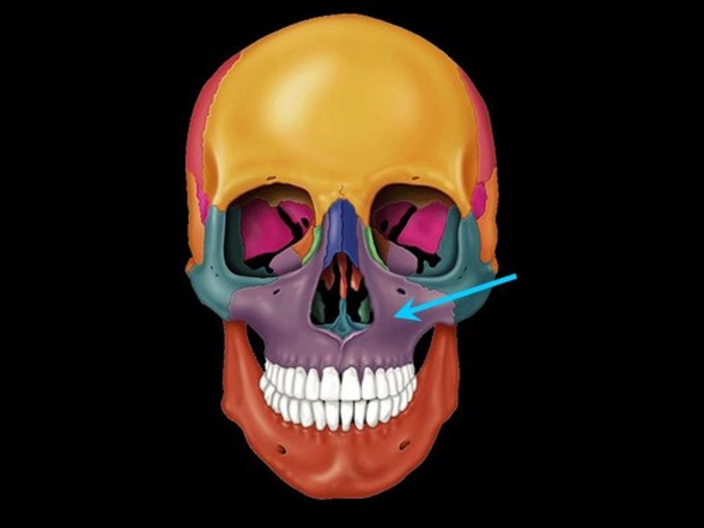
mandible
lower jaw bone
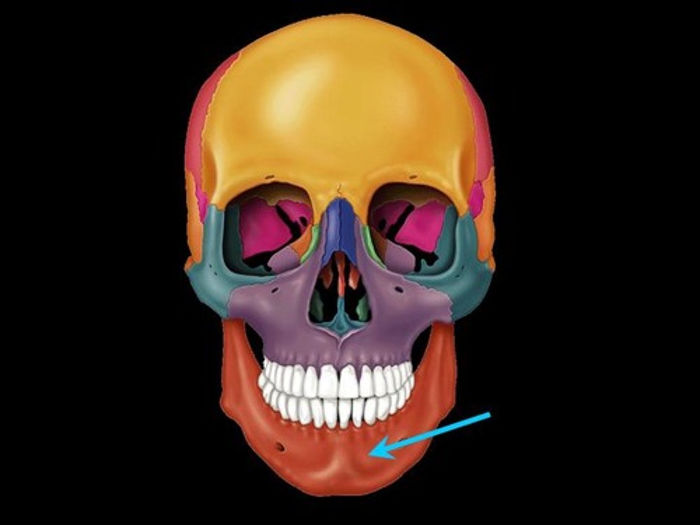
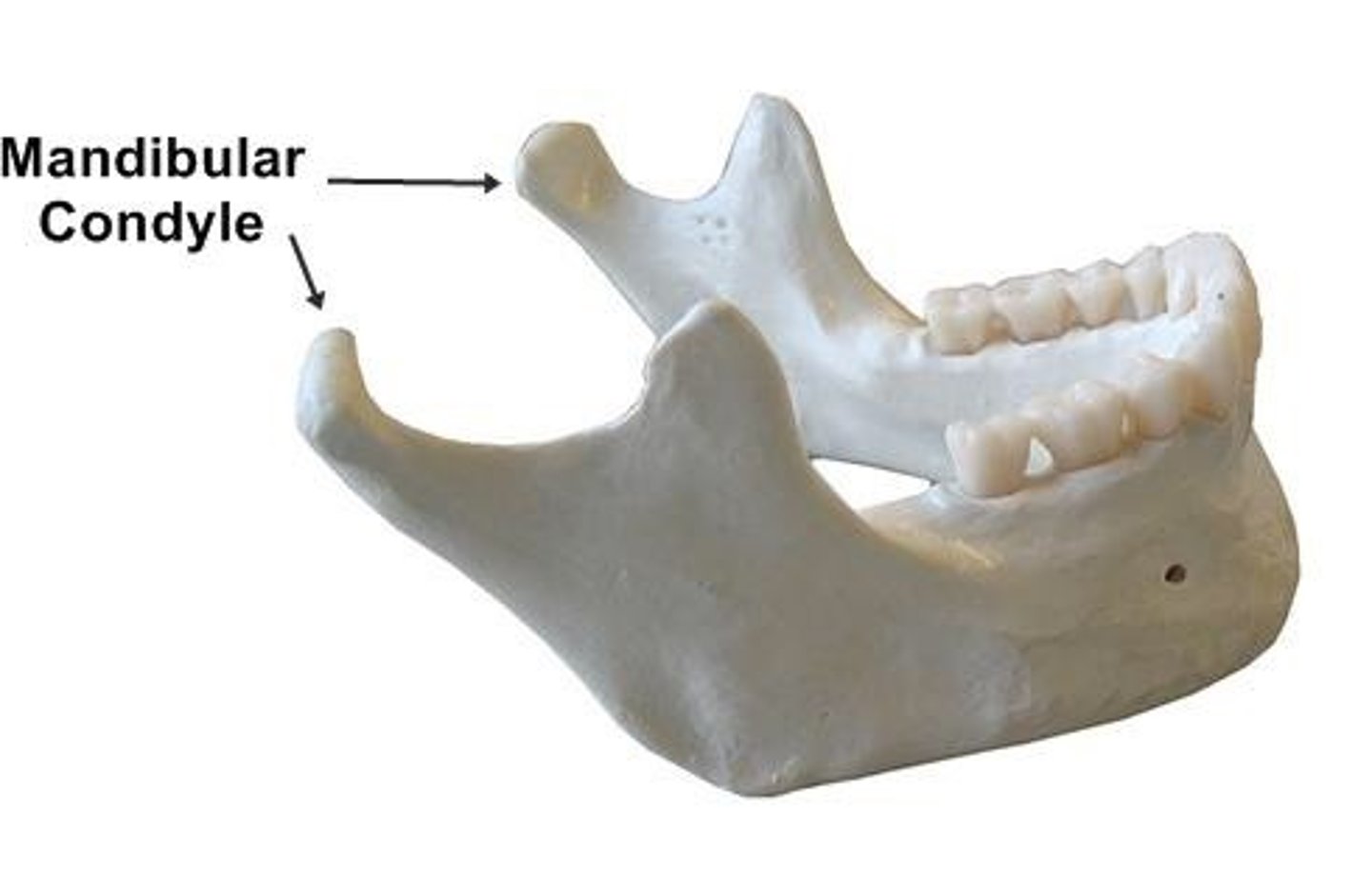
mandibular condoyle
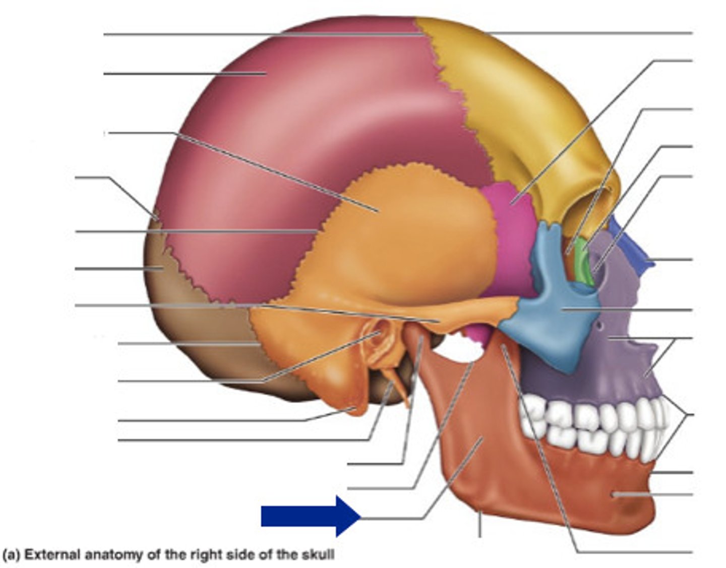
mandibular ramus
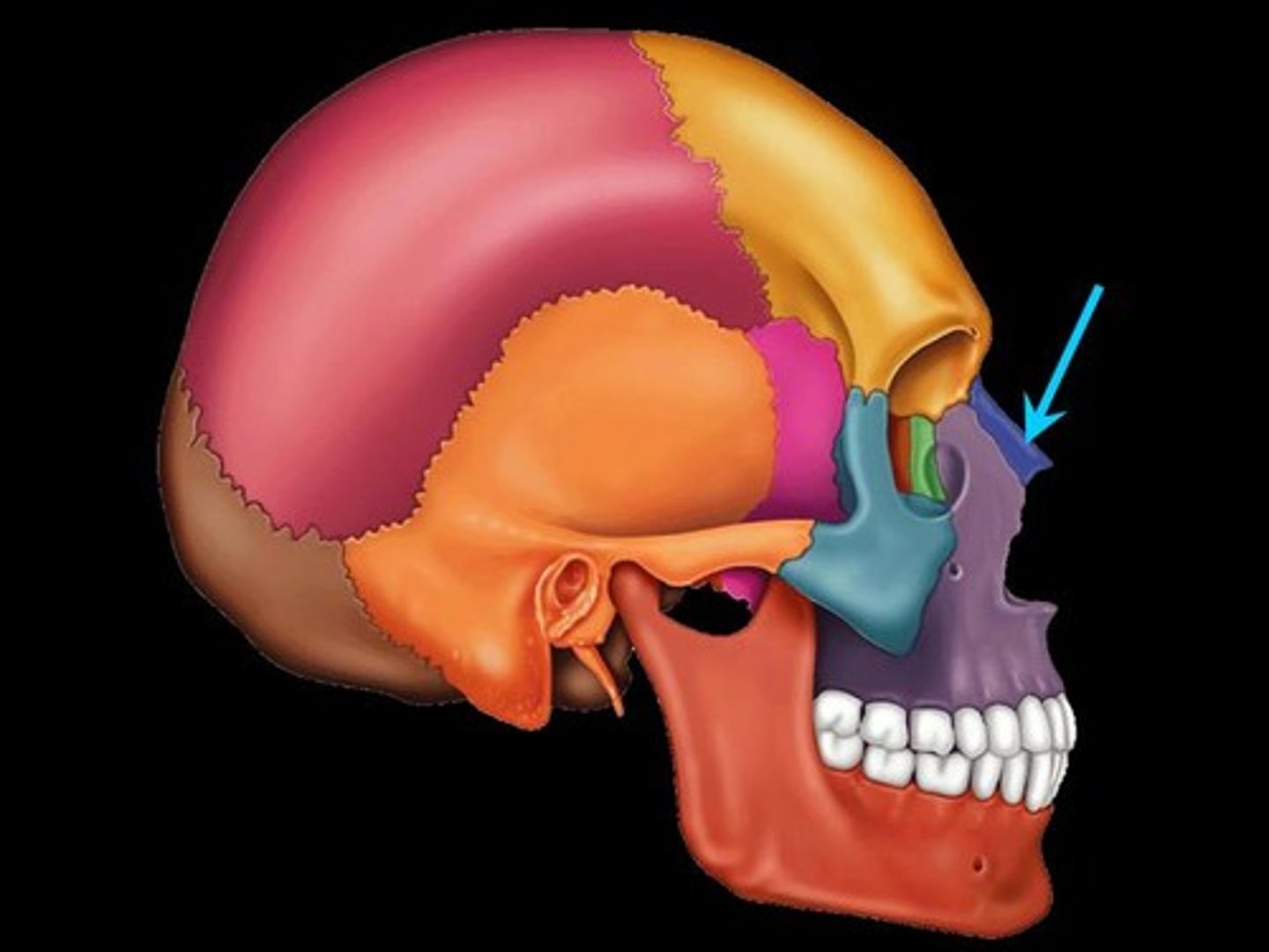
nasal bones
Bones that form the bridge of the nose.
vomer
forms the inferior portion of the nasal septum
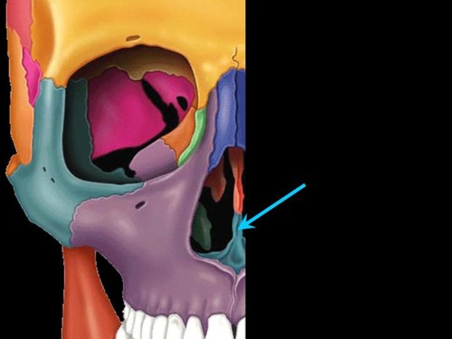
coronal suture
the suture between the parietal and frontal bones of the skull
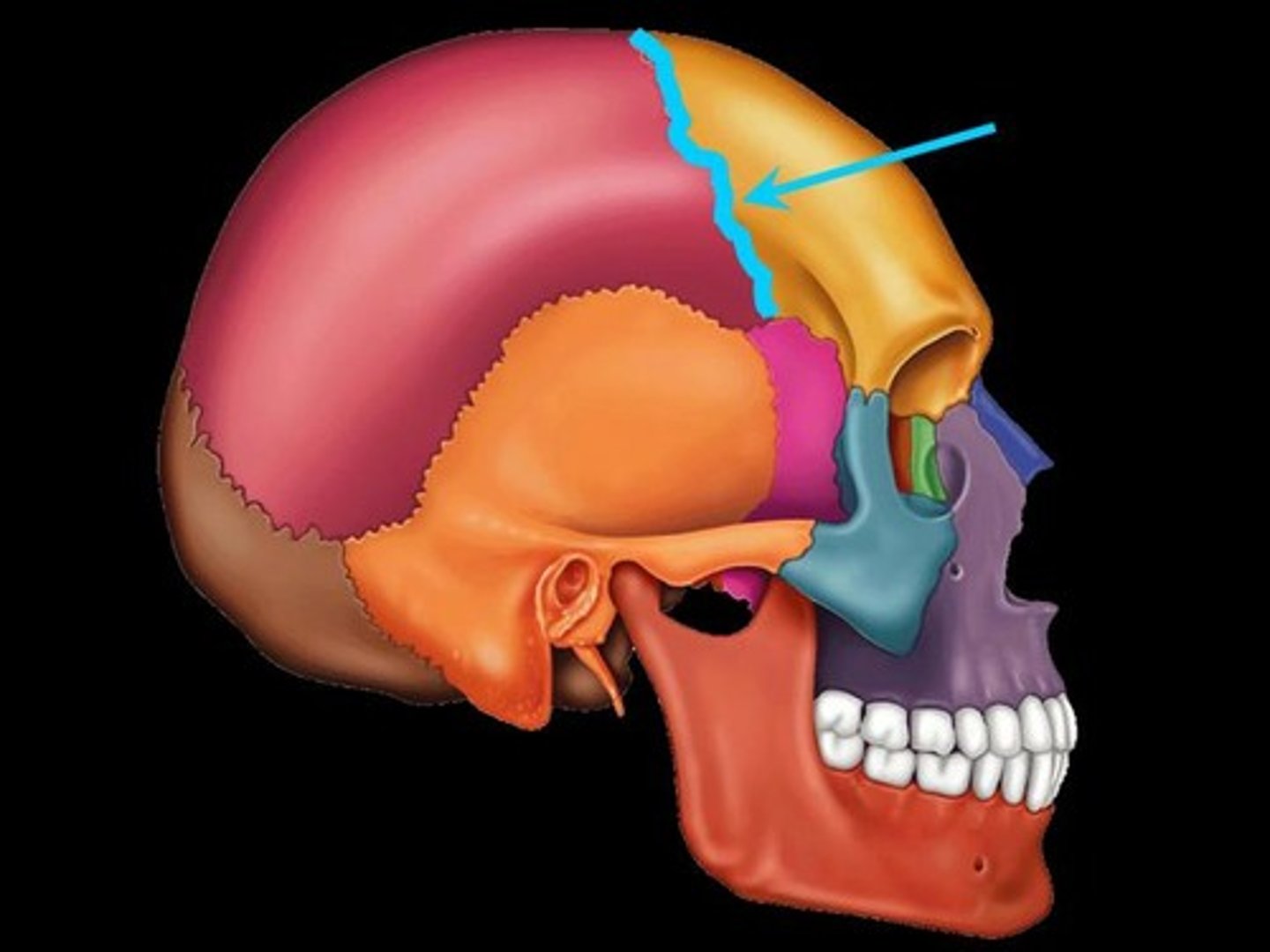
sagittal suture
the suture between the right and left parietal bones
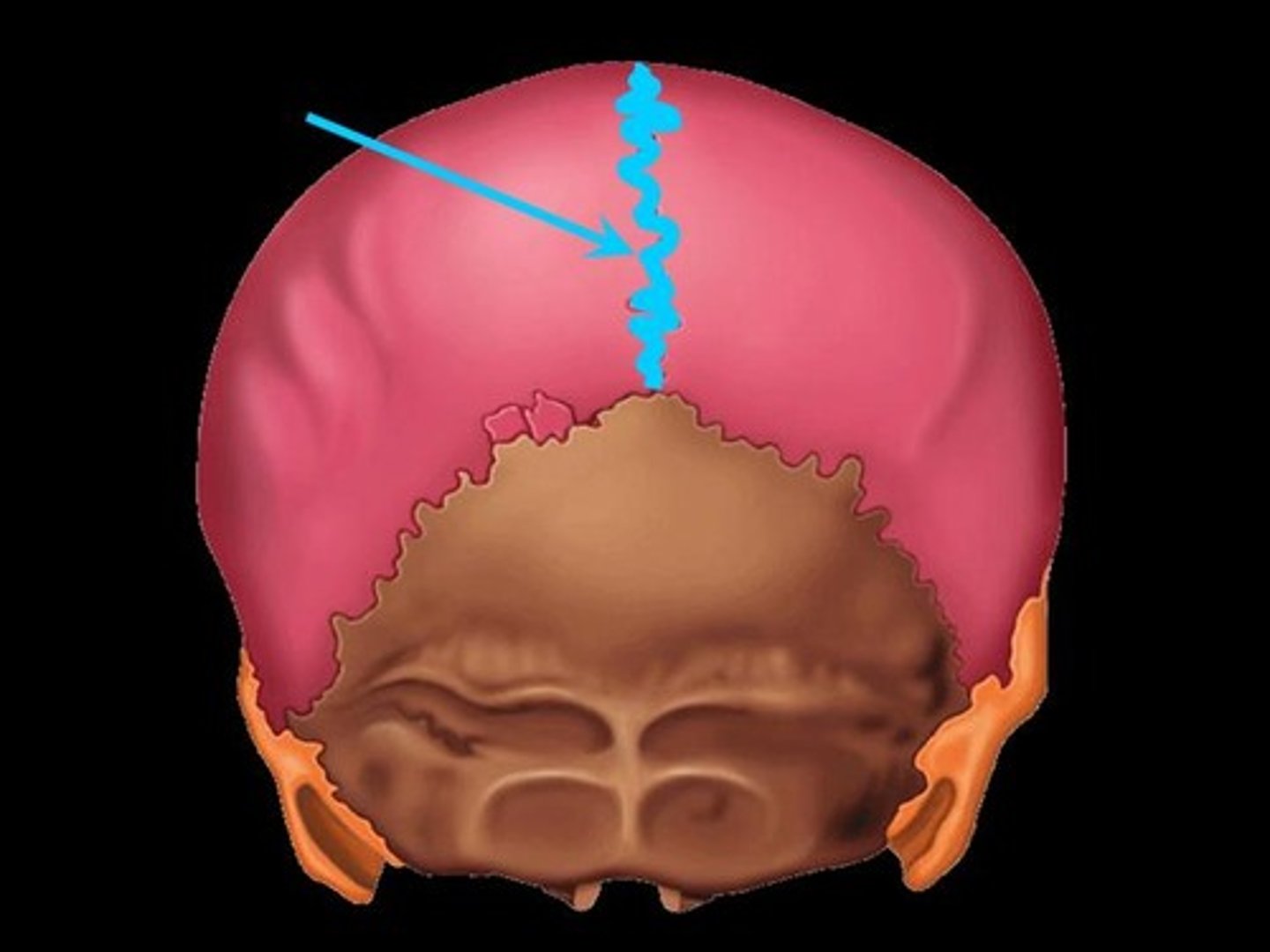
lambdoid suture
between parietal bones and occipital bone
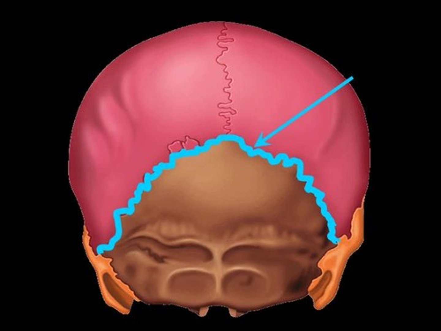
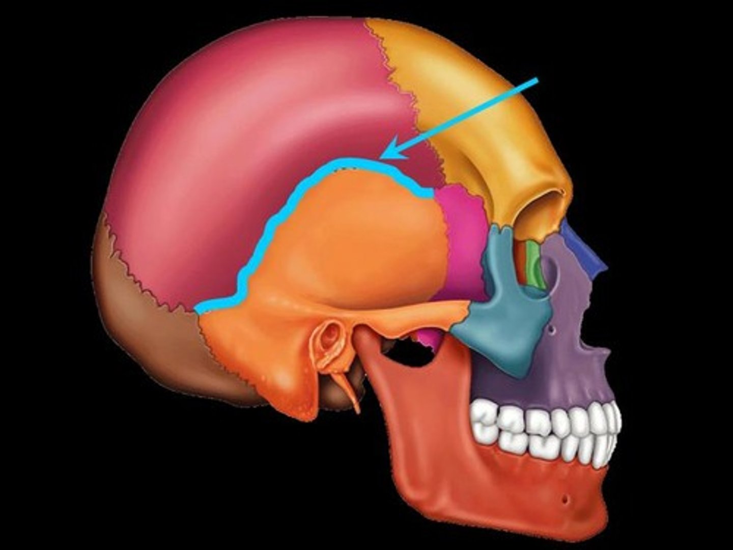
squamous (squamousal) suture
suture between the parietal and temporal bones
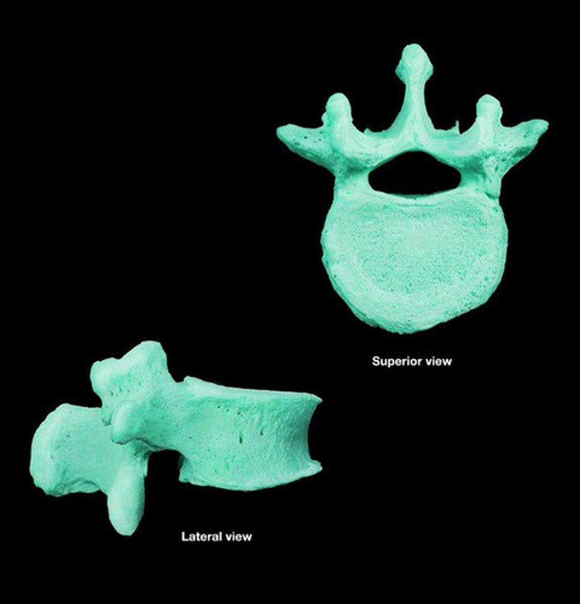
vertebrae
spine
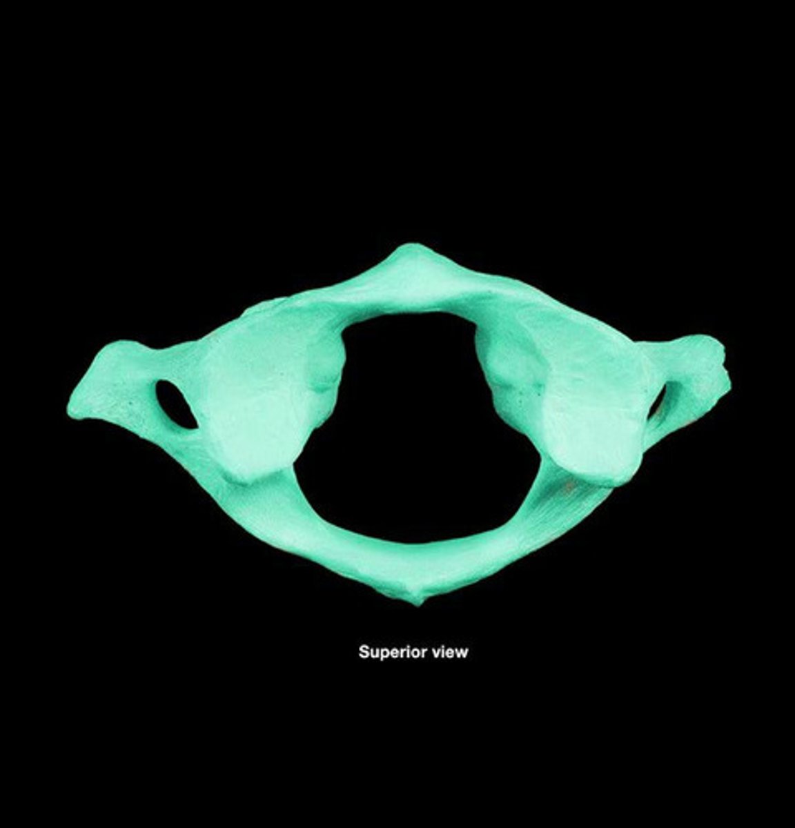
atlas
first cervical vertebra (C1), jointed with the occipital bone allowing the head to nod
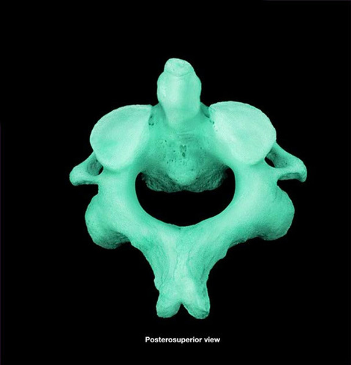
axis
the second cervical vertebrae (C2), allows the head to shake "no"
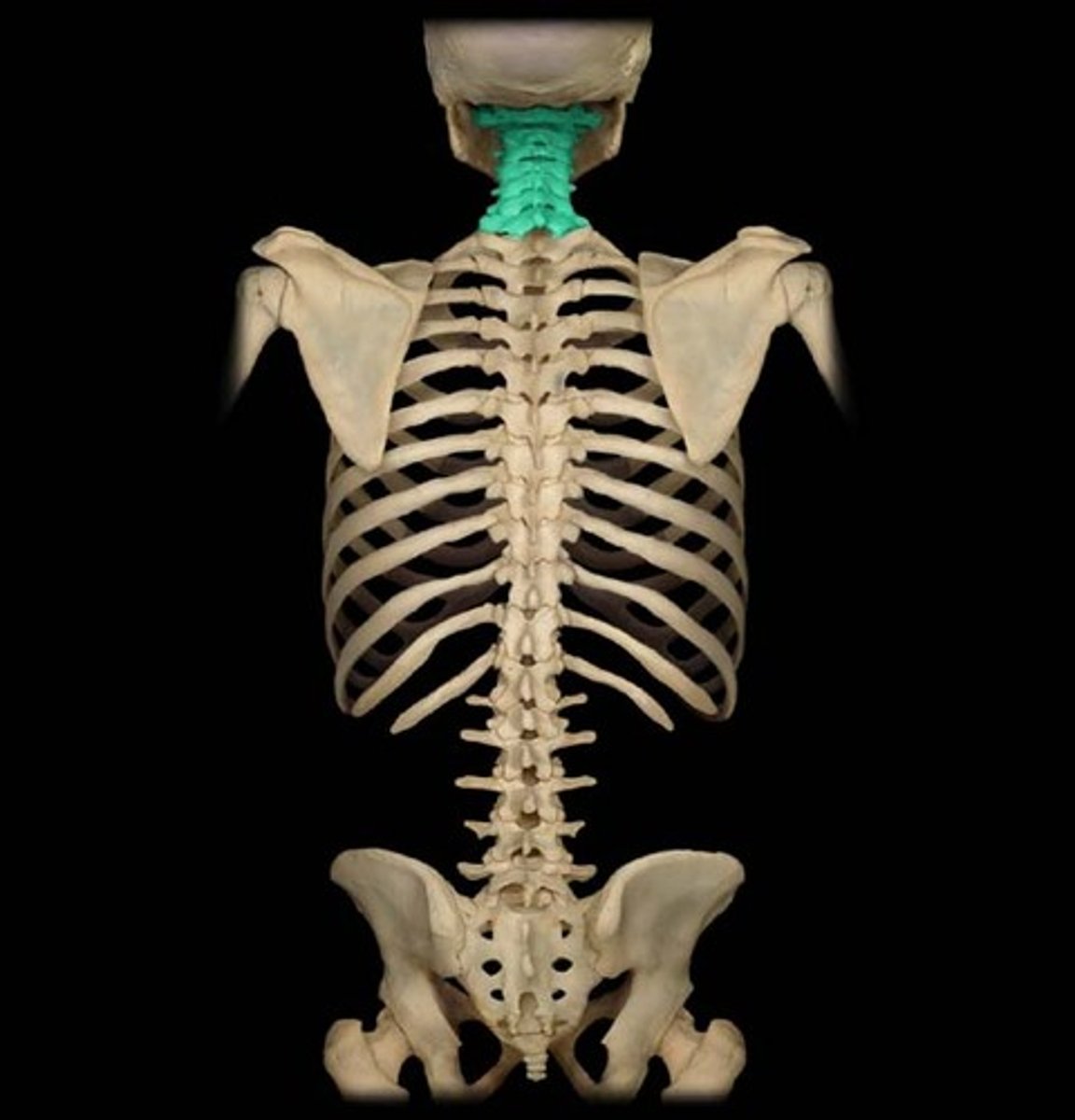
cervical
neck region of the spine (C1 - C7)
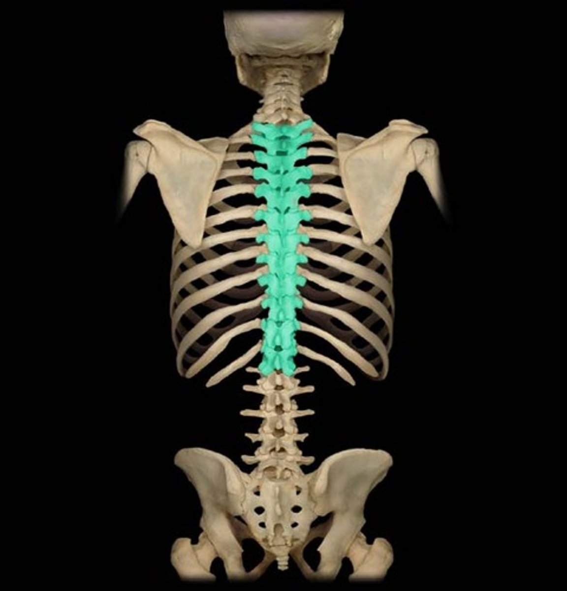
thoracic
chest region of the spine (T1 - T12). Characterized by long, sharp spinous processes and rib facets
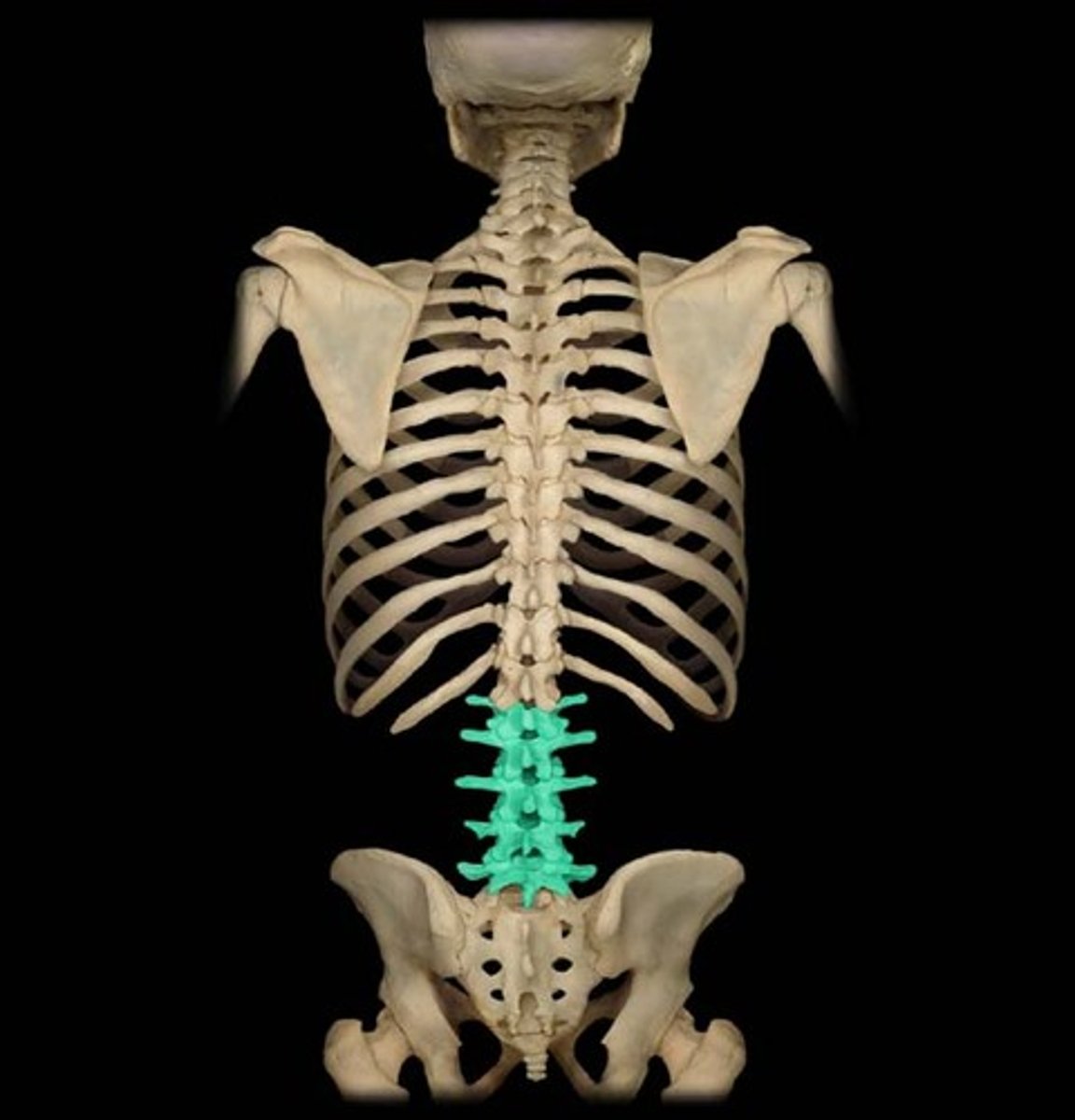
lumbar
back region of the spine (L1 - L5). Characterized by short, blunt spinous process and large body.
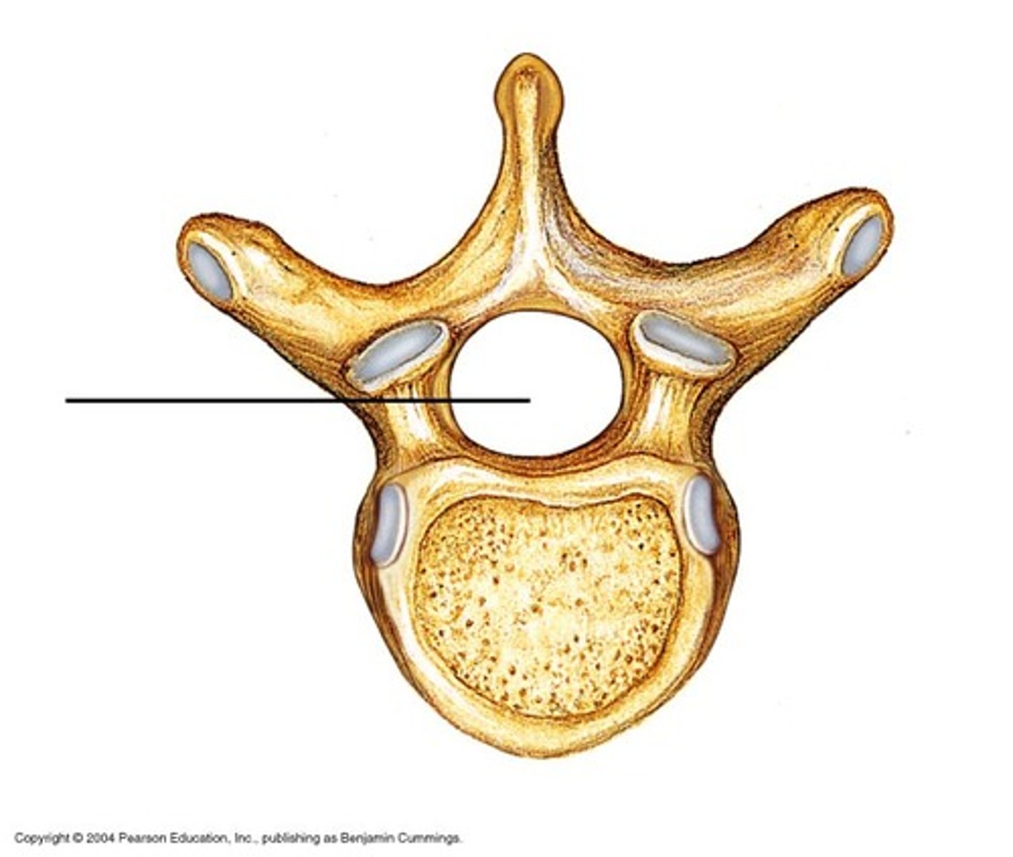
vertebral foramen
canal through which spinal cord passes
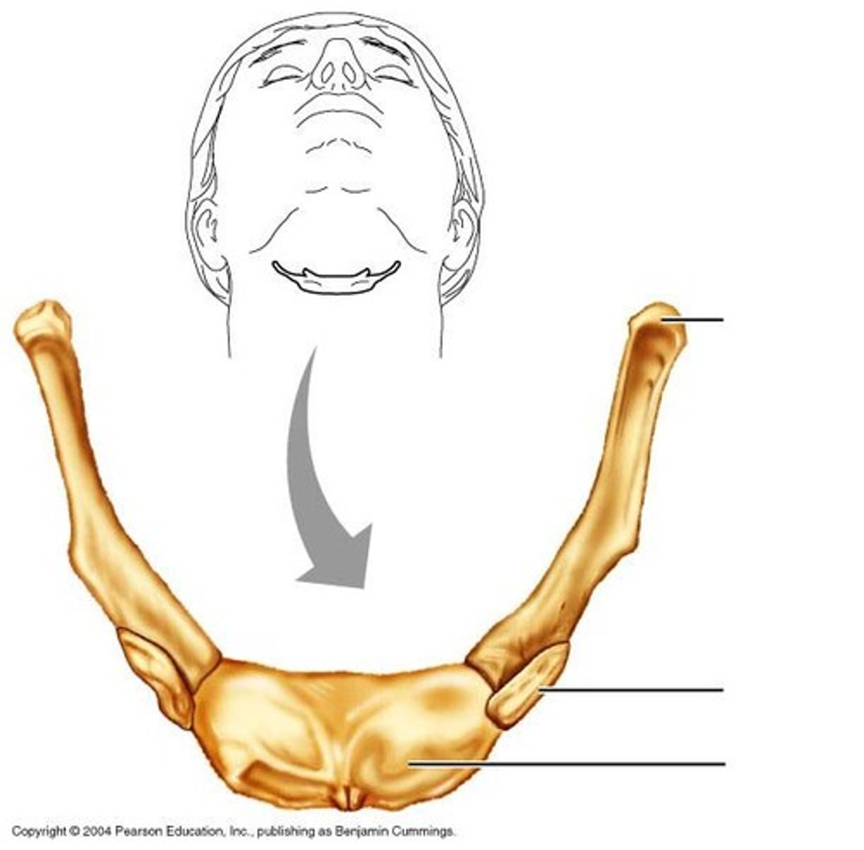
hyoid bone
U-shaped bone at the base of the tongue that supports the tongue and its muscles.
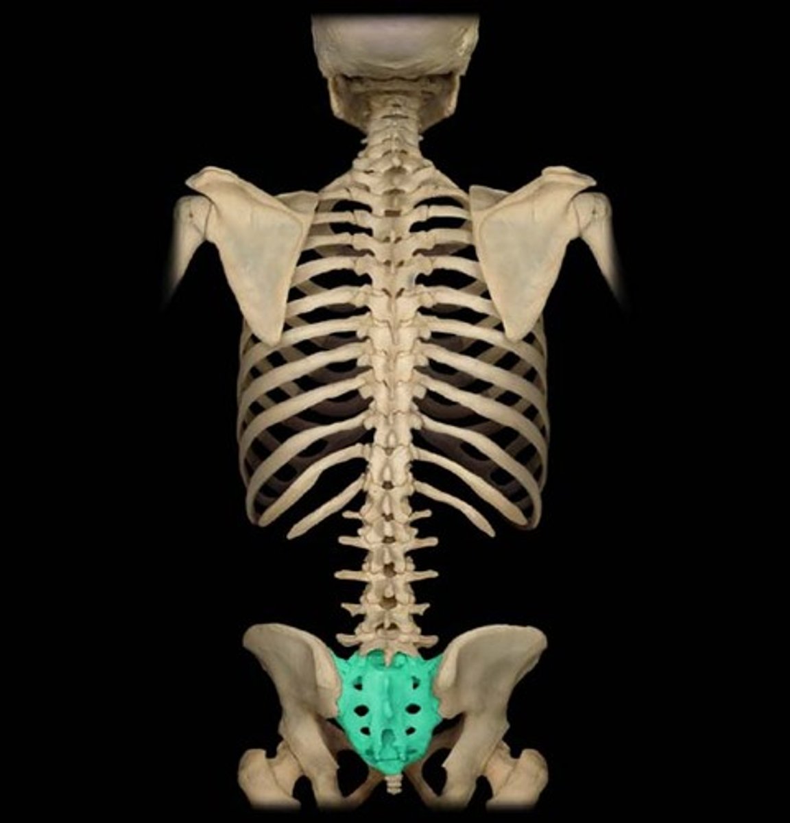
sacrum
bone formed from five vertebrae fused together near the base of the spinal column
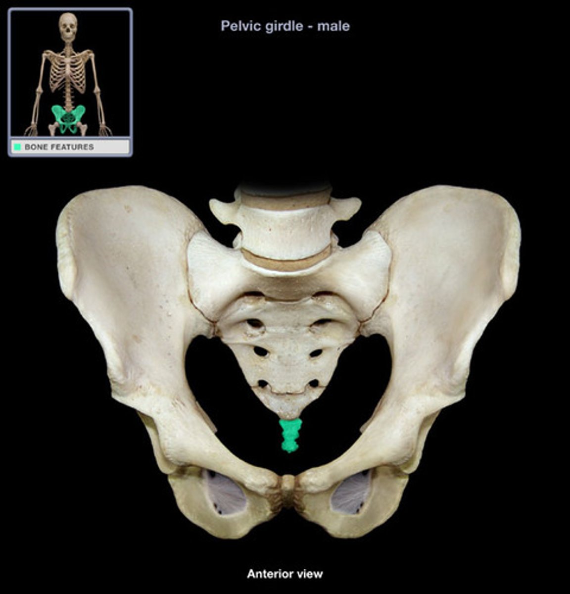
coccyx
four vertebrae fused together to form the tailbone
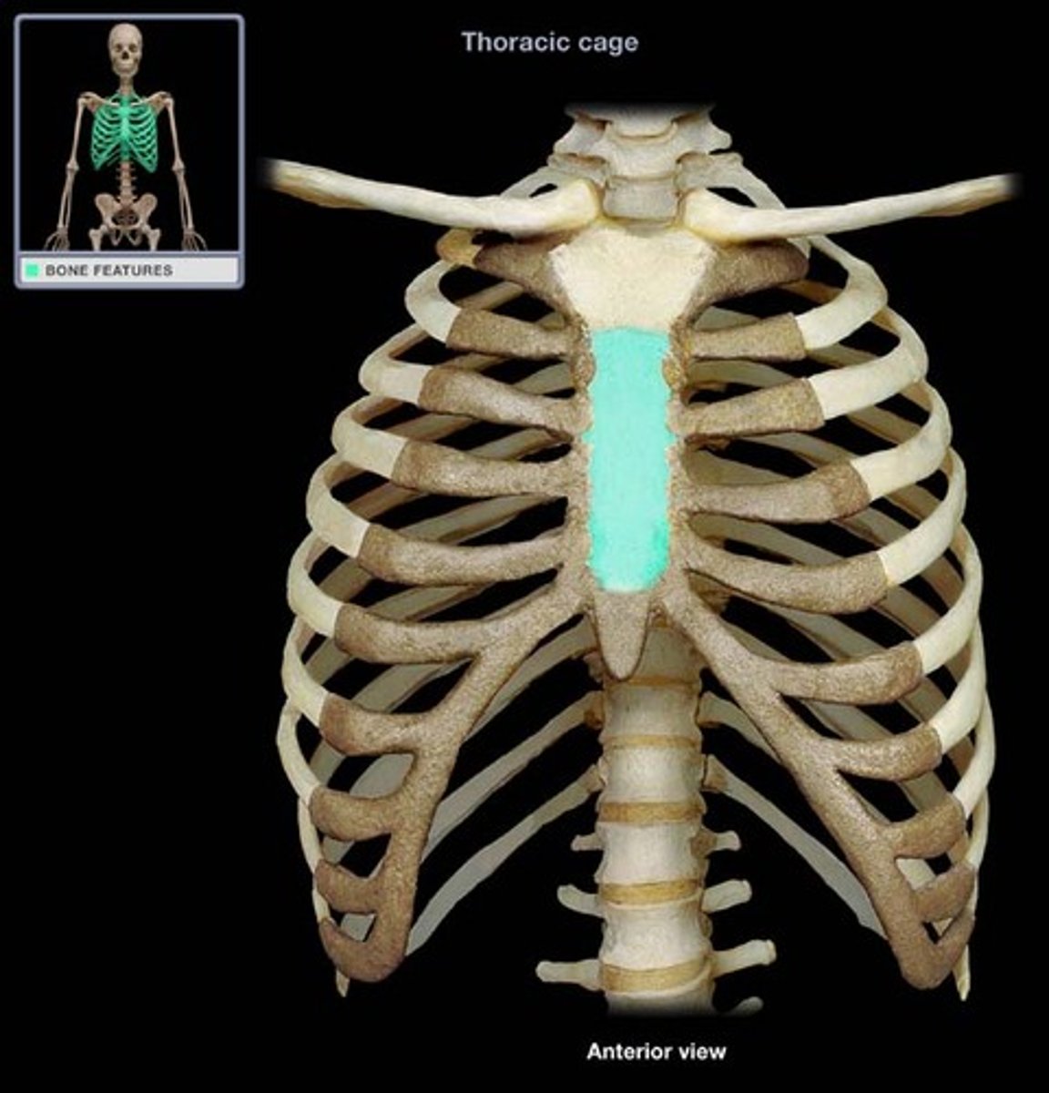
sternum
breastbone
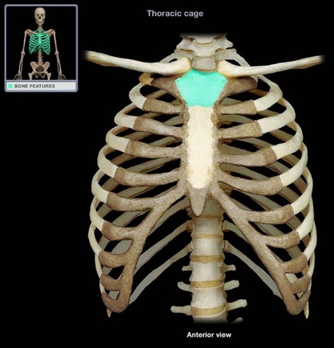
manubrium
the superior portion of the sternum
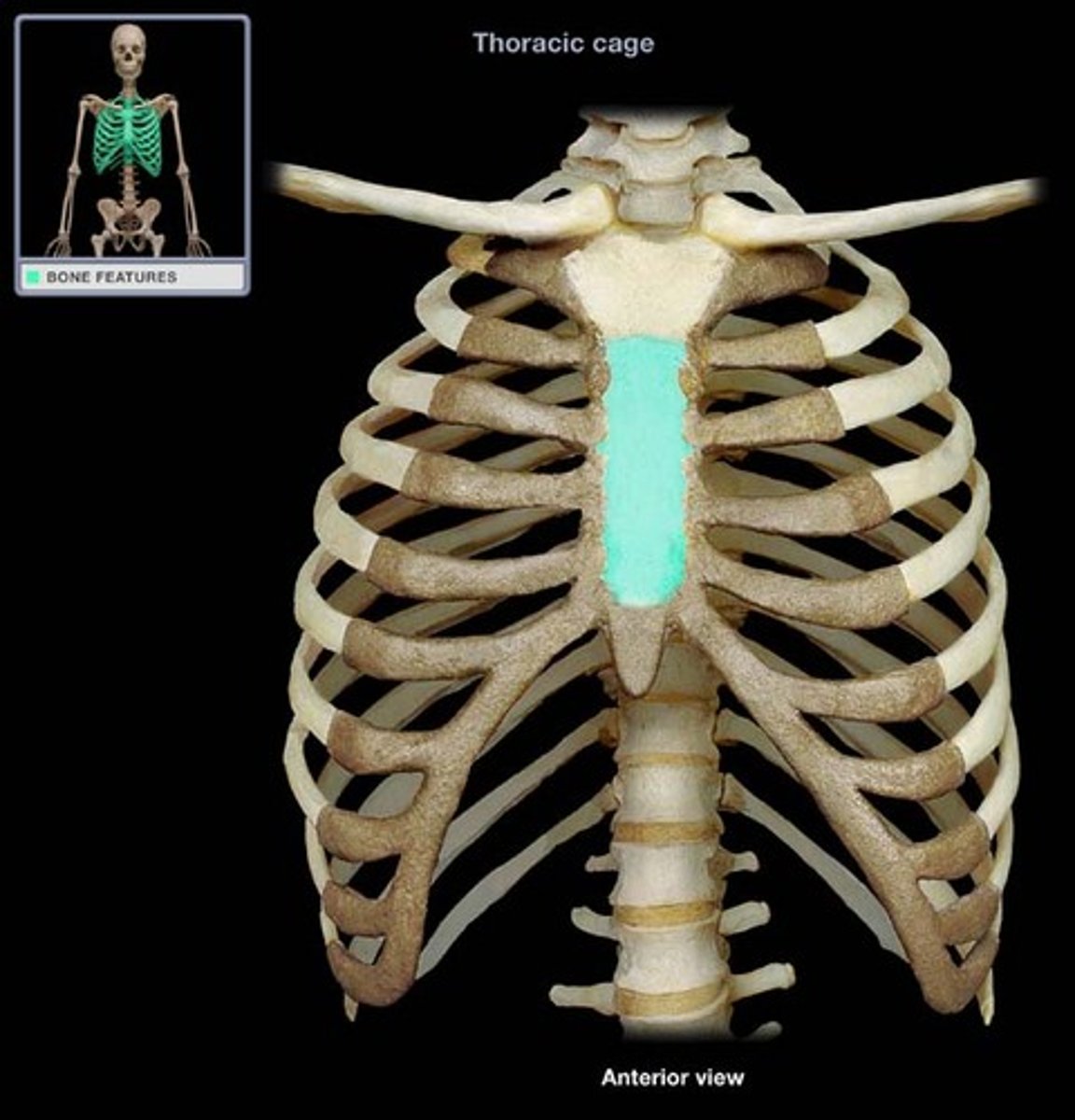
sternum body
main long part of sternum
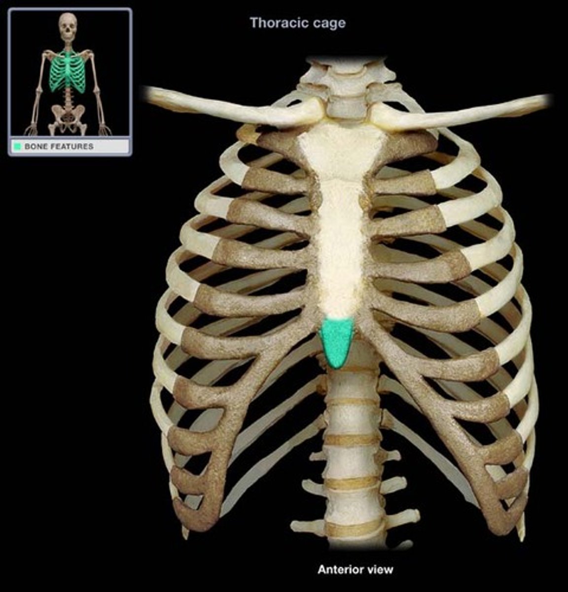
xiphoid process
inferior portion of the sternum
rib
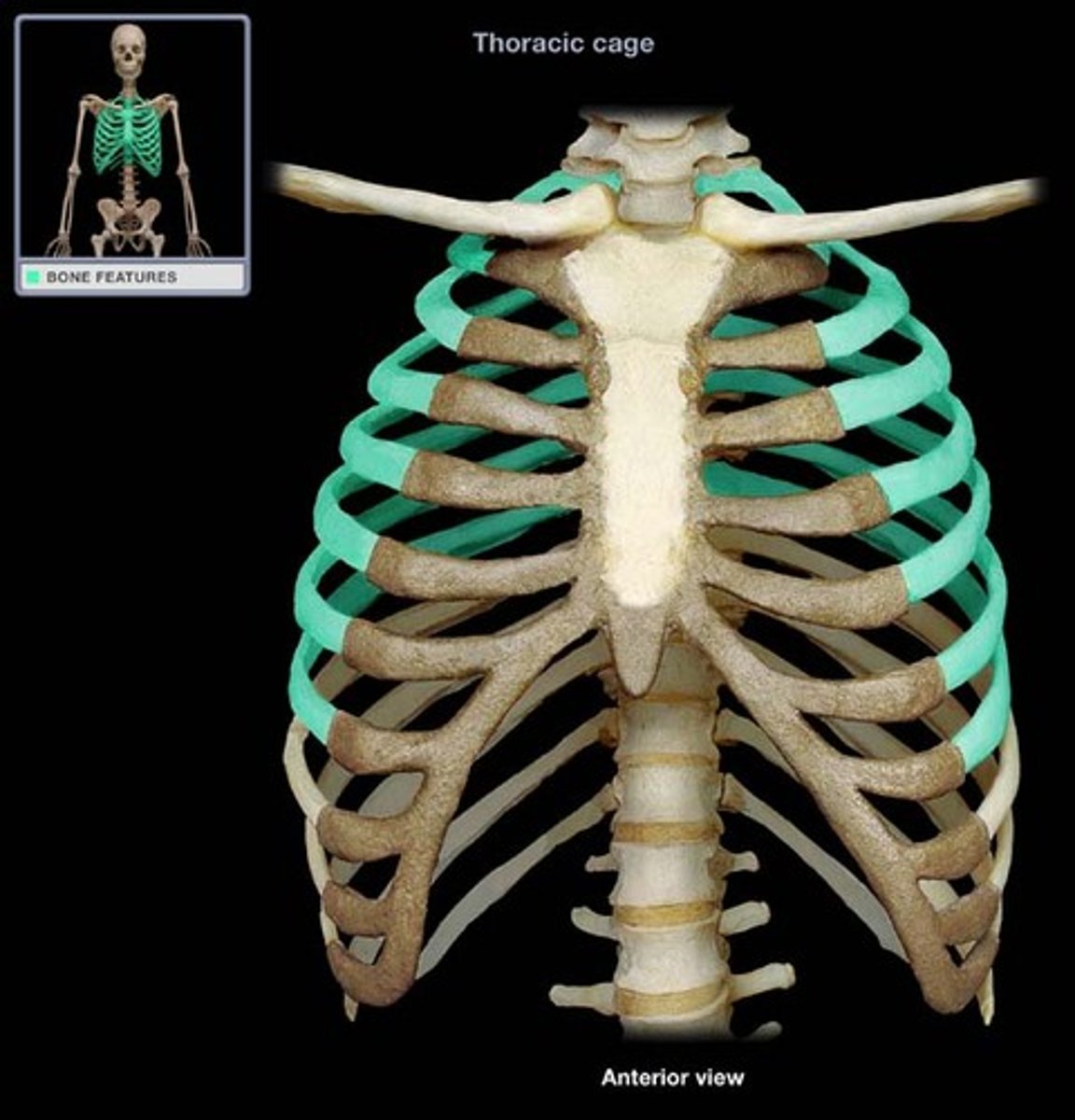
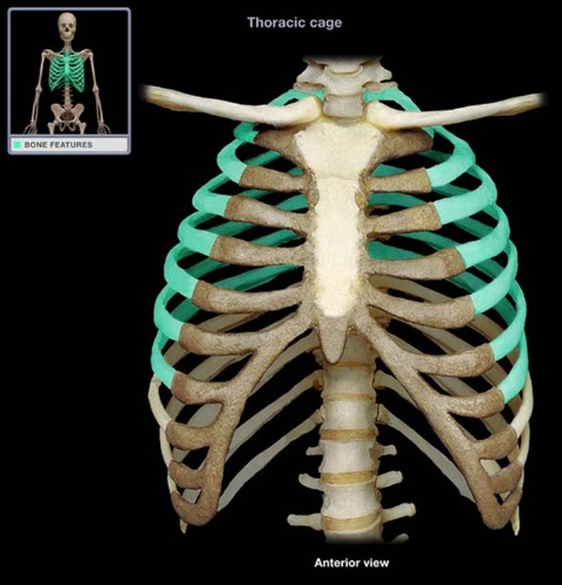
true ribs
1-7 attach directly to sternum
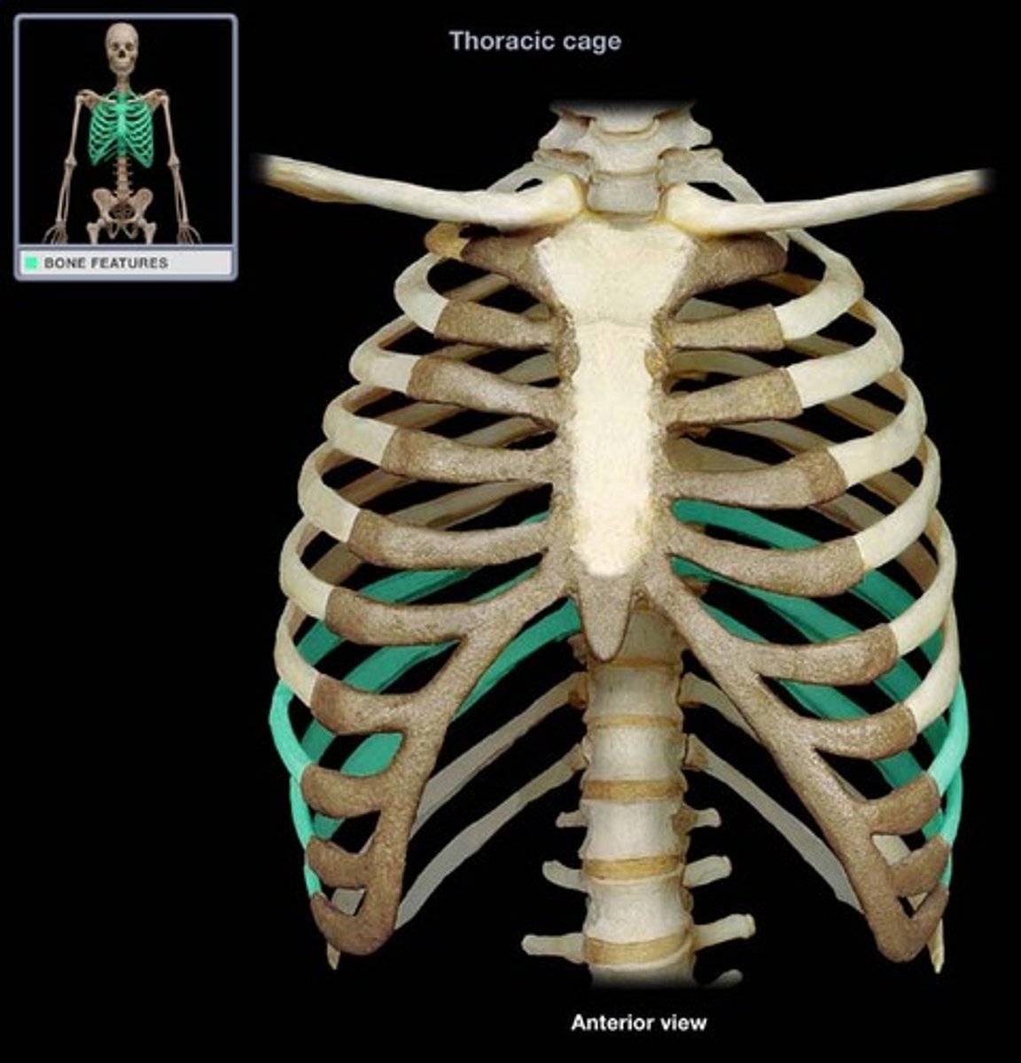
false ribs
8-12 do not attach directly to sternum
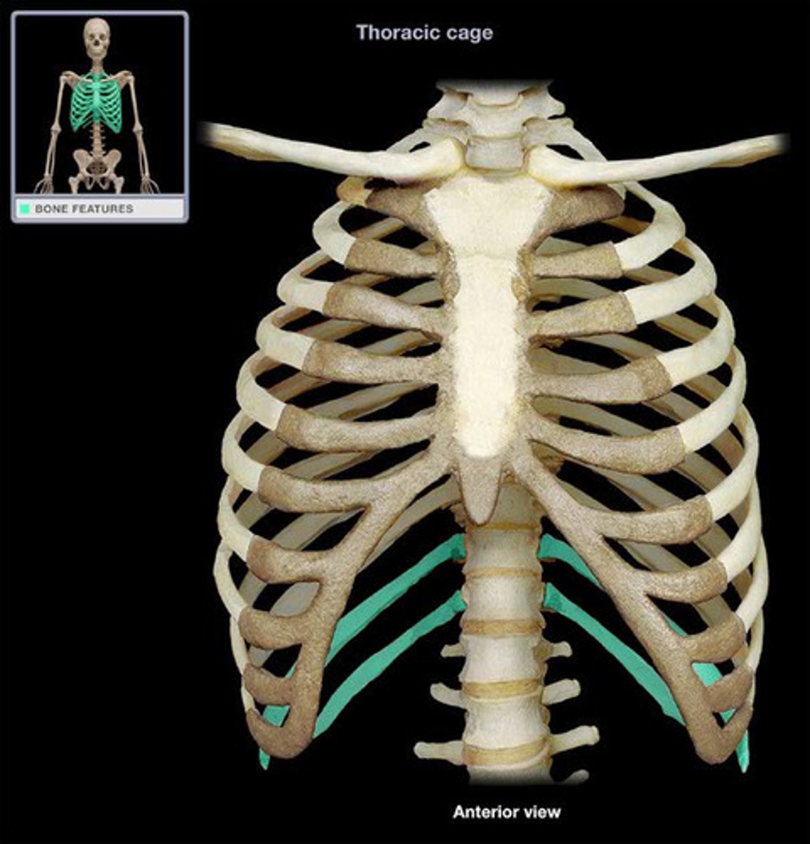
floating ribs
11-12 do not attach to sternum
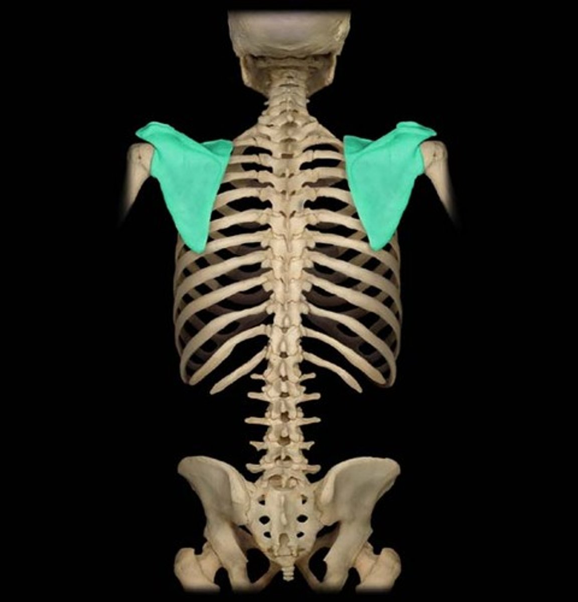
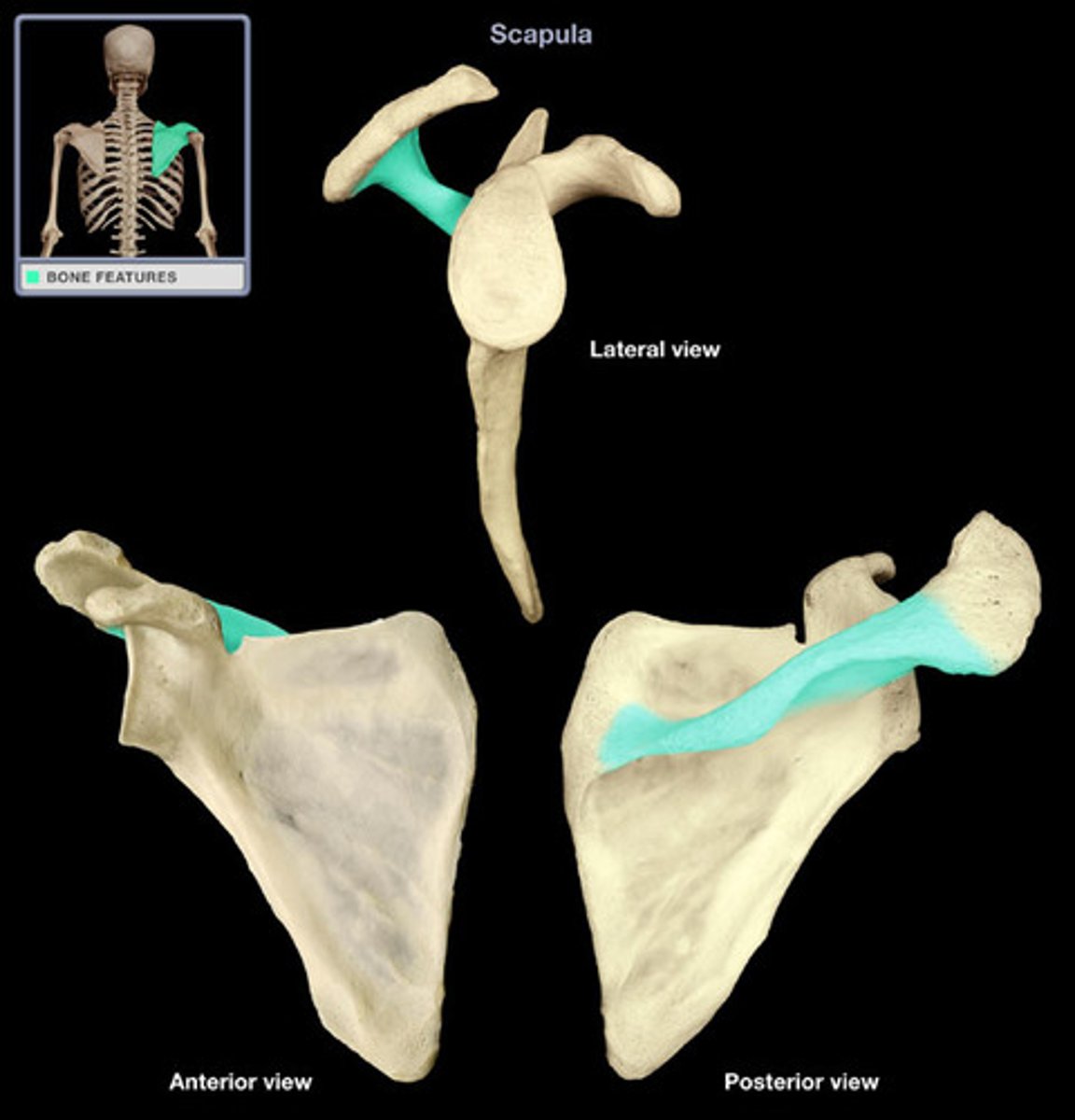
scapula
shoulder blade
spine
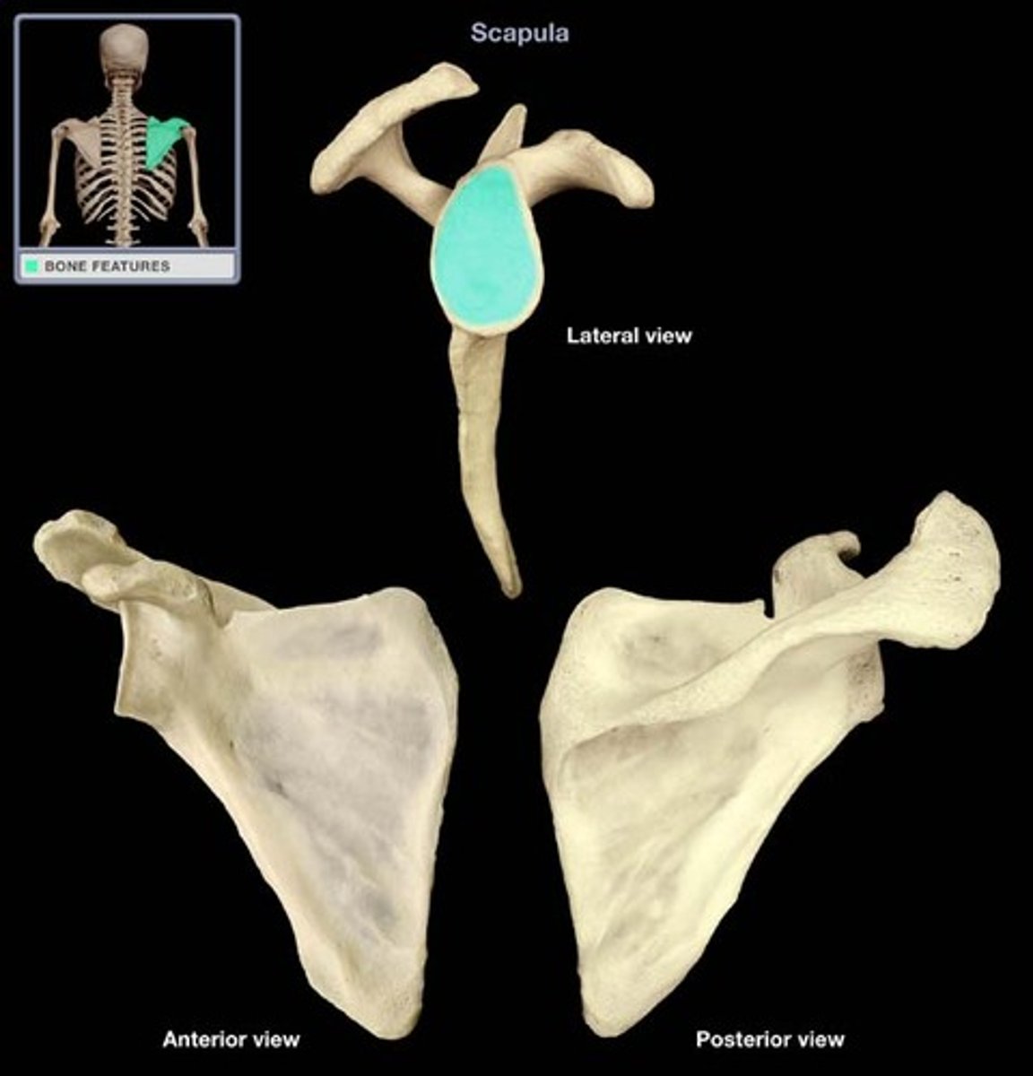
glenoid cavity
socket in scapula that receives head of humerus
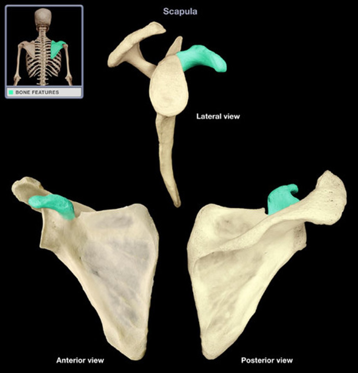
coracoid process
process above the glenoid cavity that permits muscle attachment
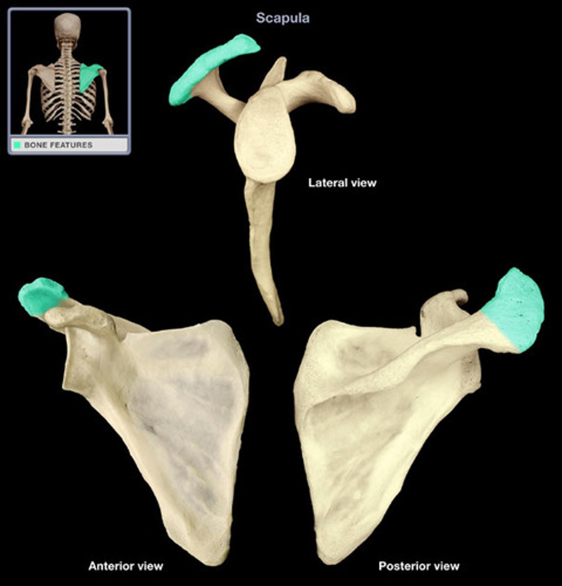
acromion
Outward extension of the shoulder blade forming the point of the shoulder.
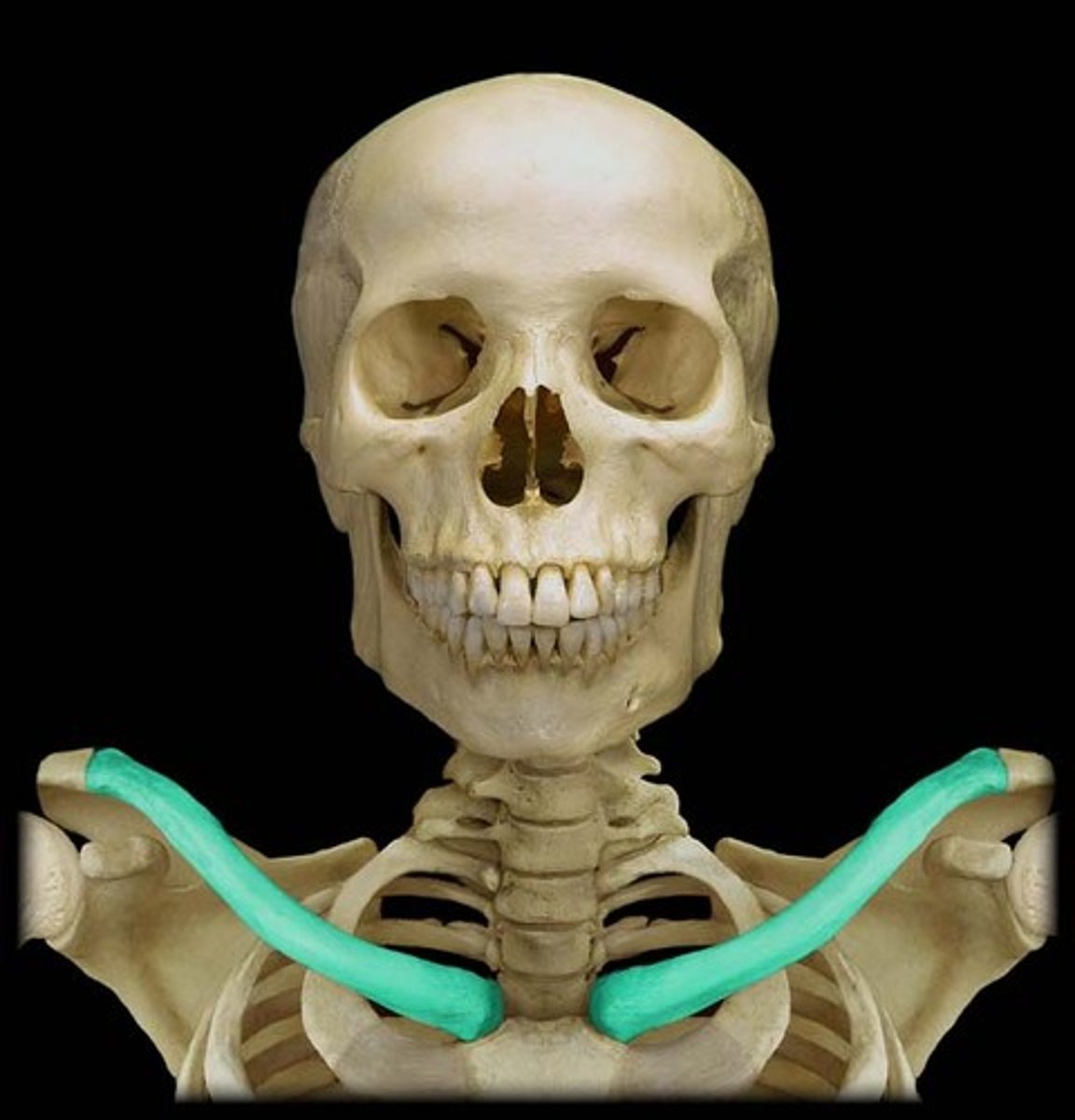
clavicle
collar bone
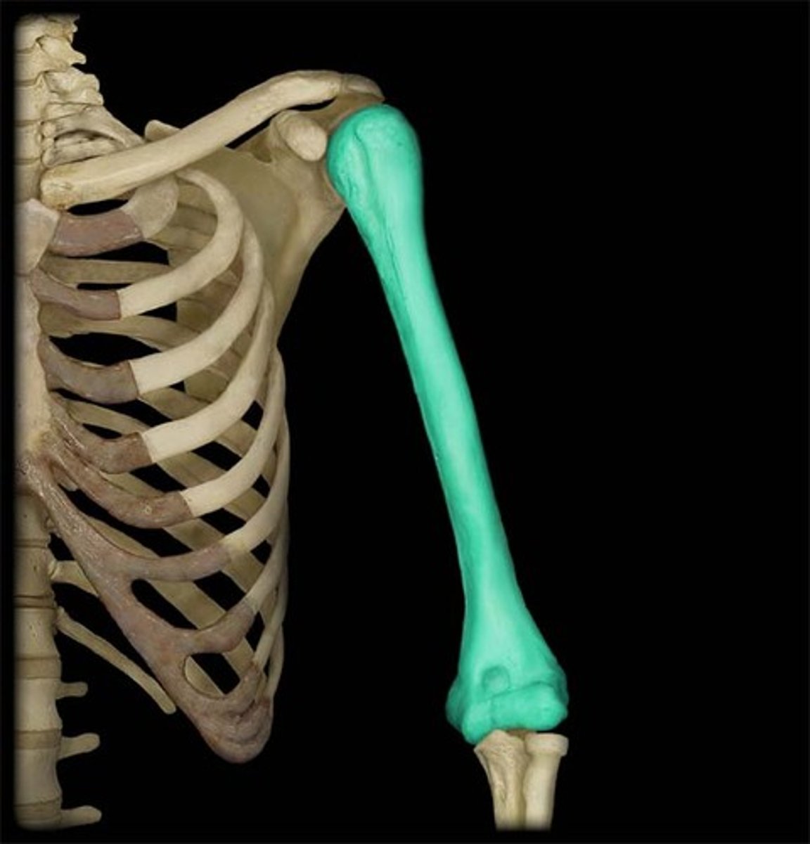
humerus
arm
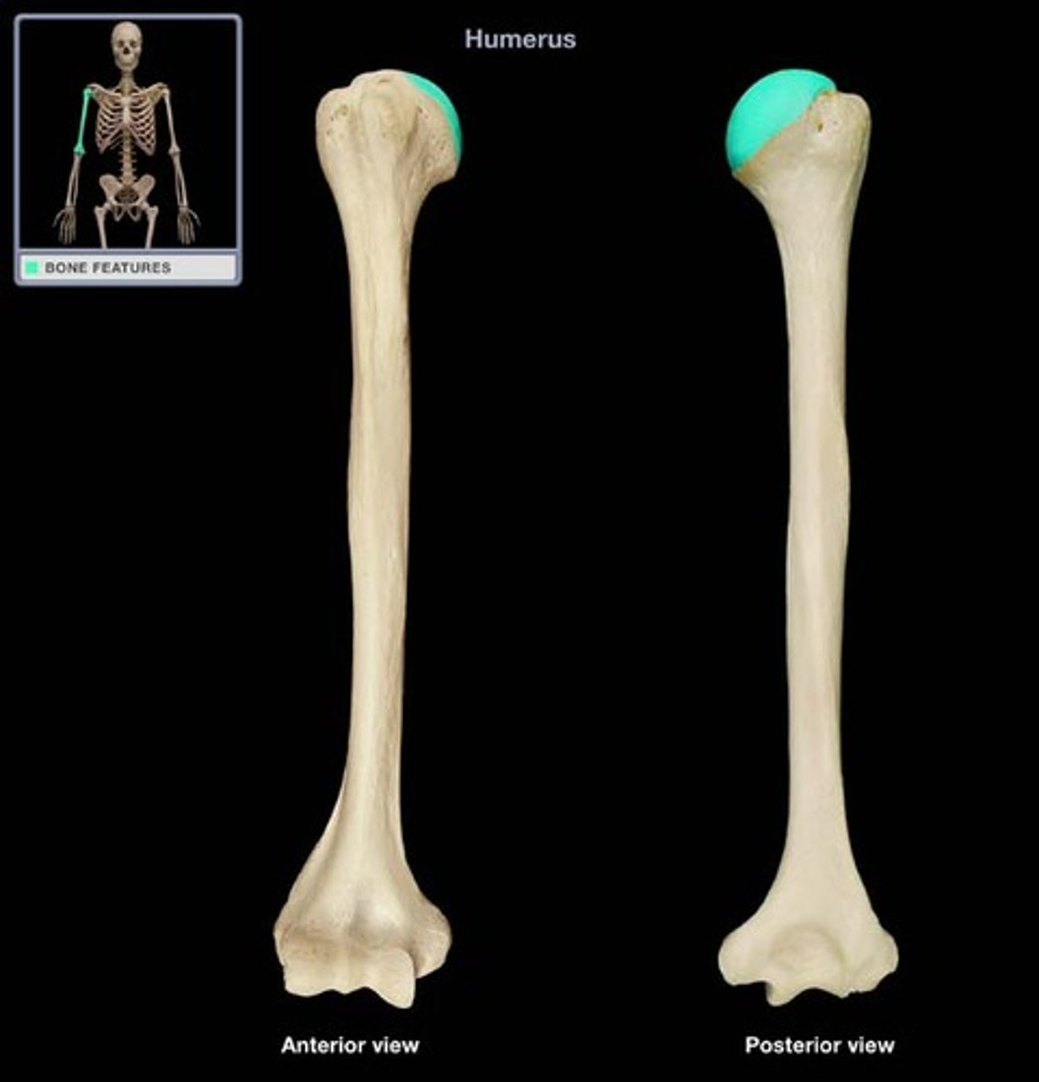
humerus head
rounded section of the humerus that articulates with the glenoid cavity of the scapula
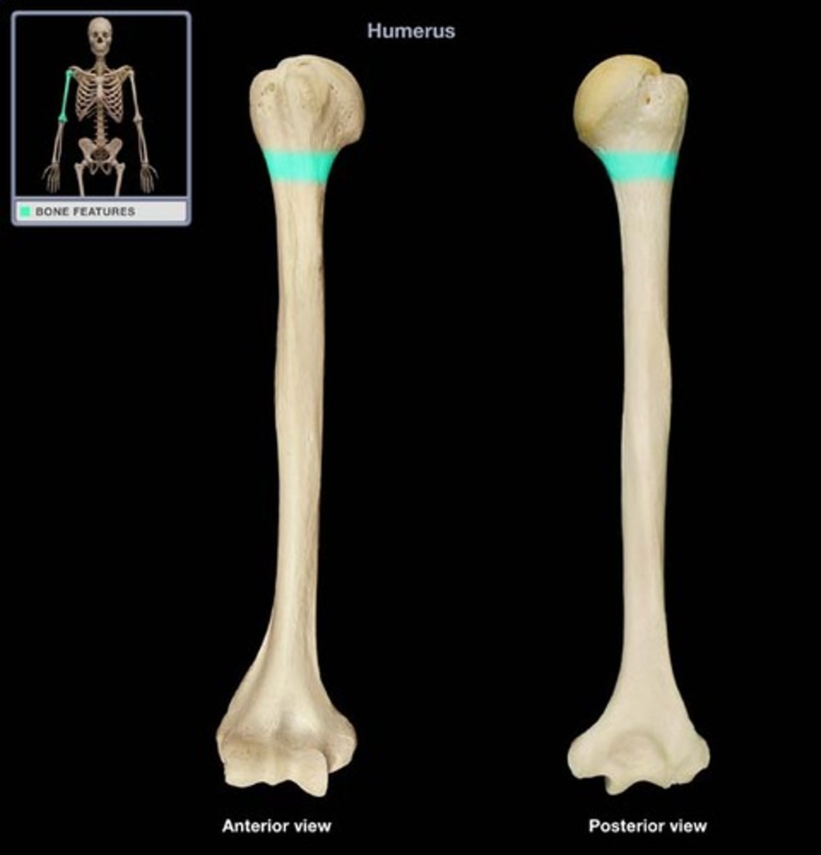
surgical neck
greater tubercle
Large lateral prominence on humerus
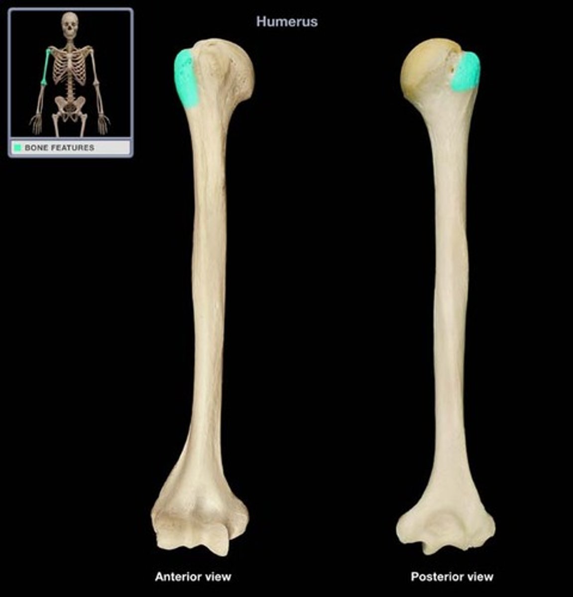
lesser tubercle
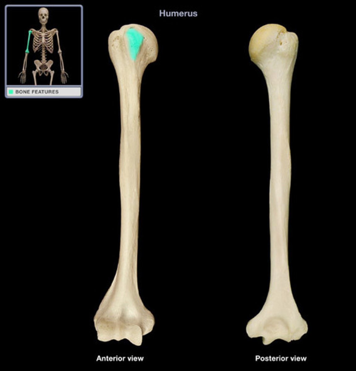
medial epicondyle (humerus)
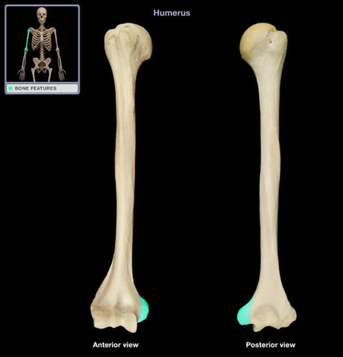
lateral epicondyle (humerus)
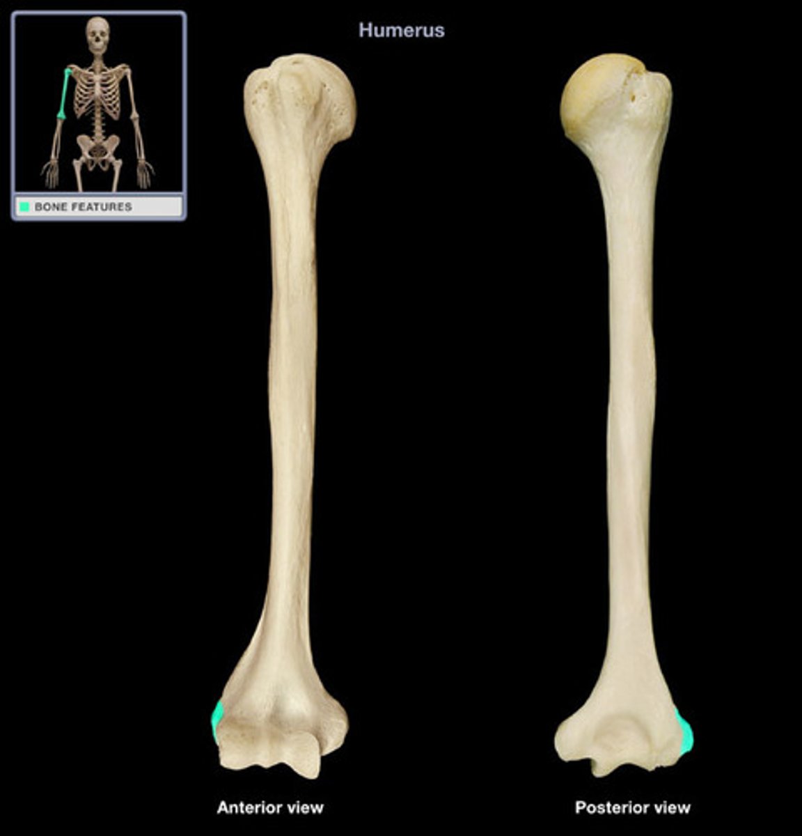
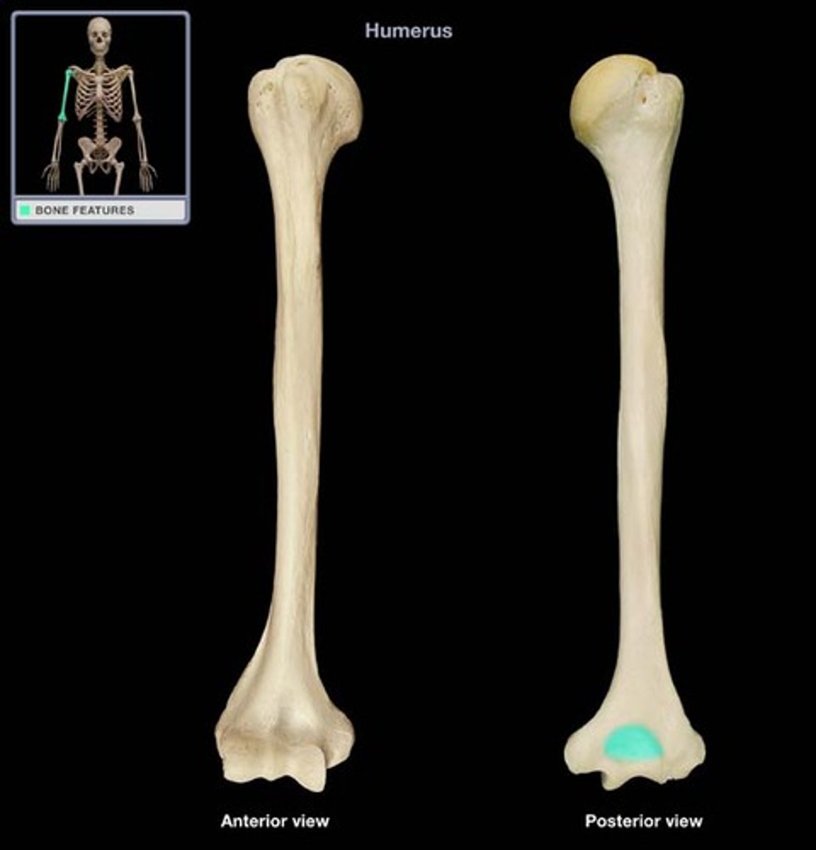
olecranon fossa
located on the posterior side of the distal end of the humerus superior to the trochlea and articulates with the olecranon process of the ulna
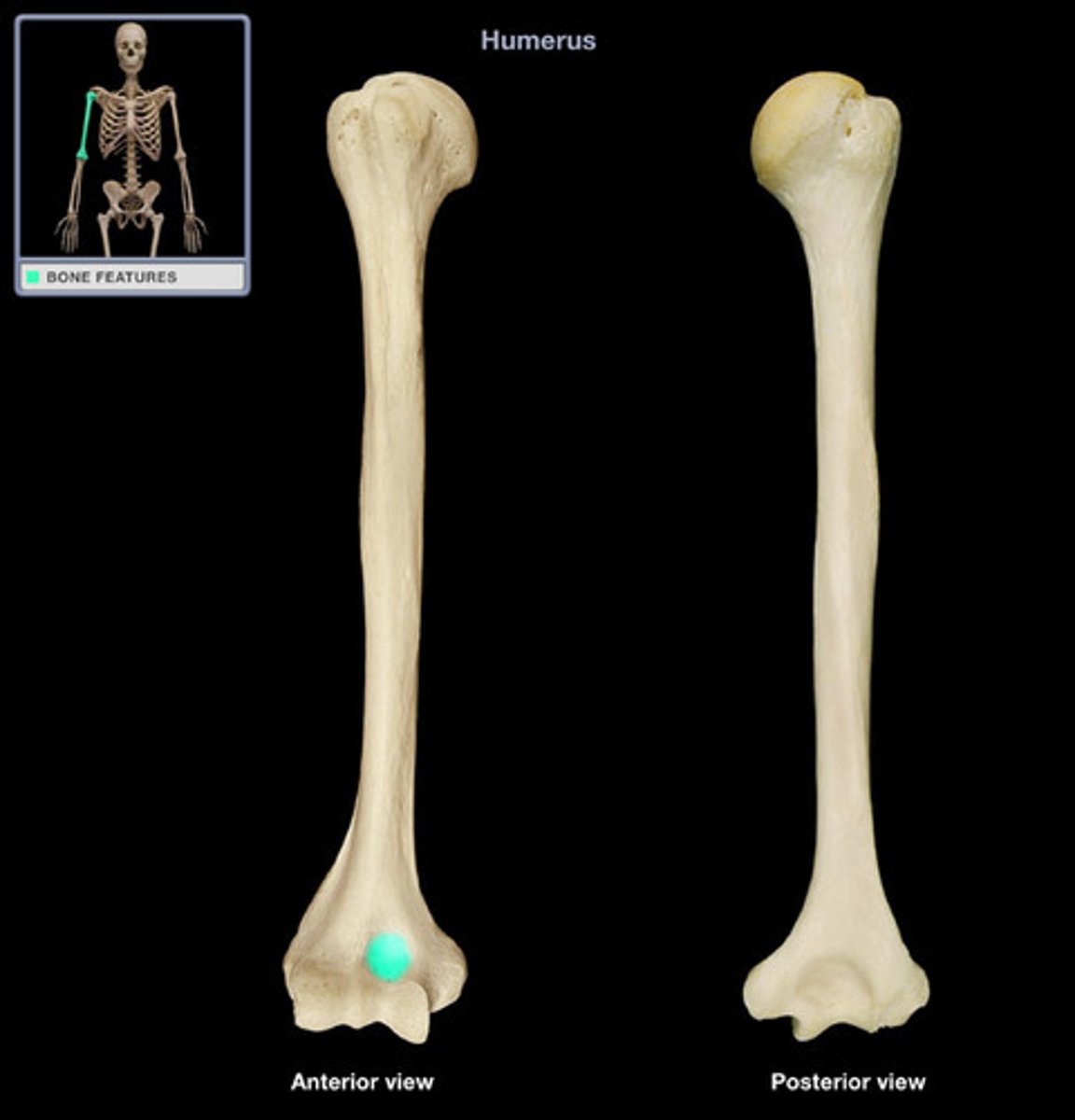
coronoid fossa
anterior depression that receives the coronoid process of the ulna
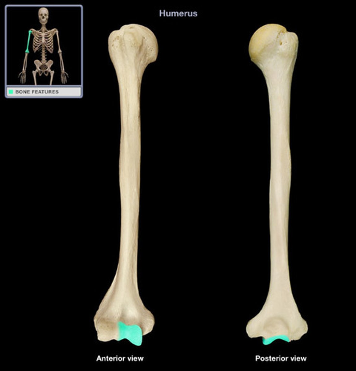
trochlea
a smooth, grooved articular process shaped like a pulley
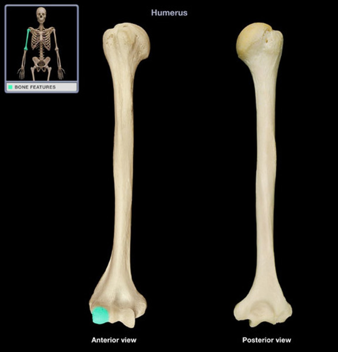
capitulum
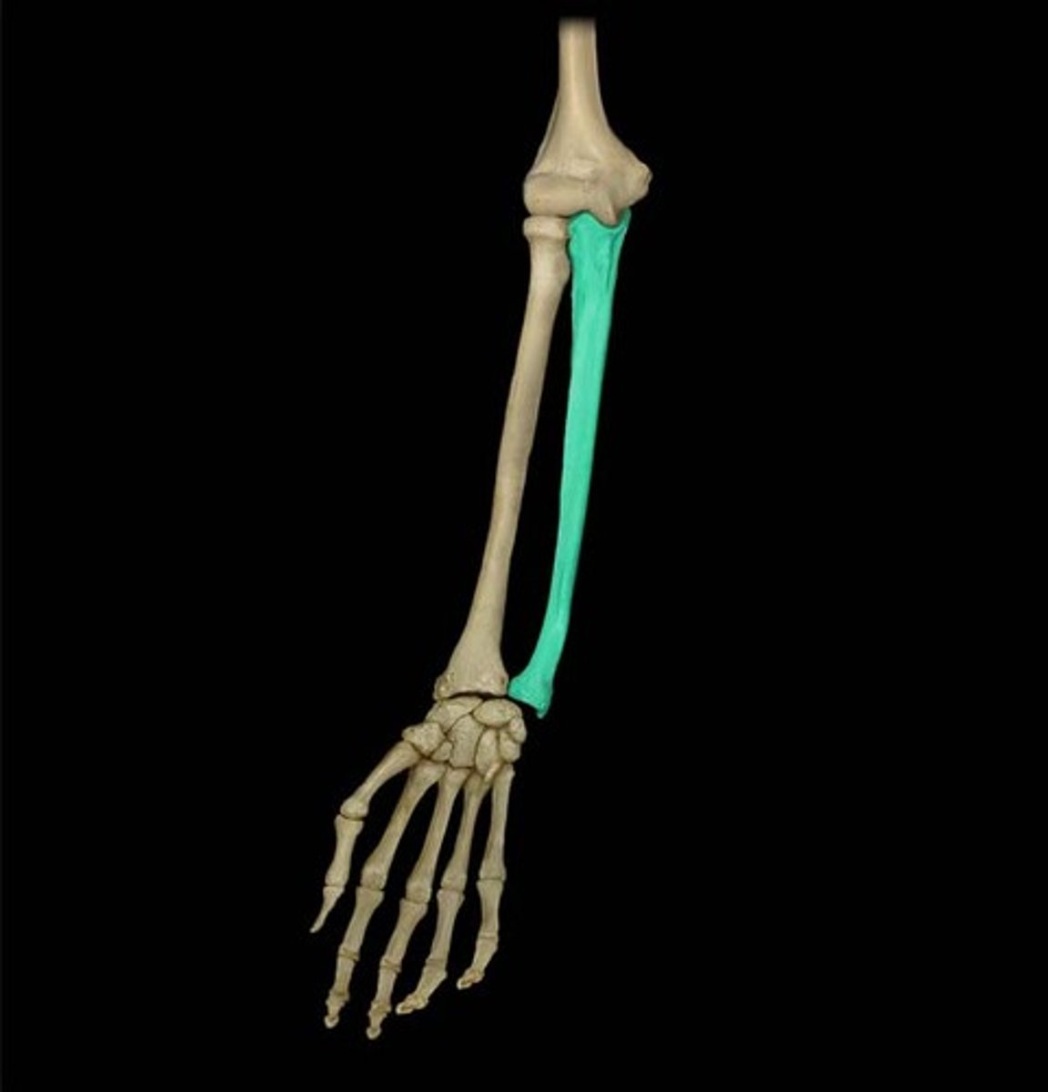
ulna
medial bone of the forearm
ulna coronoid process
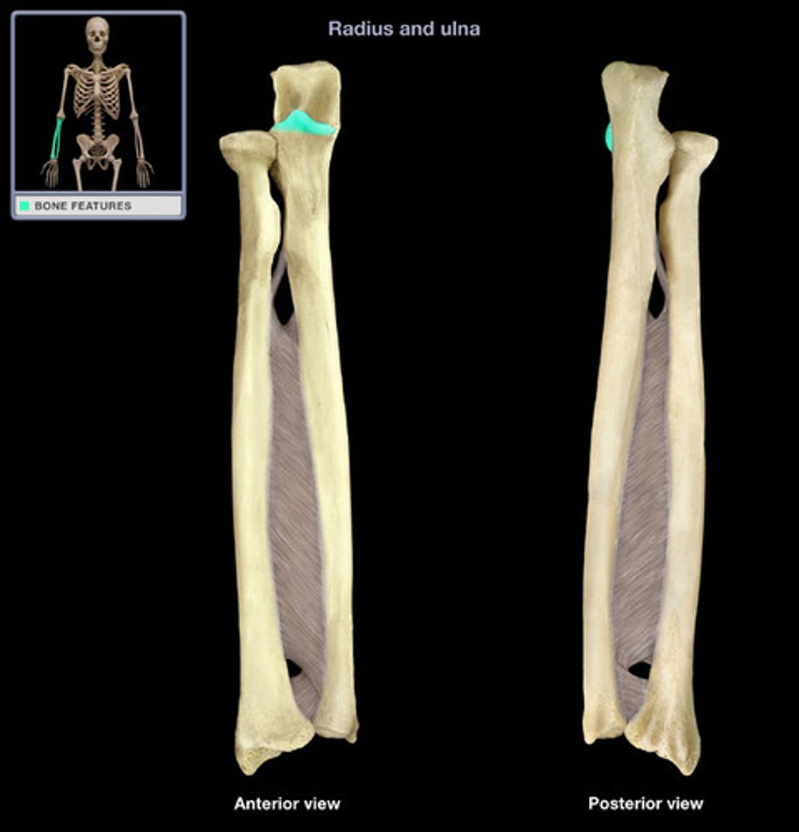
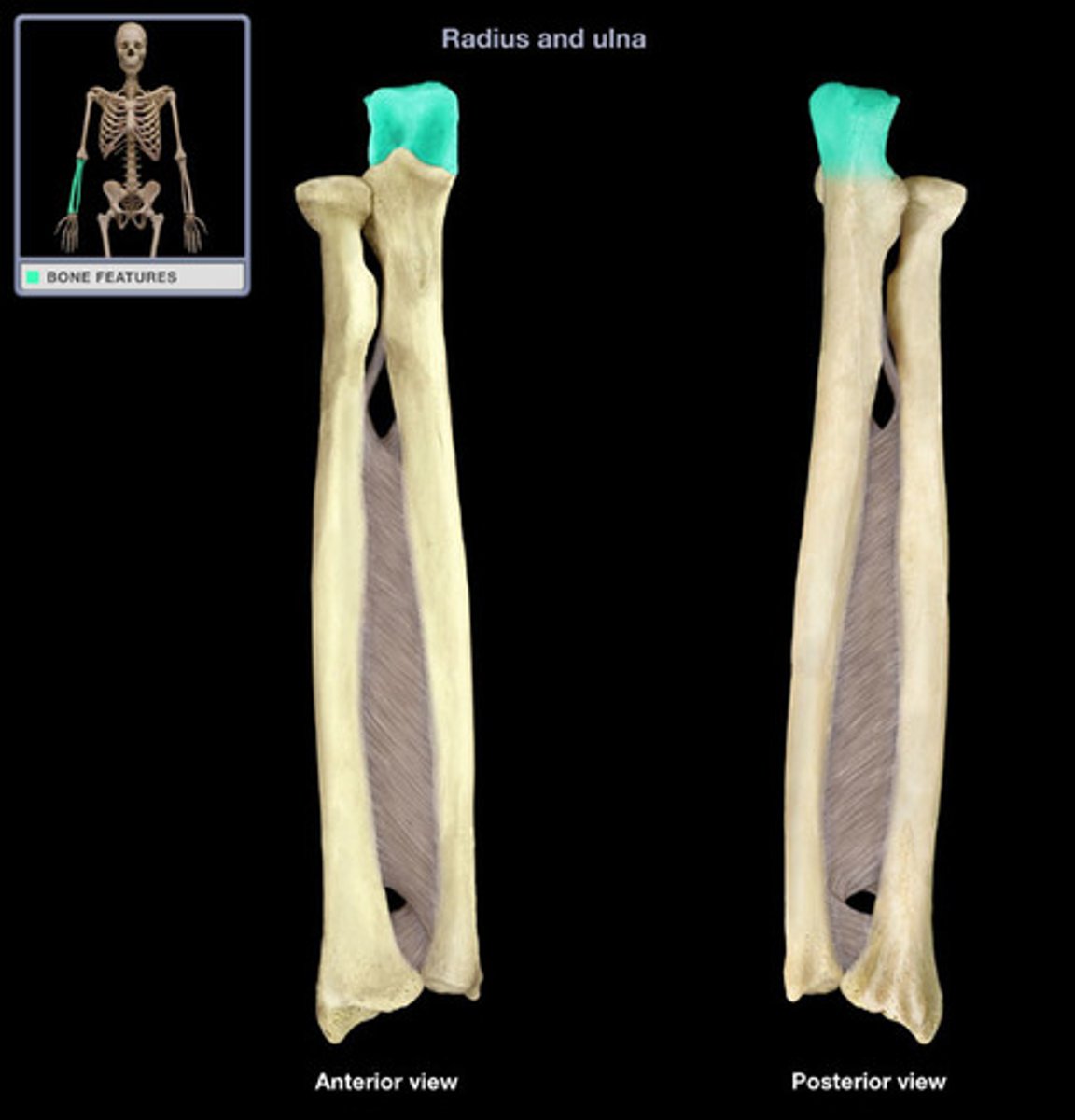
ulna olecranon process
elbow
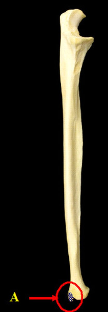
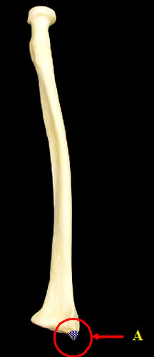
ulna styloid process
pointy projection on the distal end of ulna
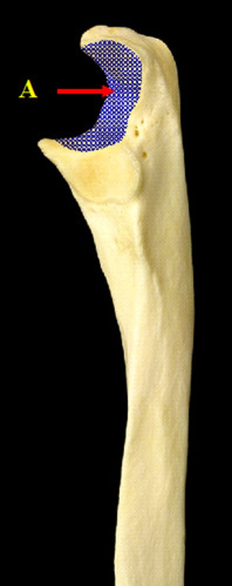
ulna trochlear notch
deep notch that separates the olecranon and the coronoid process; articulates with the trochlea of the humerus
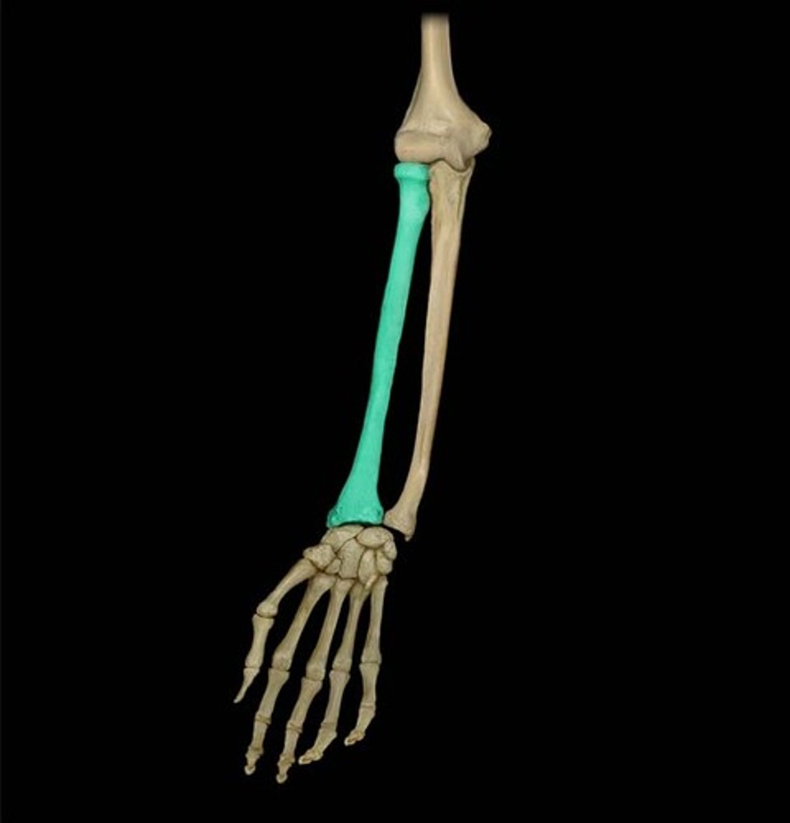
radius
lateral bone of the forearm
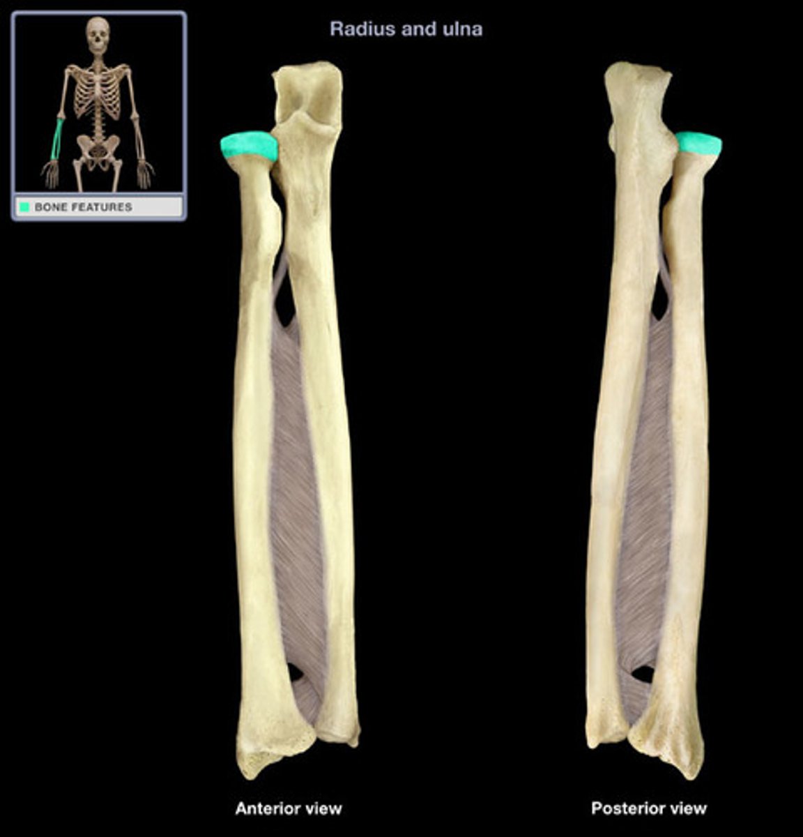
radius head
A round, articular structure on the proximal end of the radius.
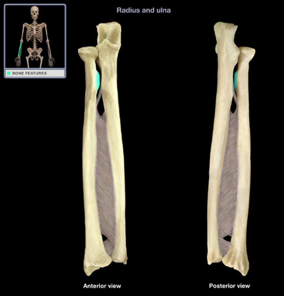
radial tuberosity
radius styloid process
projection of bone on the lateral surface of the distal radius bone
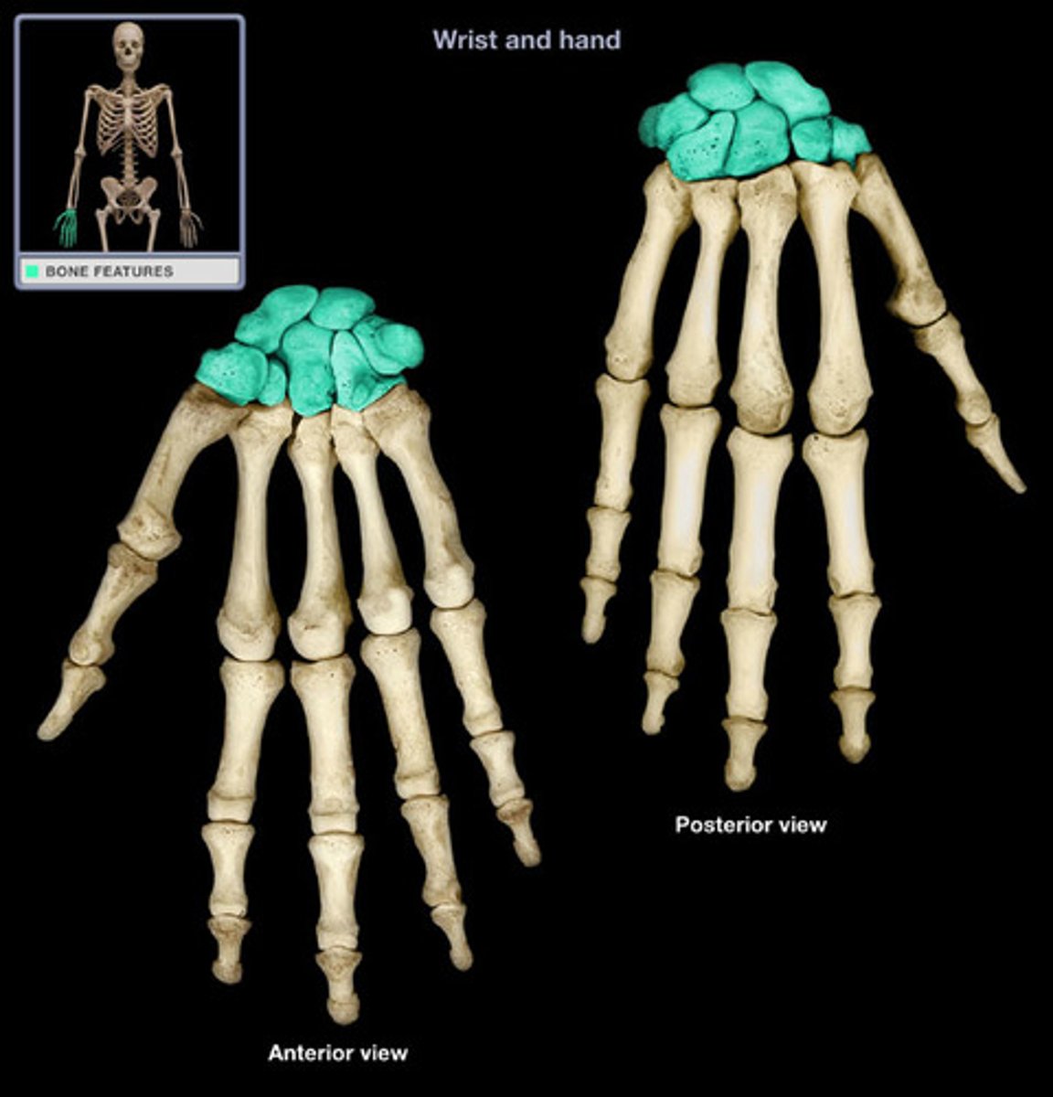
carpals
bones of the wrist
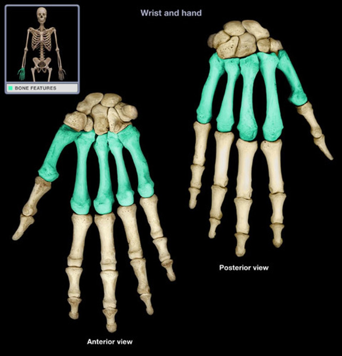
metacarpals
the five bones that form the palms of the hand
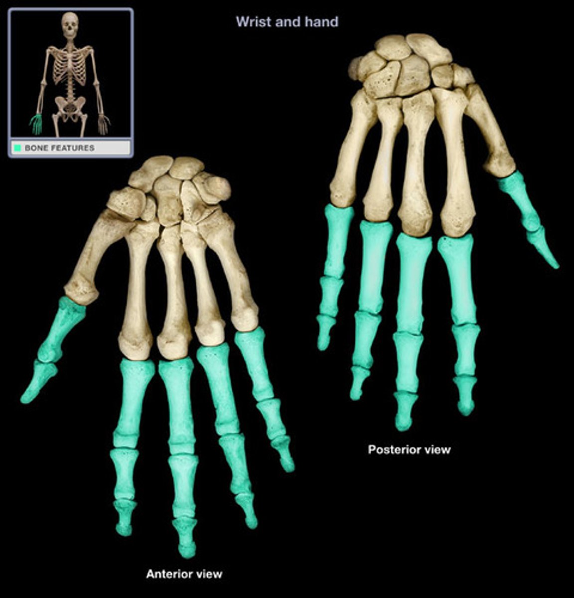
phalanges
bones of the fingers and toes
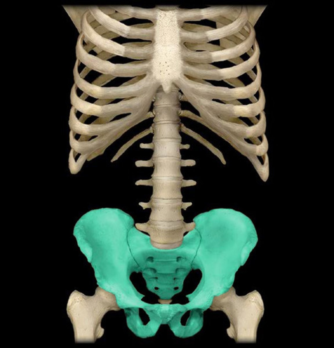
pelvic girdle
coxal bones and sacrum
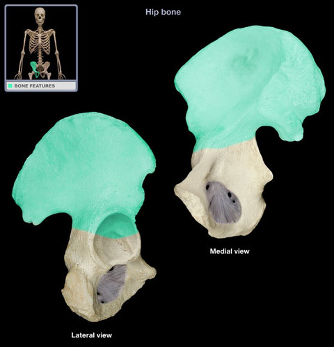
ilium
the superior and widest portion of the pelvis
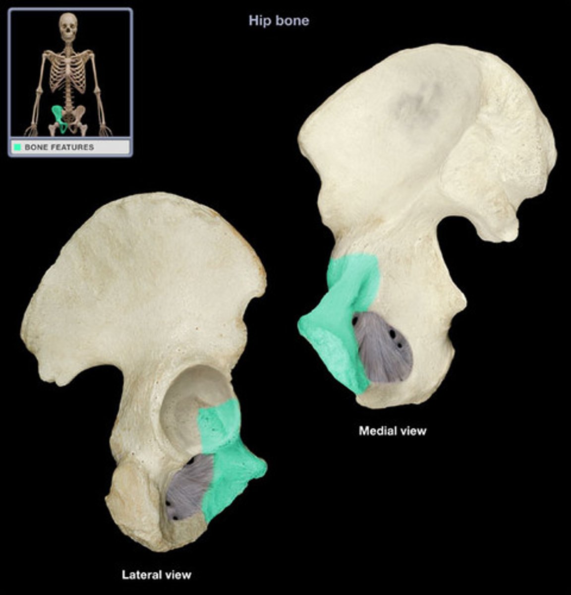
pubis
The medial anterior portion of the pelvis
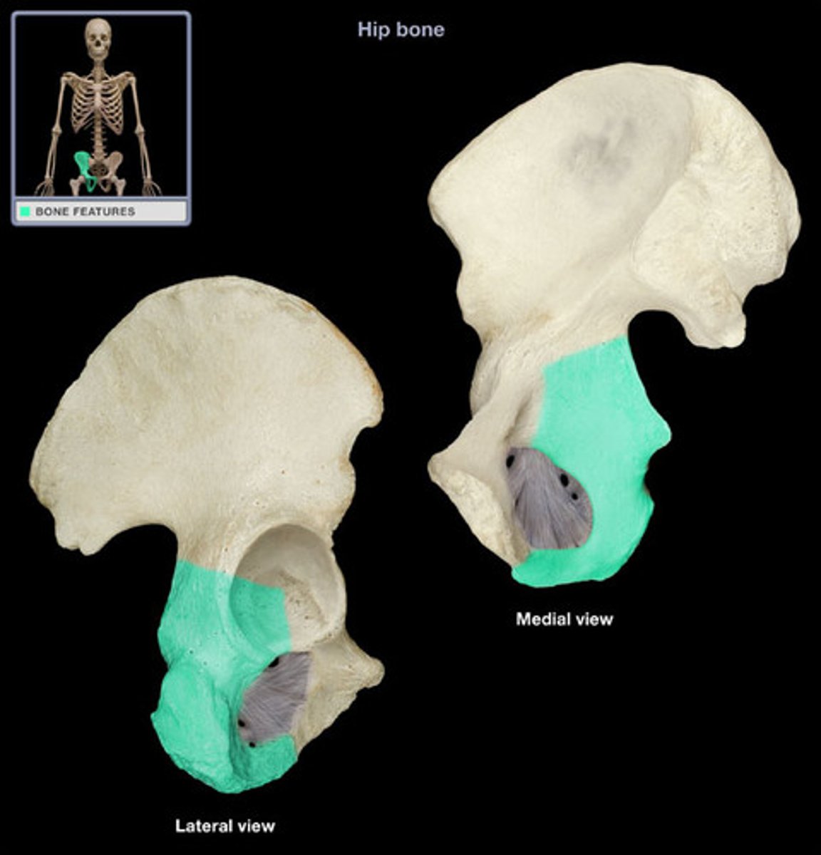
ischium
the lower, posterior portions of the pelvis
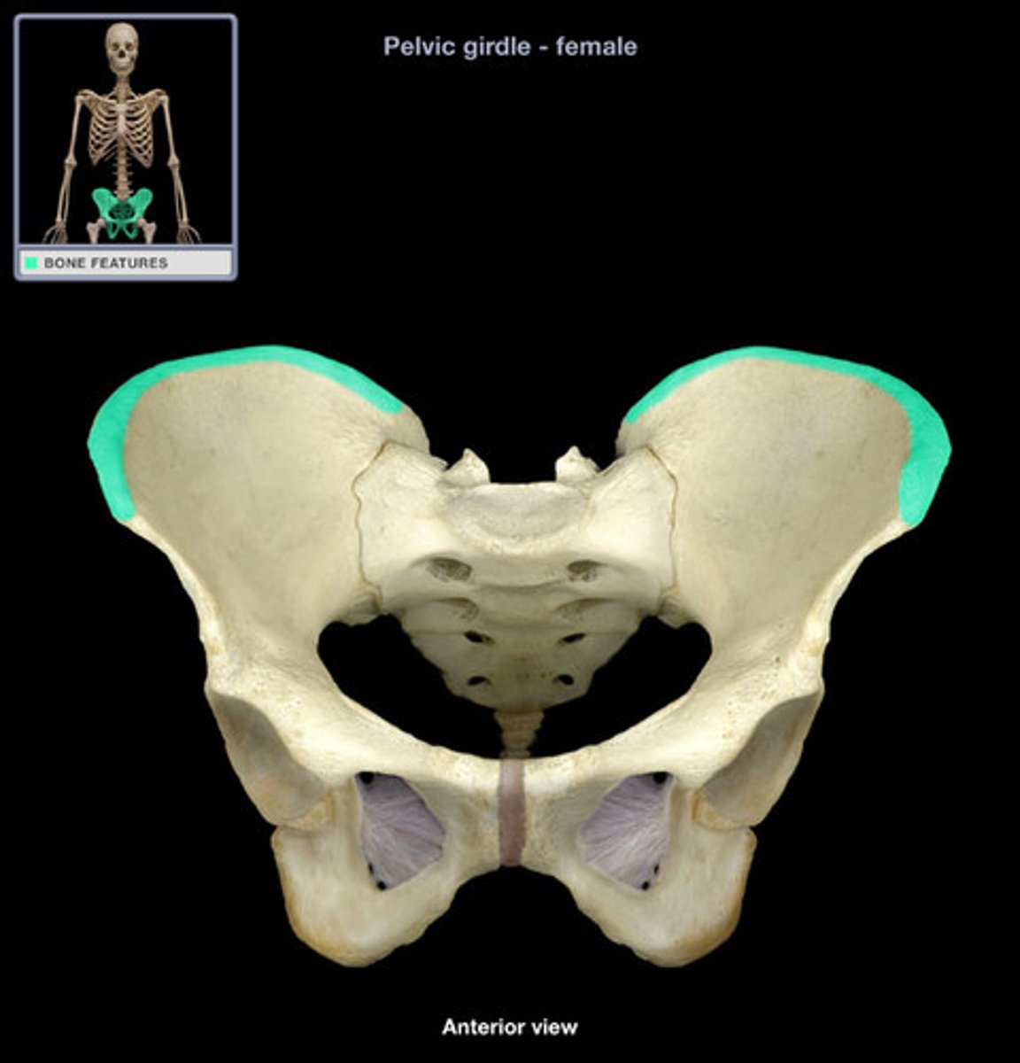
iliac crest
superior border of ilium
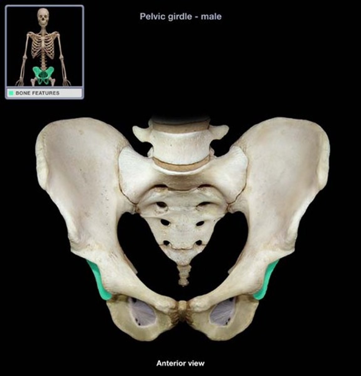
acetabulum
where the ischium, ilium, and pubis unite, creating the socket for the head of the femur
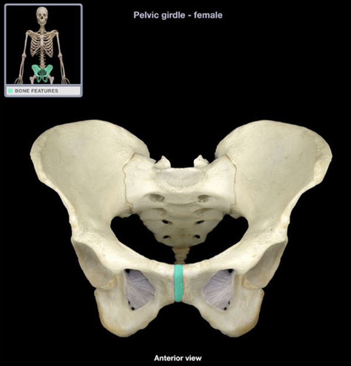
pubic symphysis (symphyseal surface)
the anterior joint where the two pubic bones meet
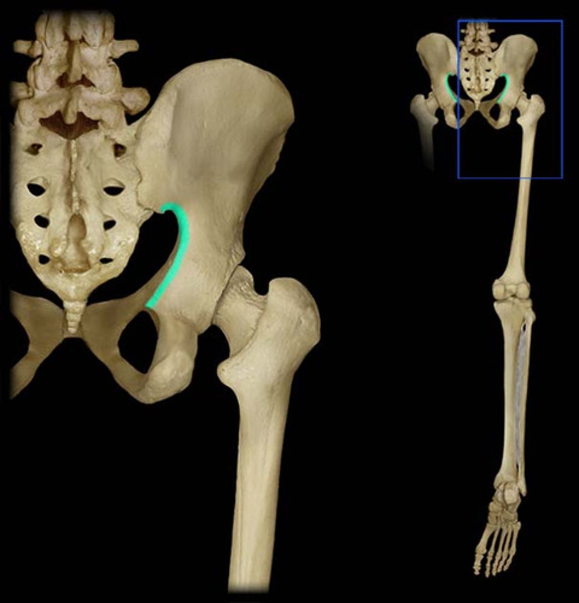
greater sciatic notch
allows blood vessels and the large sciatic nerve to pass from the pelvis posteriorly into the thigh
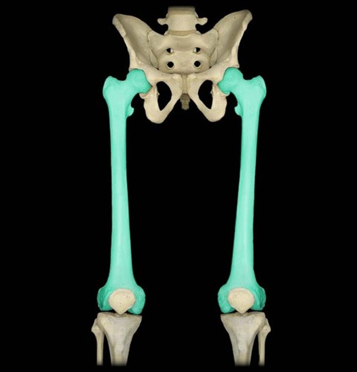
femur
thigh bone
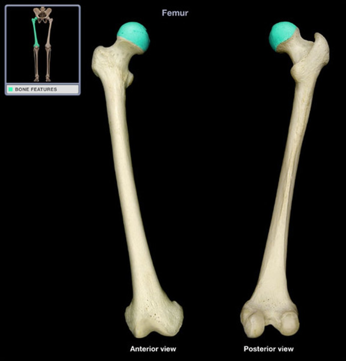
head (femur)
articulates with the acetabulum (hip socket)
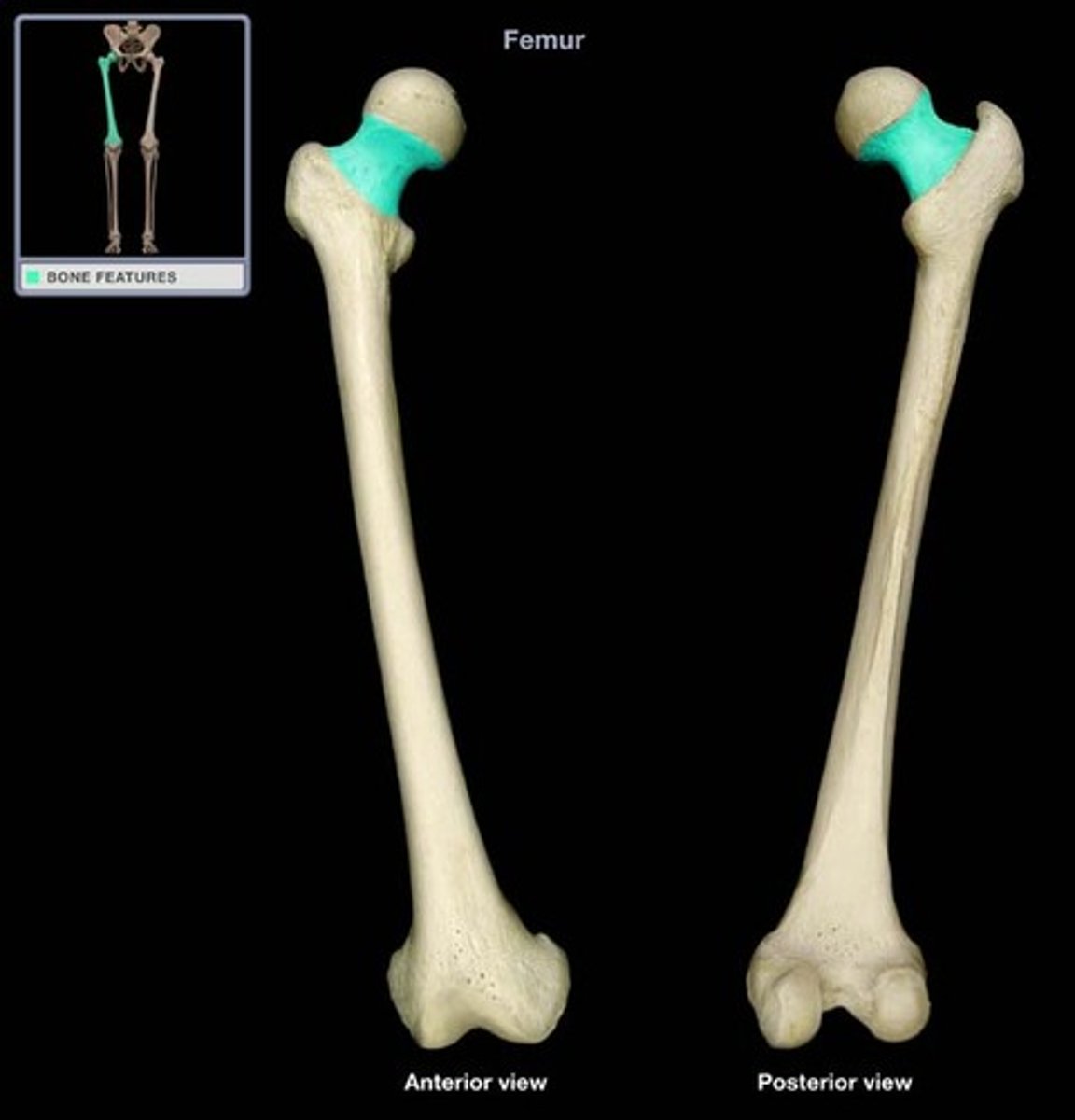
neck (femur)
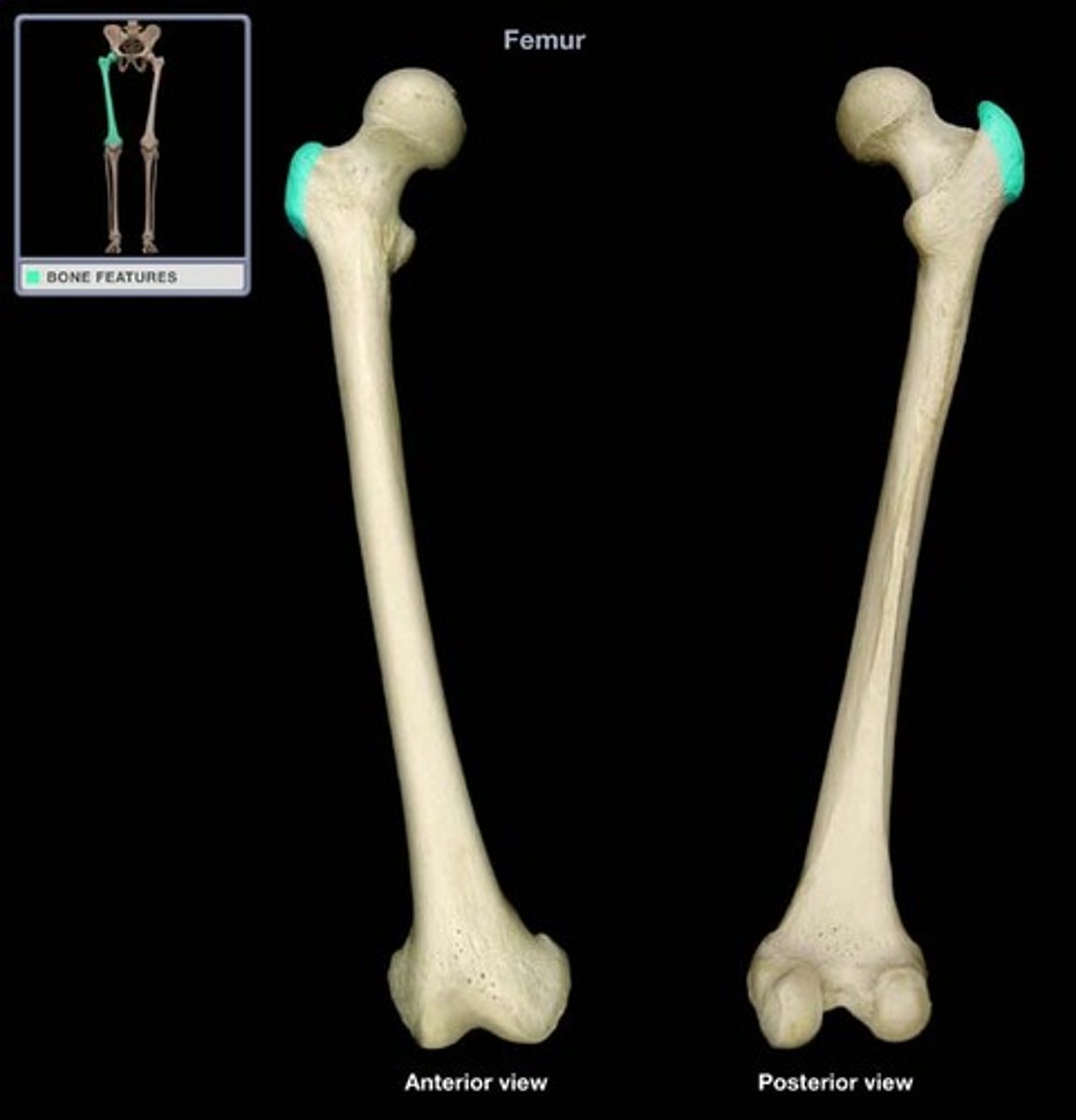
greater trochanter
A bony prominence on the proximal lateral side of the thigh, just below the hip joint.
lesser trochanter
The projection on the medial/superior portion of the femur.
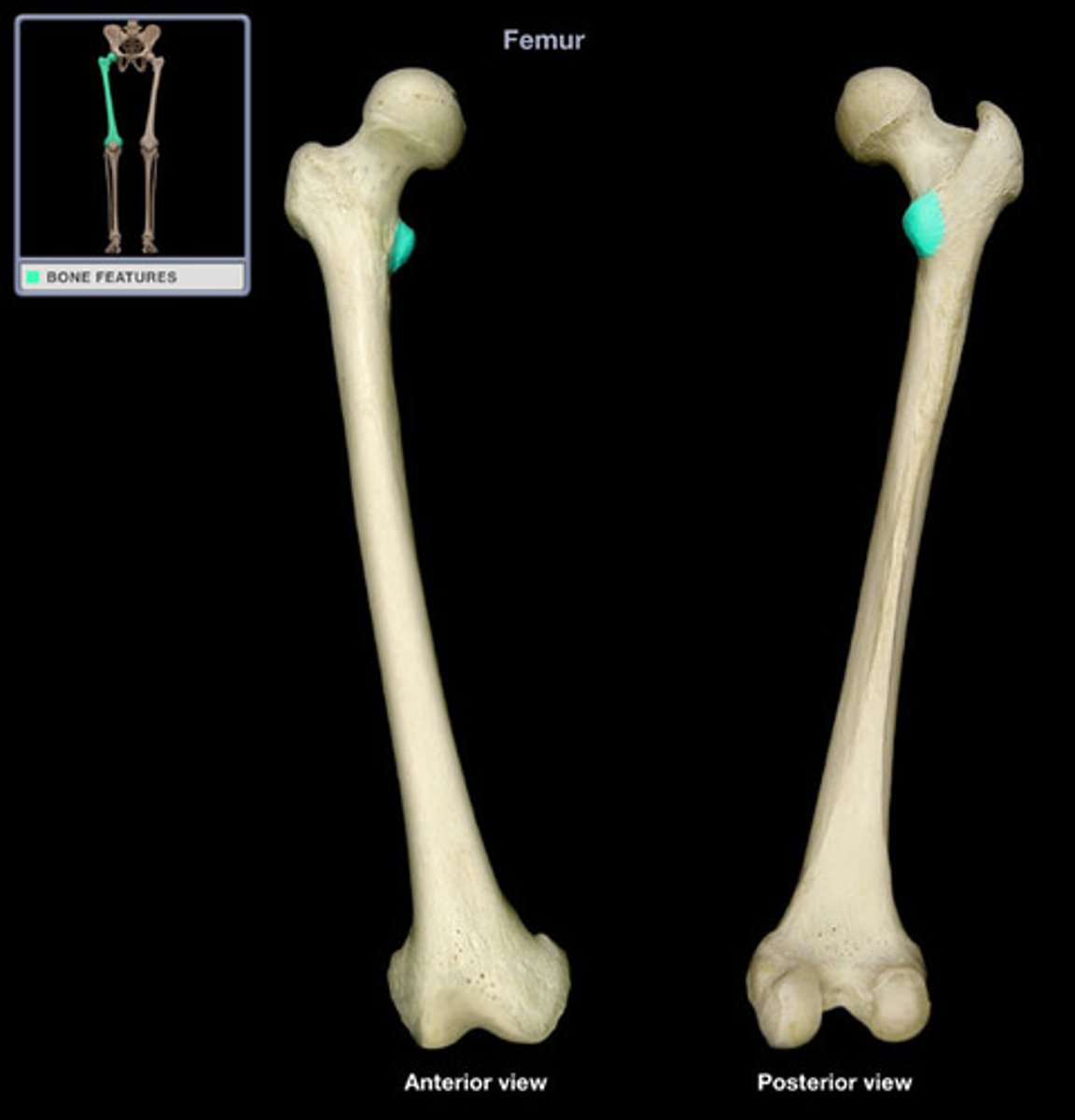
medial condyle (femur)
articulates with the medial condyle of the tibia
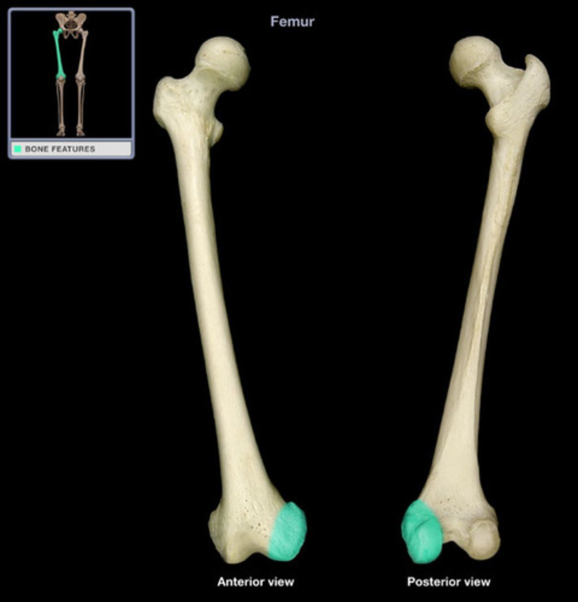
lateral condyle (femur)
articulates with the lateral condyle of the tibia
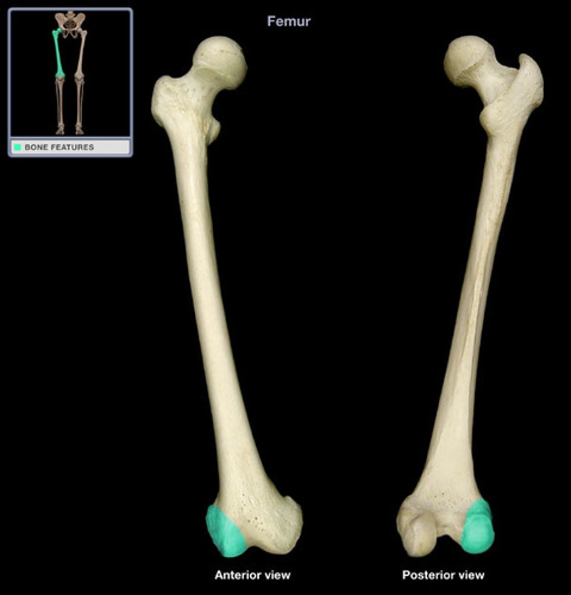
tibia
The shin bone, the larger of the two bones of the lower leg.
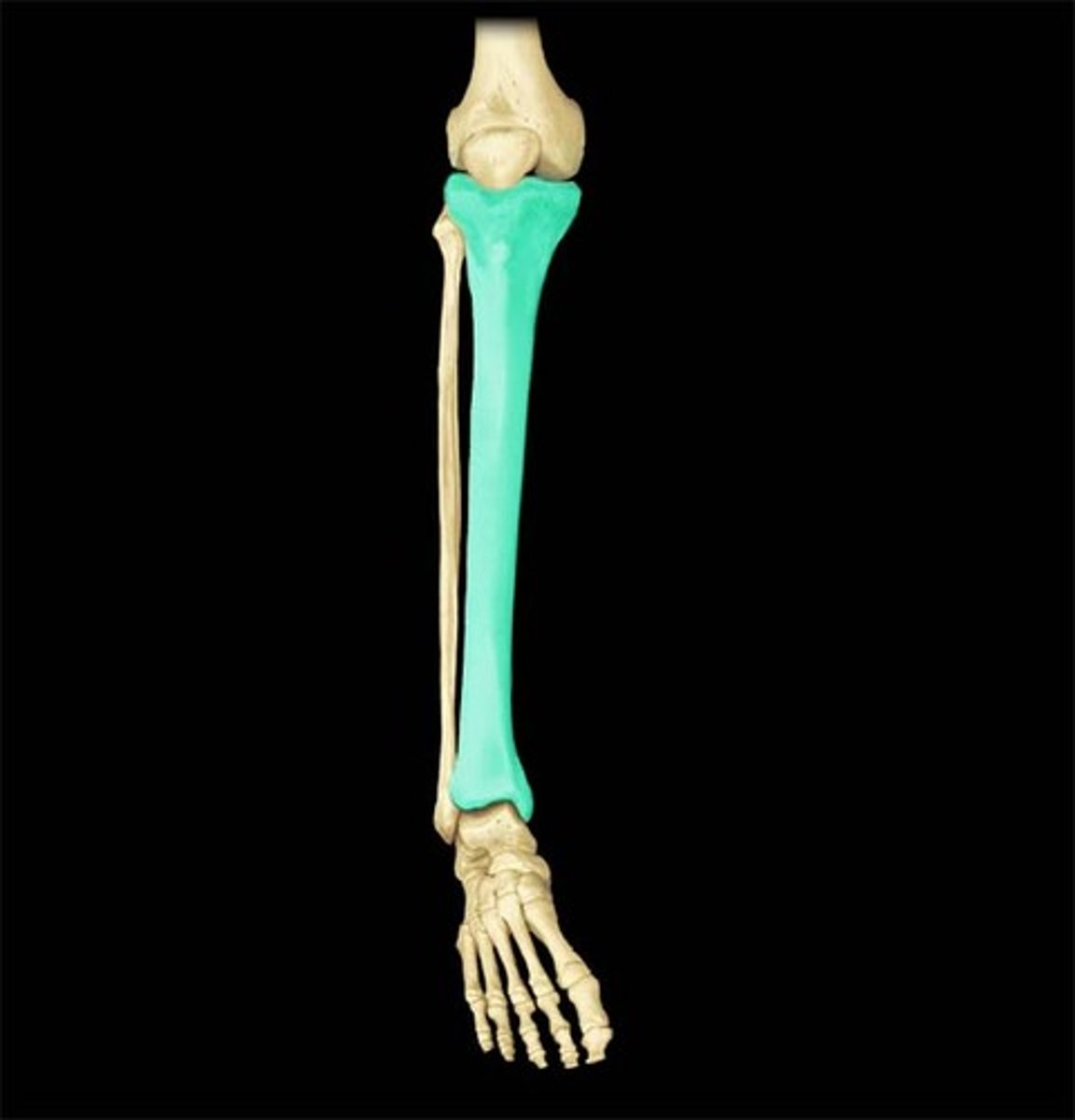
medial condyle (tibia)
articulates with medial condyle of femur
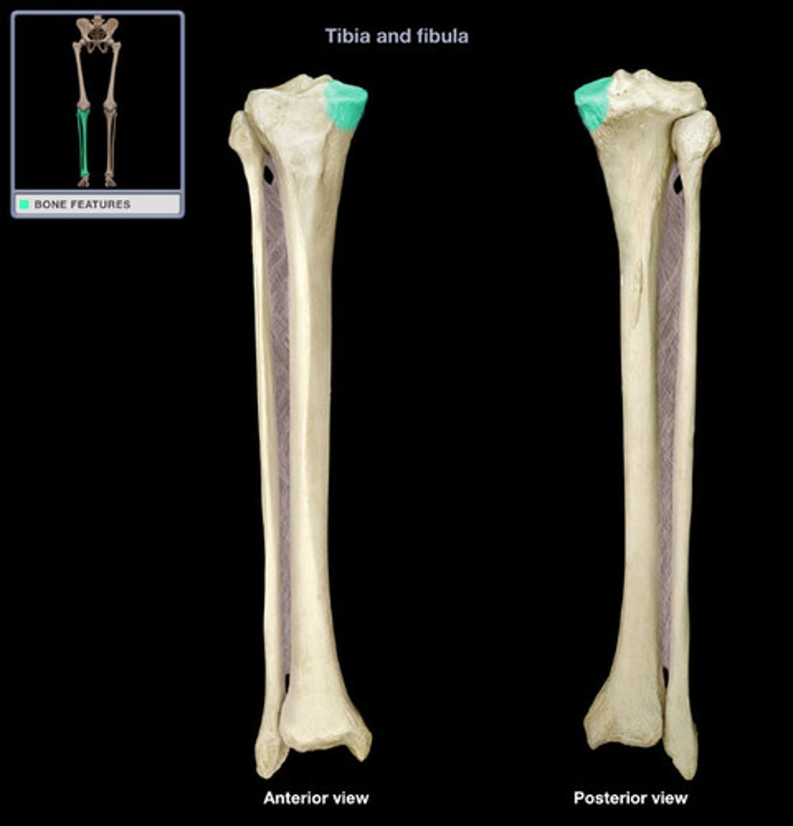
lateral condyle (tibia)
articulates with lateral condyle of femur
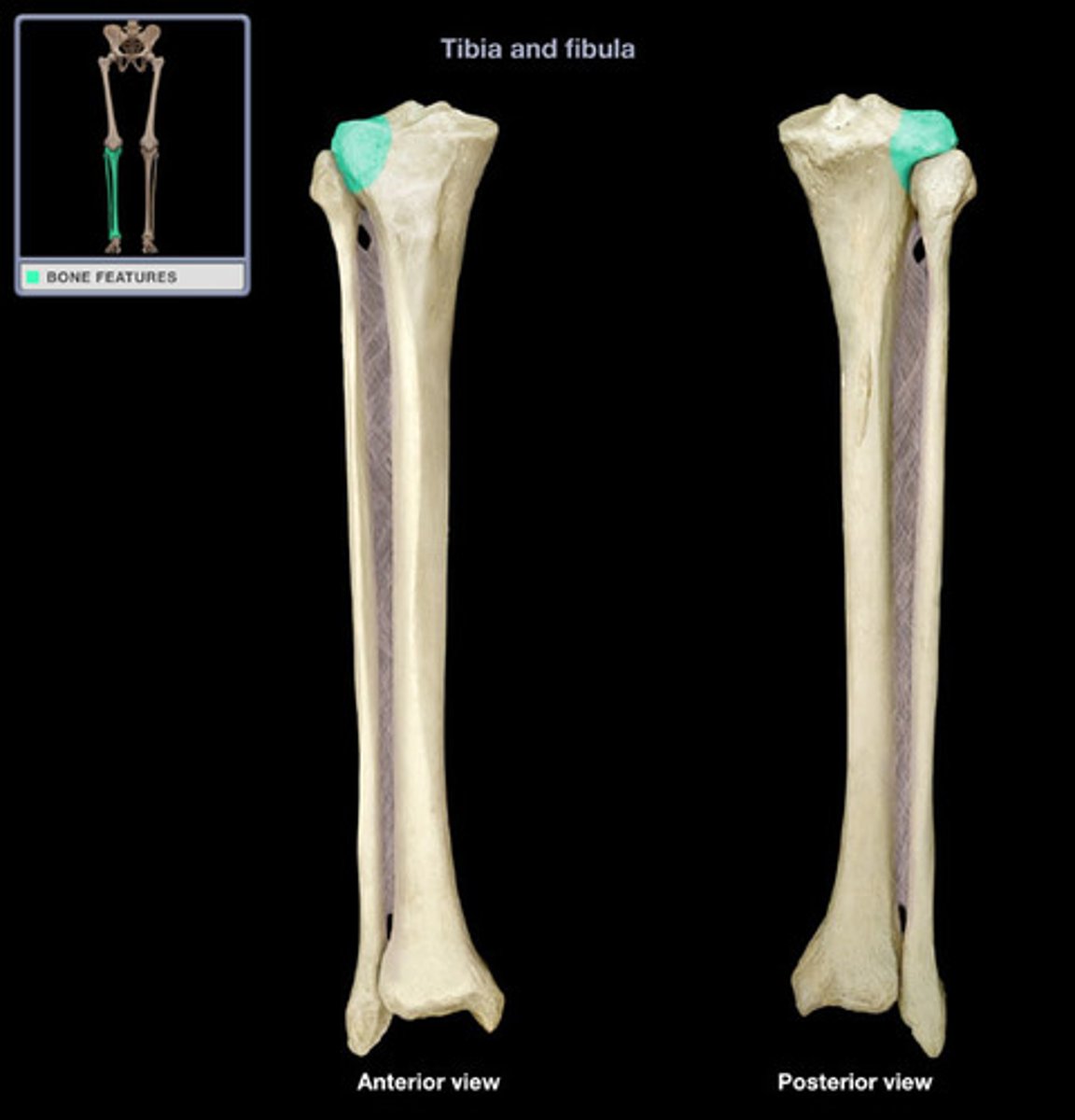
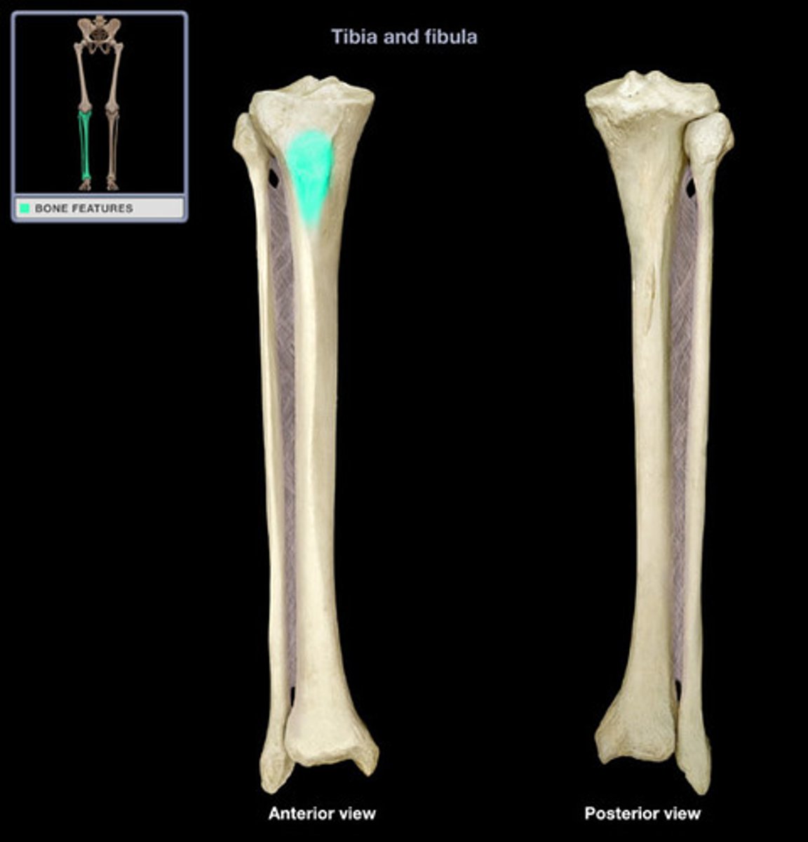
tibial tuberosity
point where the patellar ligament attaches
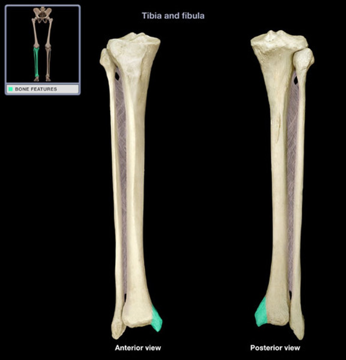
medial malleolus
distal process on medial tibial surface
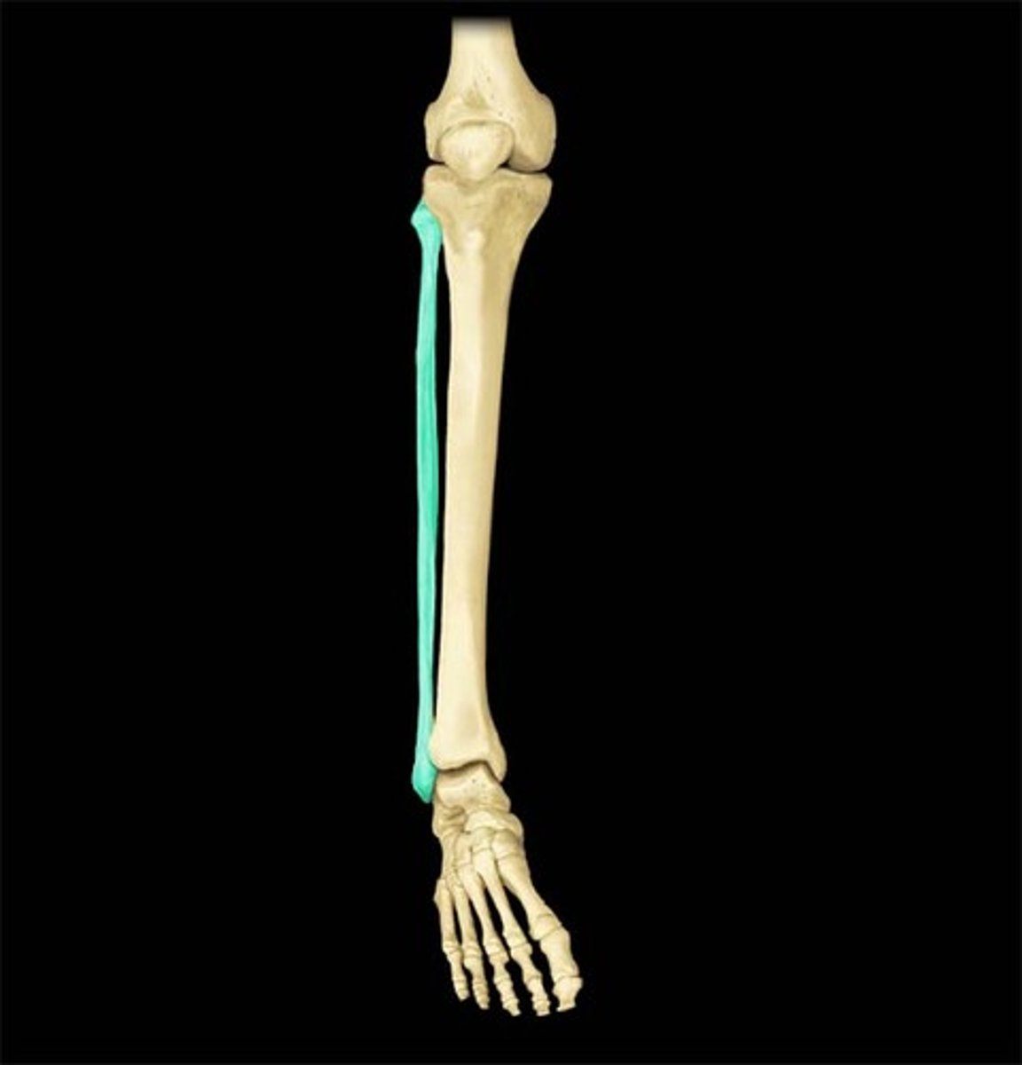
fibula
calf bone; the lateral and smaller bone of the lower leg
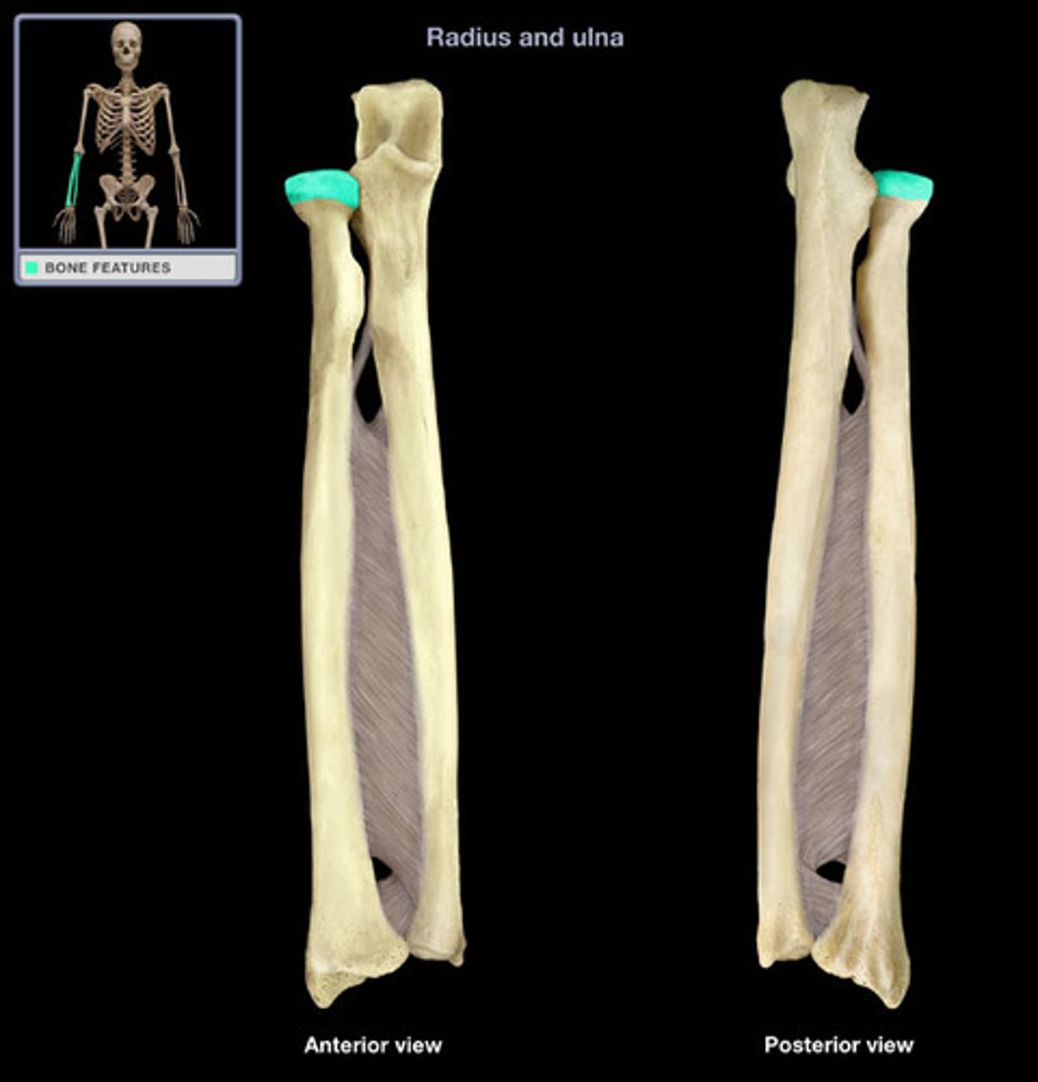
head (radius)
articulates with the capitulum and radial notch
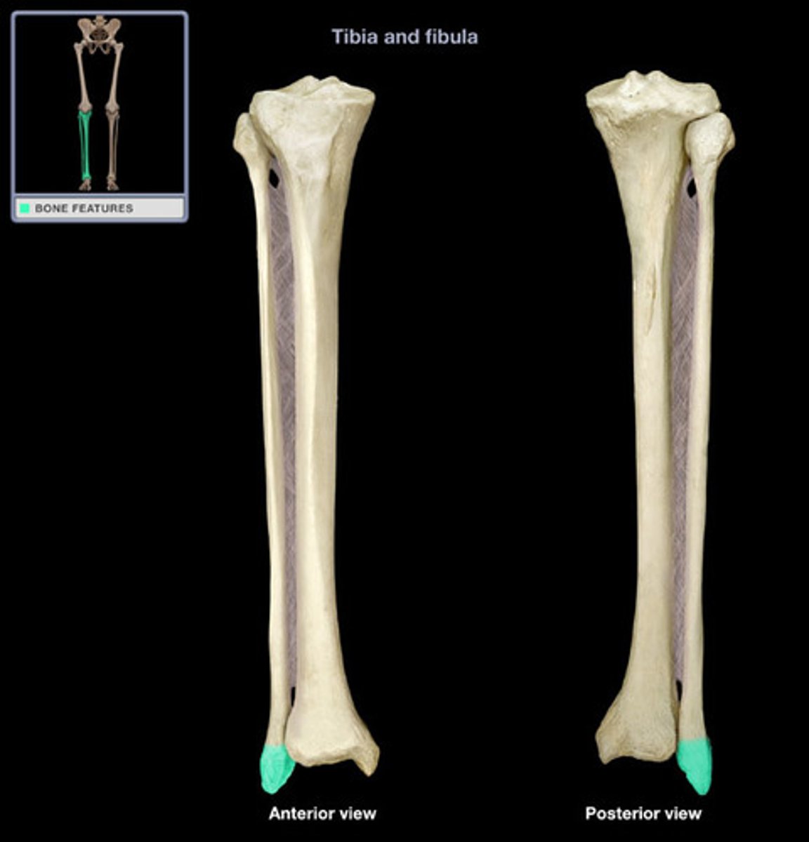
lateral malleolus
process forming the outer (lateral) ankle
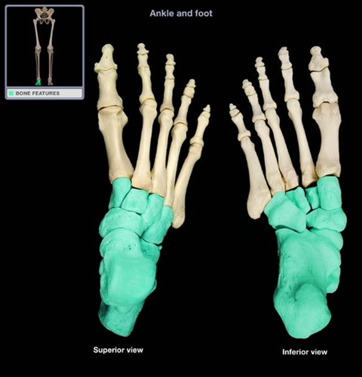
tarsals
bones of the ankle
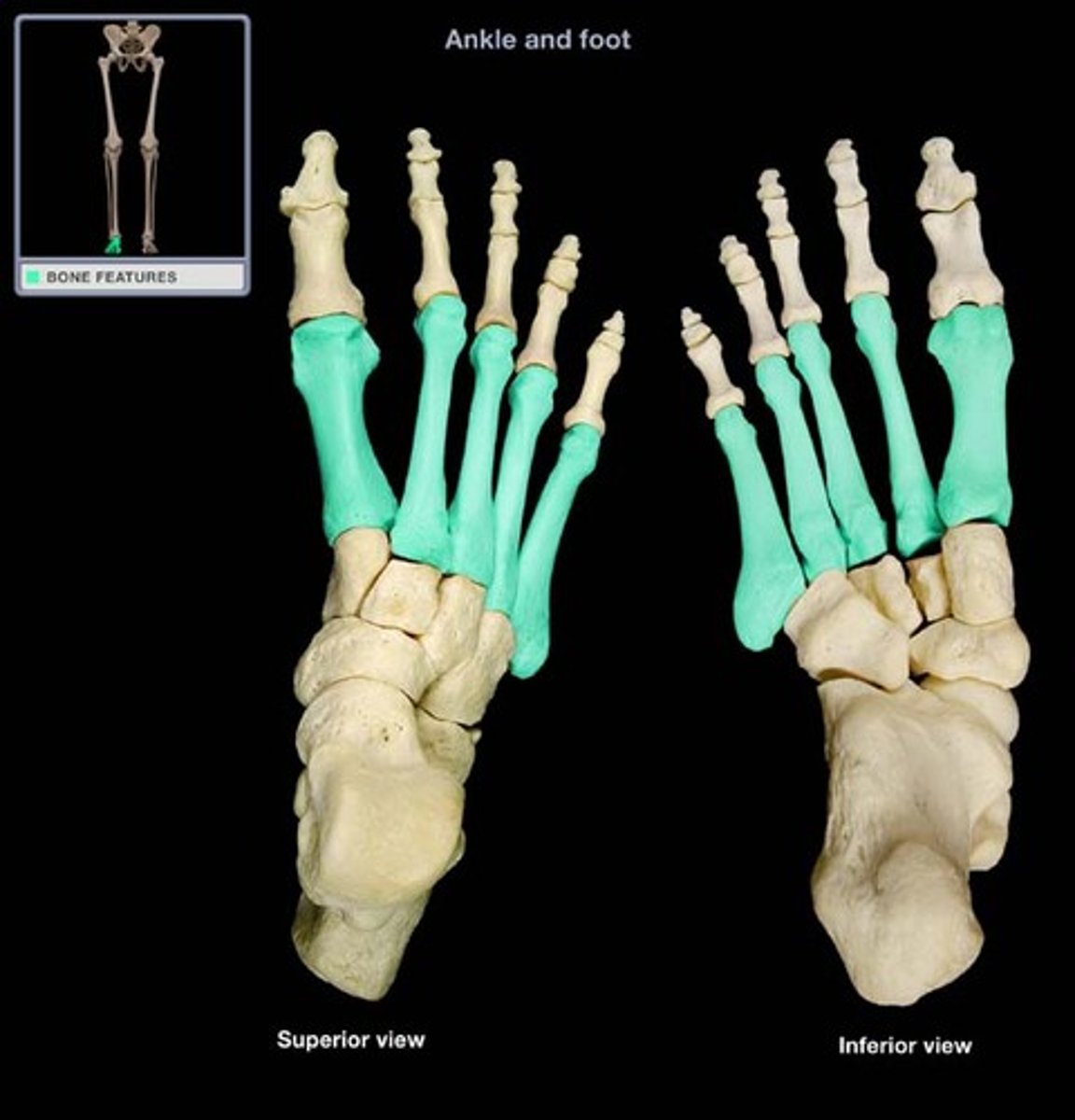
metatarsals
bones of the foot between ankle and toes
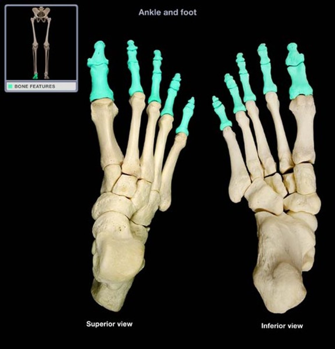
phalanges
bones of the fingers and toes
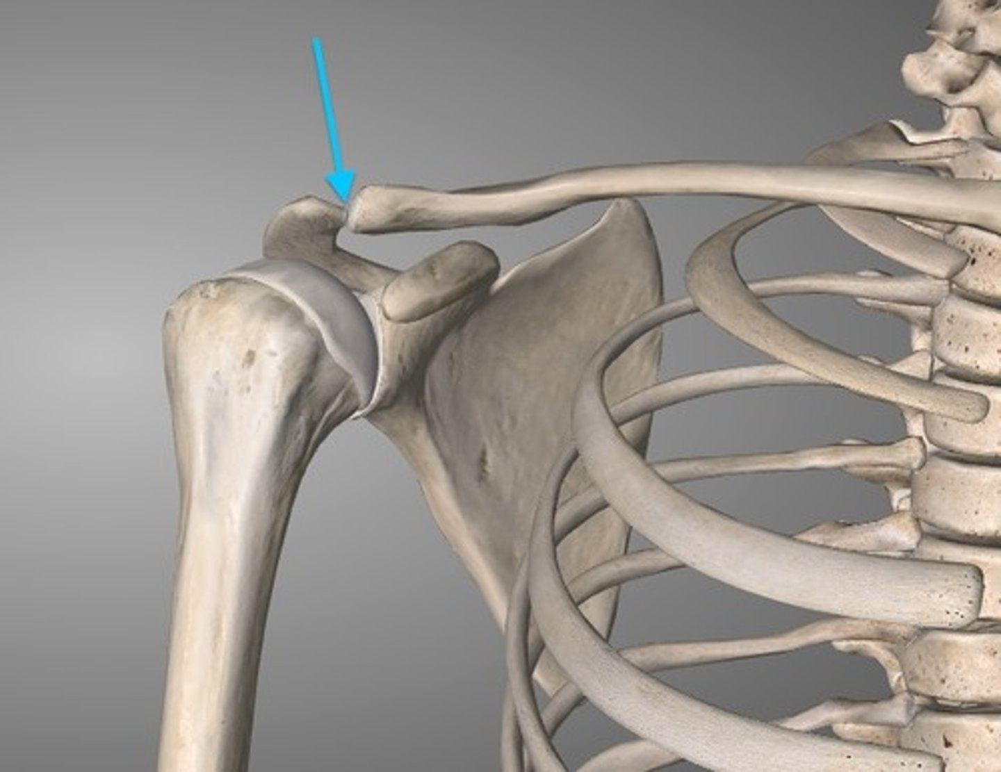
acrominoclavicular joint
the area where the clavicle and scapula connect
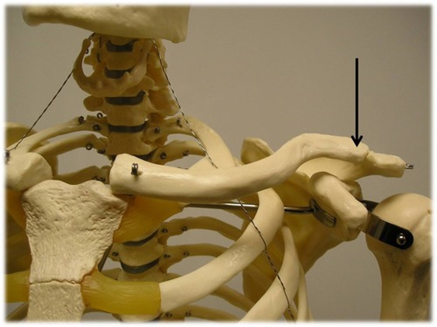
acromial end (lateral)
articulates with scapula
sternal end (medial)
articulates with the manubrium of the sternum
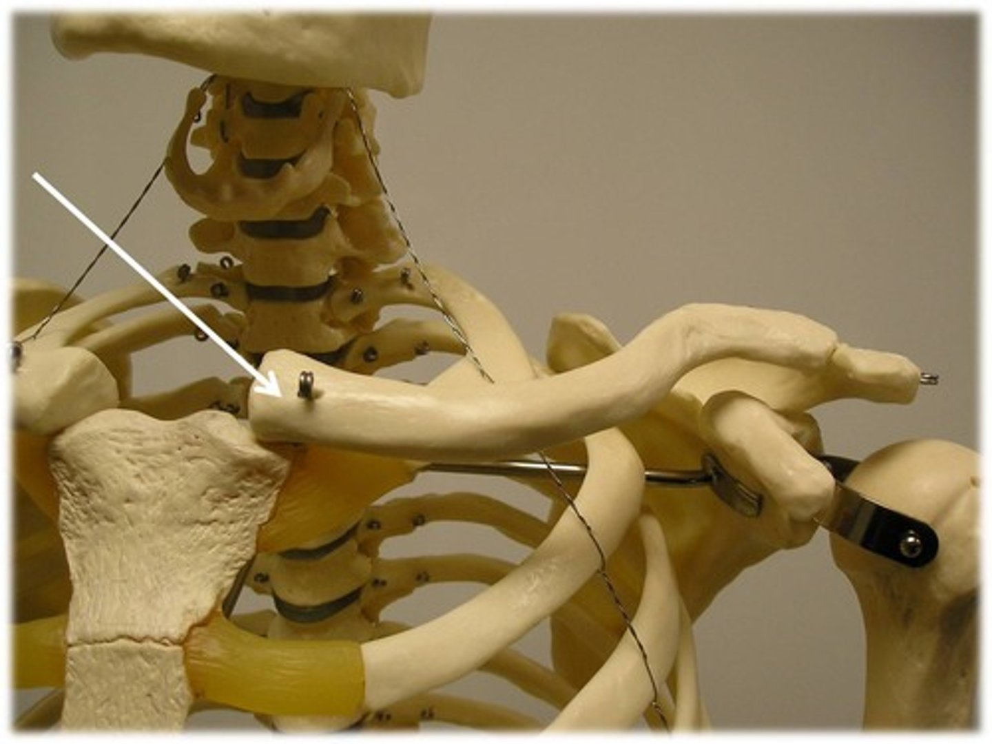
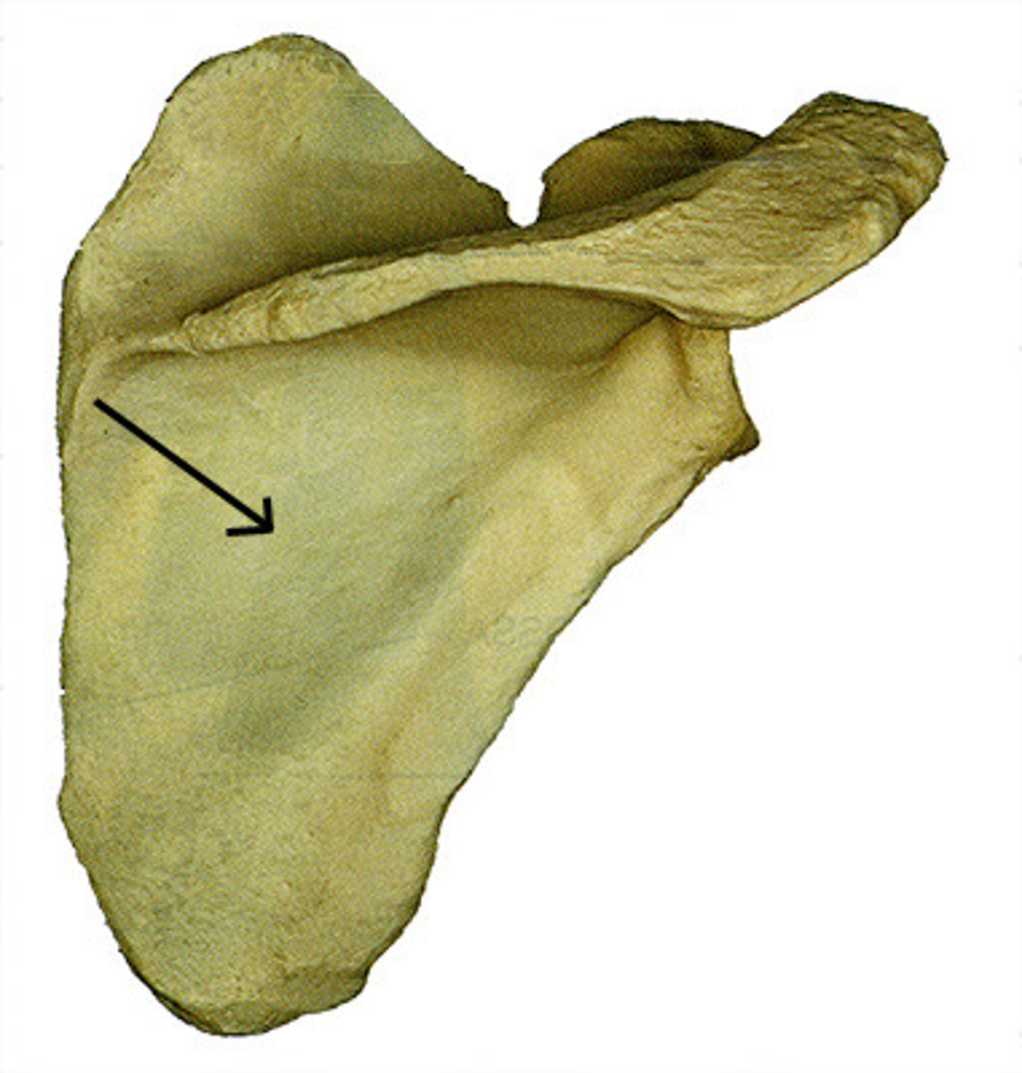
subscapular fossa
anterior depression of scapula
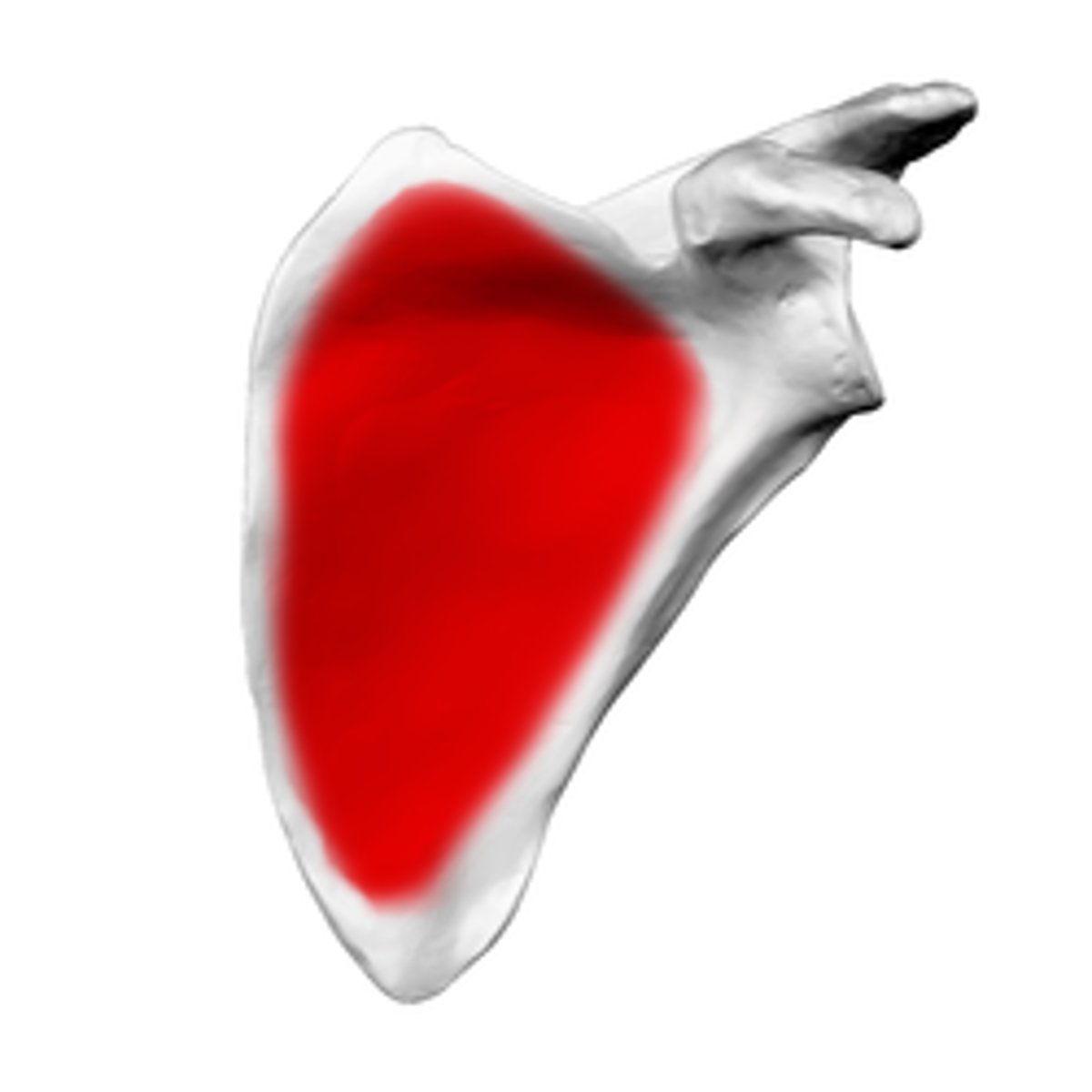
supraspinous fossa
posterior concavity superior to the scapular spine
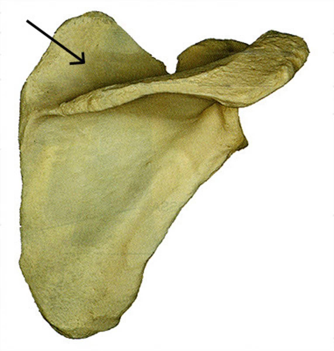
infraspinous fossa
posterior concavity inferior to the scapular spine