Brain stem
1/29
Earn XP
Description and Tags
Name | Mastery | Learn | Test | Matching | Spaced |
|---|
No study sessions yet.
30 Terms
brainstem divisions
tectum (green): DORSAL, consist of superior and inferior colliculi
basis (purple): MOST VENTRAL, contains corticospinal and corticobulbar tracts
tegmentum (blue): contains main bulk of brainstem nuclei and reticular formation
corticobulbar tract
travel w the corticospinal tract → terminates on cranial nuclei in brainstem → cranial nuclei sends axons (cranial nerves) → control muscles of head, neck, face
control movements from neck and above
superior border of brainstem
mammillary bodies - posterior commissure
inferior border of brainstem
pyramidal decussation
which cranial nerve is derived from brainstem?
CN 3 to CN 12
which main part of the brain does the brainstem belong to?
the mesencephalon
corticospinal tract
motor fibers carry movement related info from primary motor cortex to spinal cord
control movement of whole body
sphenoid bone
contain sella turnica, optic groove, optic canal, superior orbital fissure, foramen rotundum, foramen ovale
optic canal
transmits optic nerve and ophthalmic artery
optic groove
houses optic chiasm
sella turcica
houses pitutitary gland
superior orbital fissure
transmist CN 3, 4, V1 (ophthalmic division), 6, ophthalmic veins
petrous portion of temporal bone
pg 14
occipital bone (inferior 2/3)
add picture
sphenoid bone (superior 1/3)
internal acoustic meatus
in the posterior fossa
CN 7 and 8 exit from the auditory canal
middle cranial fossa foraminas
optic canal
superior orbital fissure
foramen rotundum
foramen ovale
foramen spinosum
foramen lacerum
optic canal
CN 2
ophthalmic artery
superior orbital fissure
CN 3-4-5(1)-6
ophthalmic veins
V(1) - ophthalmic nerve
foramen rotundum
CN V2 - maxillary nerve
foramen ovale
CN V3- mandibualr nerve
foramen spinosum
middle meningeal artery
meningeal nerve V3
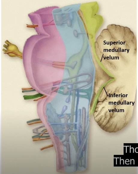
mid-sagittal section
add picture from lecture pg 18
identify structures on a mid sagittal MRI
sphenoid sinus, pituitary gland, clivus, optic chiasm, mammillary body, midbrain, pons, medulla, 4th ventricle, cerebellum
reticular formation
the portion of brainstem tegmentum where nuclei are not visible w staining
rostral reticular formation: alertness
caudal reticular formation: respiration and circulation
neurotransmitters
2 types:
neurotransmission: mediate communication between neurons - Glutamate (excitatory) and GABA (inhibitory)
neuromodulation: facilitate or inhibit the communication between neurons - NE, serotonin, histamine, dopamine, Ach
Norepinephrine (NE)
Effects:
excite/inhibit the cortex, mostly excite the thalamus
modulate attention, sleep-wake states and mood
serotonin
in 2 regions of raphe nucleus
rostral : sleep-wake cycle, mood, contribute to depression, anxiety, OCD
caudal : pain modulation, reathing, temperature regulation
dopamine
in 3 different regions:
mesostriatal: dysfunction causes Parkinson’s disease (treat w dopaminergic agonists)
mesolimbic: plays a role in reward and addiction, overactivity relates to hallucination in schizophrenia (treat w dopaminergic agonists)
mesocortical: plays a role in working memory, damage may cause cognitive deficits in Parkinson’s
histamine
most is in mast cells outside the nervous system for immune and allergic responses
may be important for alert state
antihistamine meds cause drowsiness by blocking CNS histamine receptors