Lab Practical III
 Call Kai
 Learn
Learn Practice Test
 Spaced Repetition
 Match
Match Flashcards
 Knowt Play
1/64

There's no tags or description
Looks like no tags are added yet.
Name | Mastery | Learn | Test | Matching | Spaced | Call with Kai | Chat |
|---|
No analytics yet
Send a link to your students to track their progress
65 Terms
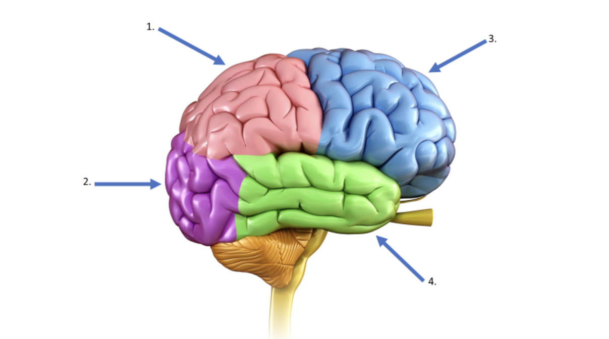
What is the name of the lobe indicated by #1?
parietal
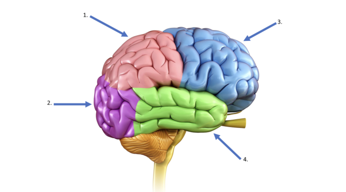
What is the name of the lobe indicated by #2?
occipital
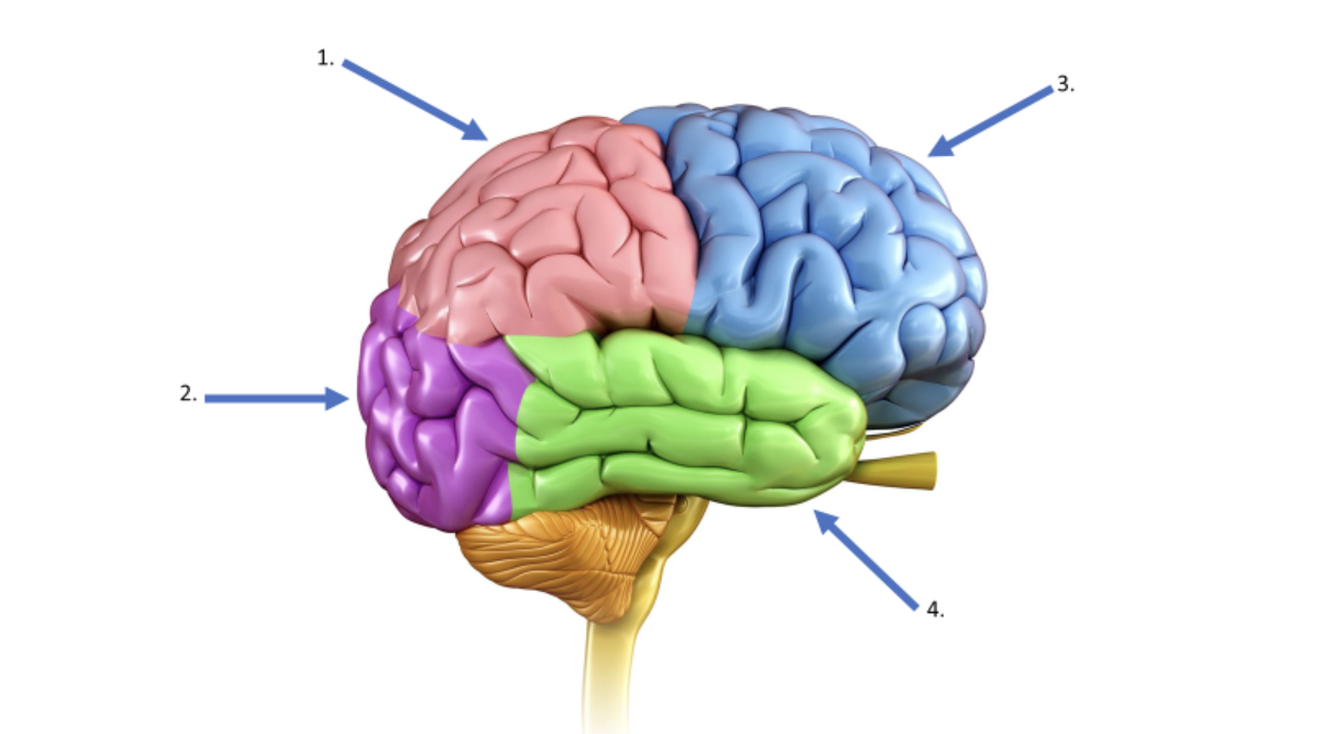
What is the name of the lobe indicated by #3?
frontal
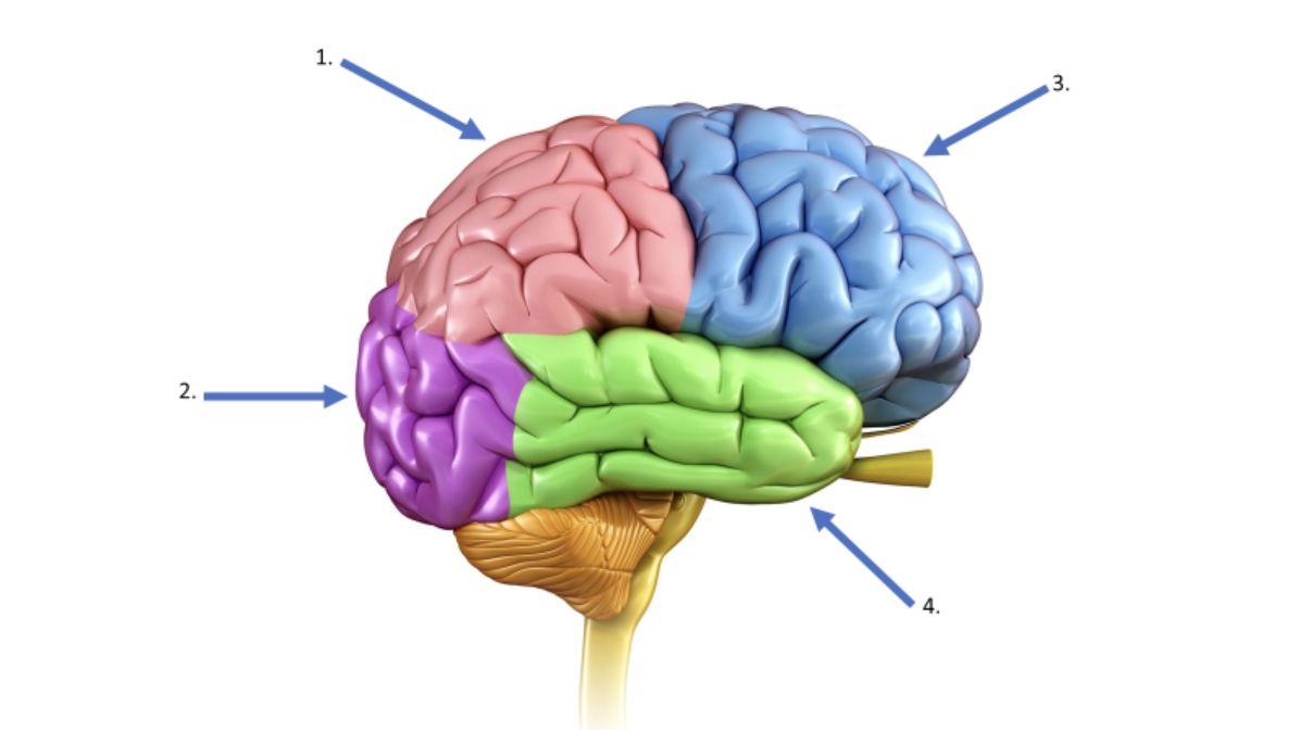
What is the name of the lobe indicated by #4?
temporal
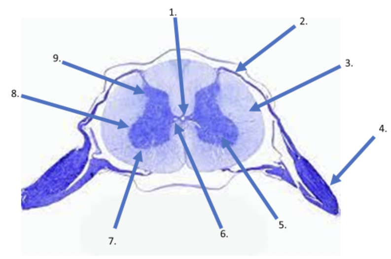
What is the name of the canal in #1?
central
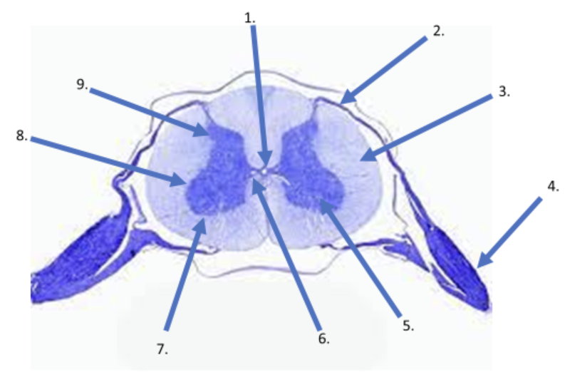
What is the name of the root indicated in #2?
dorsal
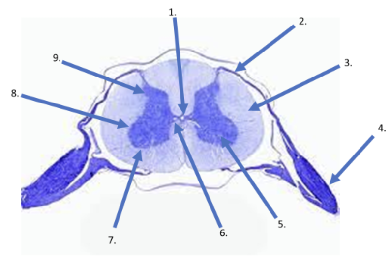
What type of matter is indicated by #3?
white
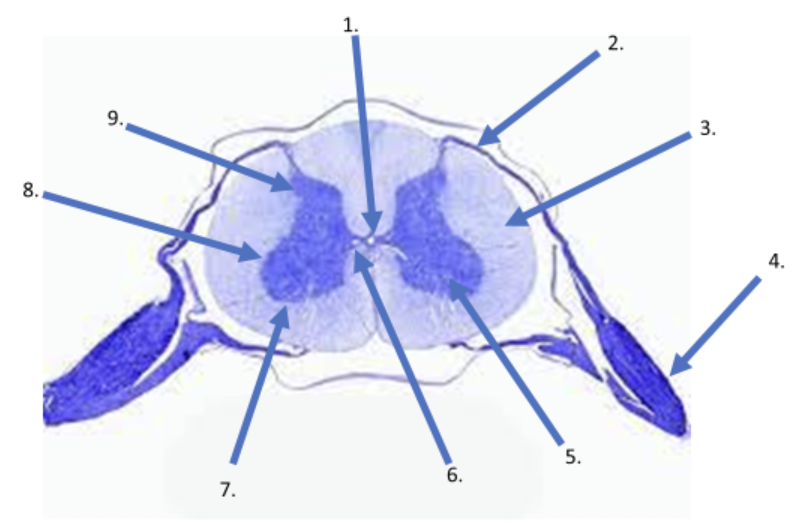
What is the name of the structure indicated by #4?
dorsal root ganglion
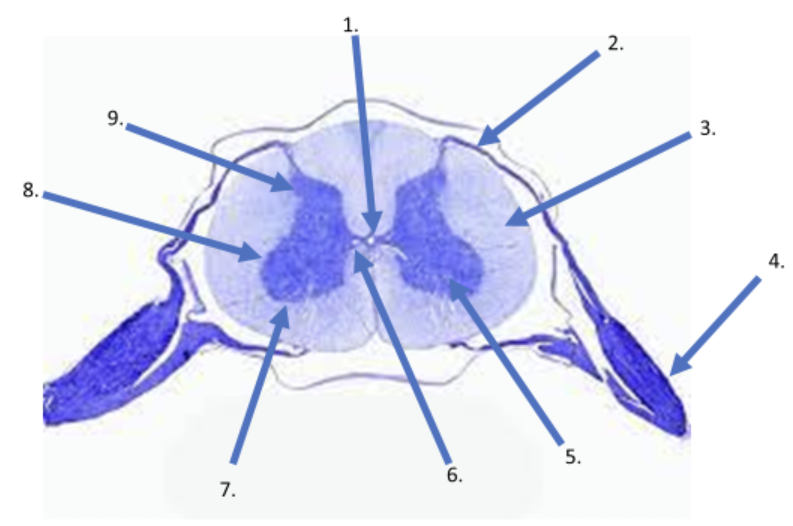
What type of matter is indicated by #5?
gray
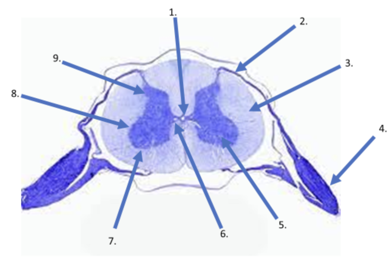
What is the name of the "bridge" indicated by #6?
gray commissure
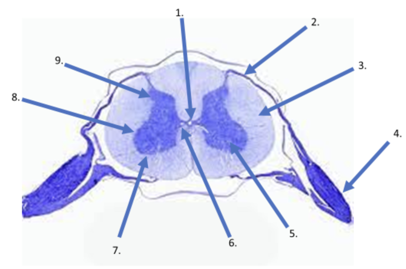
What is the name of the "horn" indicated by #7?
anterior horn
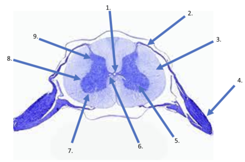
What is the name of the "horn" indicated by #8?
lateral horn
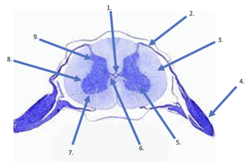
What is the name of the "horn" indicated by #9?
posterior horn
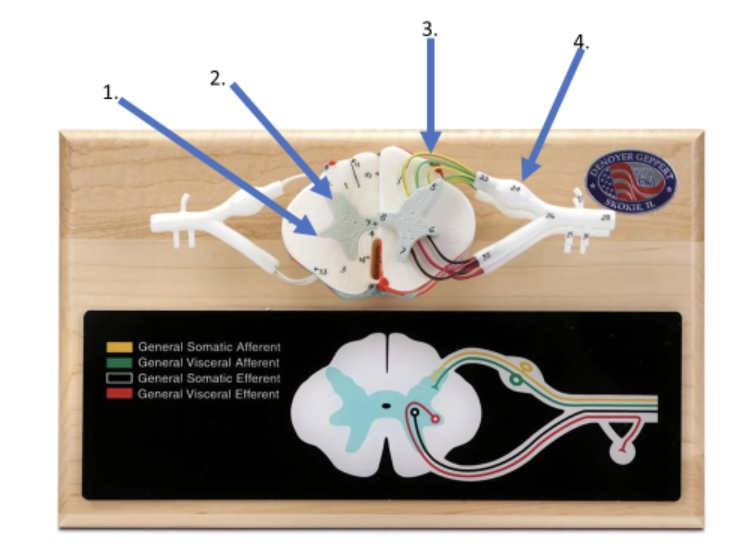
What is the name of the "horn" indicated by #1?
lateral horn
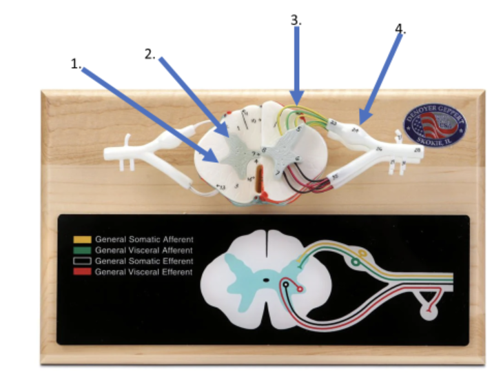
What is the name of the "horn" indicated by #2?
posterior horn
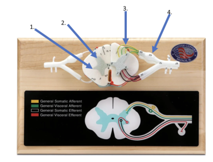
What is the name of the roots indicated in #3?
dorsal roots
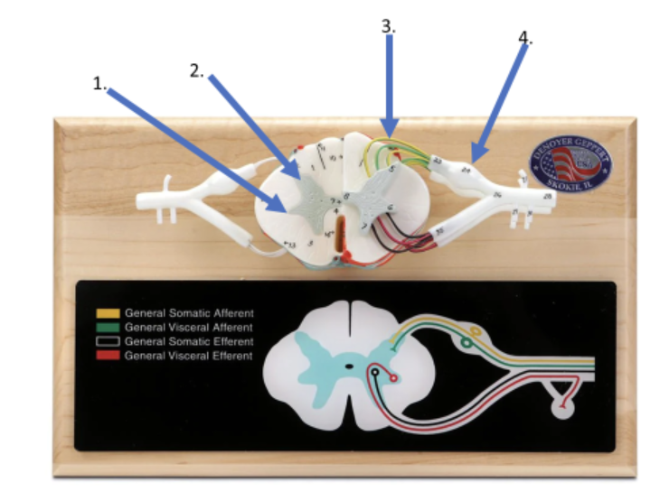
What is the name of the structure indicated by #4?
dorsal root ganglion
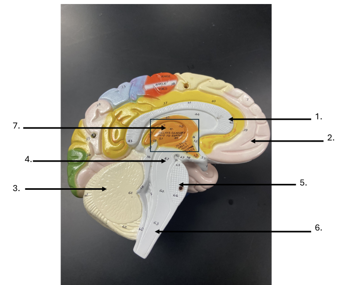
What is the name of the strucuture indicated by #1 in the image below?
corpus callosum
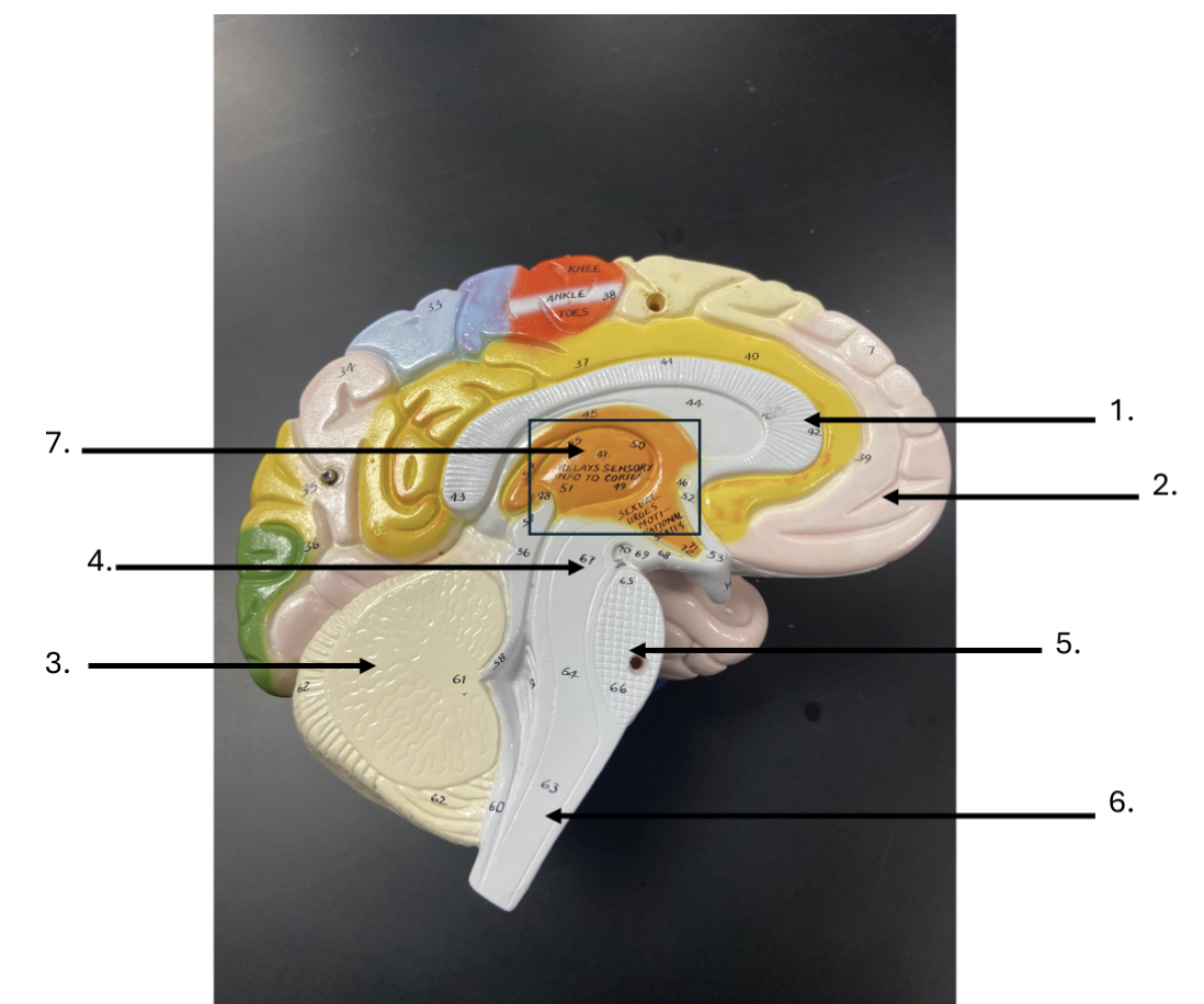
What is the name of the region of the brain indicated by #2?
cerebrum
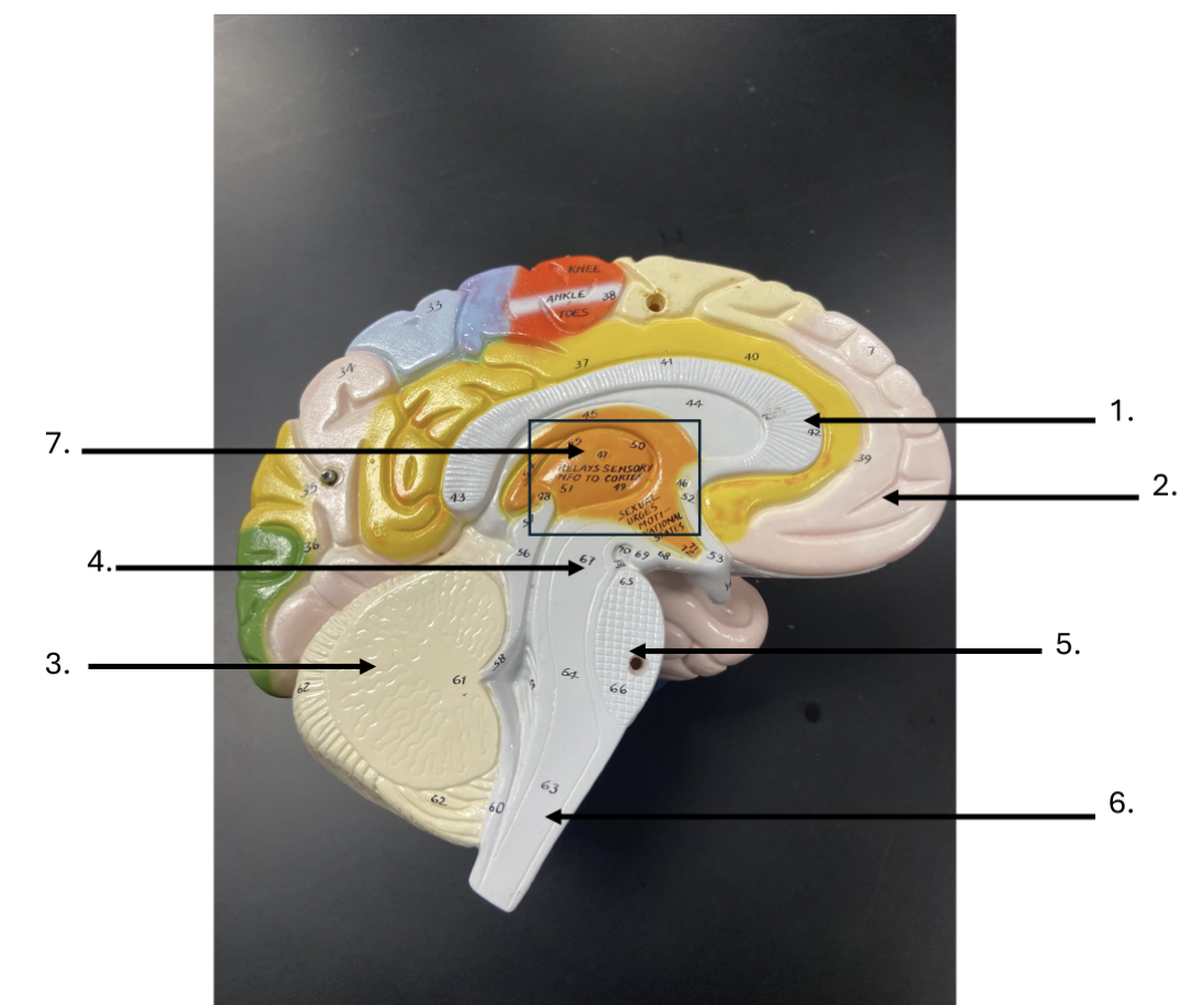
What is the name of the structure indicated by #3 in the image?
cerebellum
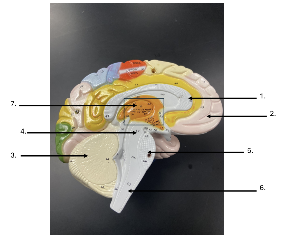
What is the region of the brainstem indicated by #4 in the image?
midbrain
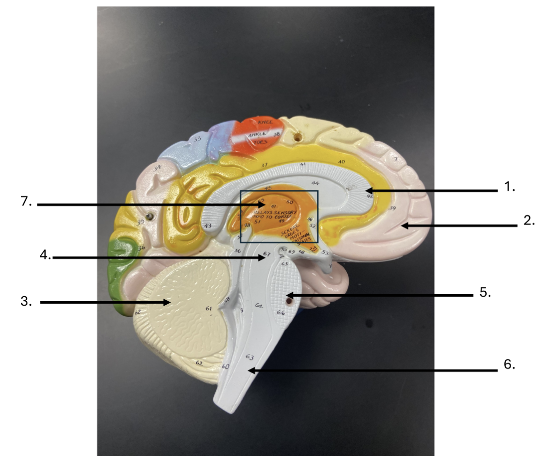
What is the name of the region of the brainstem indicated by #5?
pons
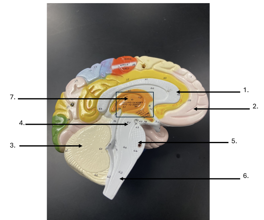
What is the name of the region of the brain stem indicated by #6?
medulla oblongata
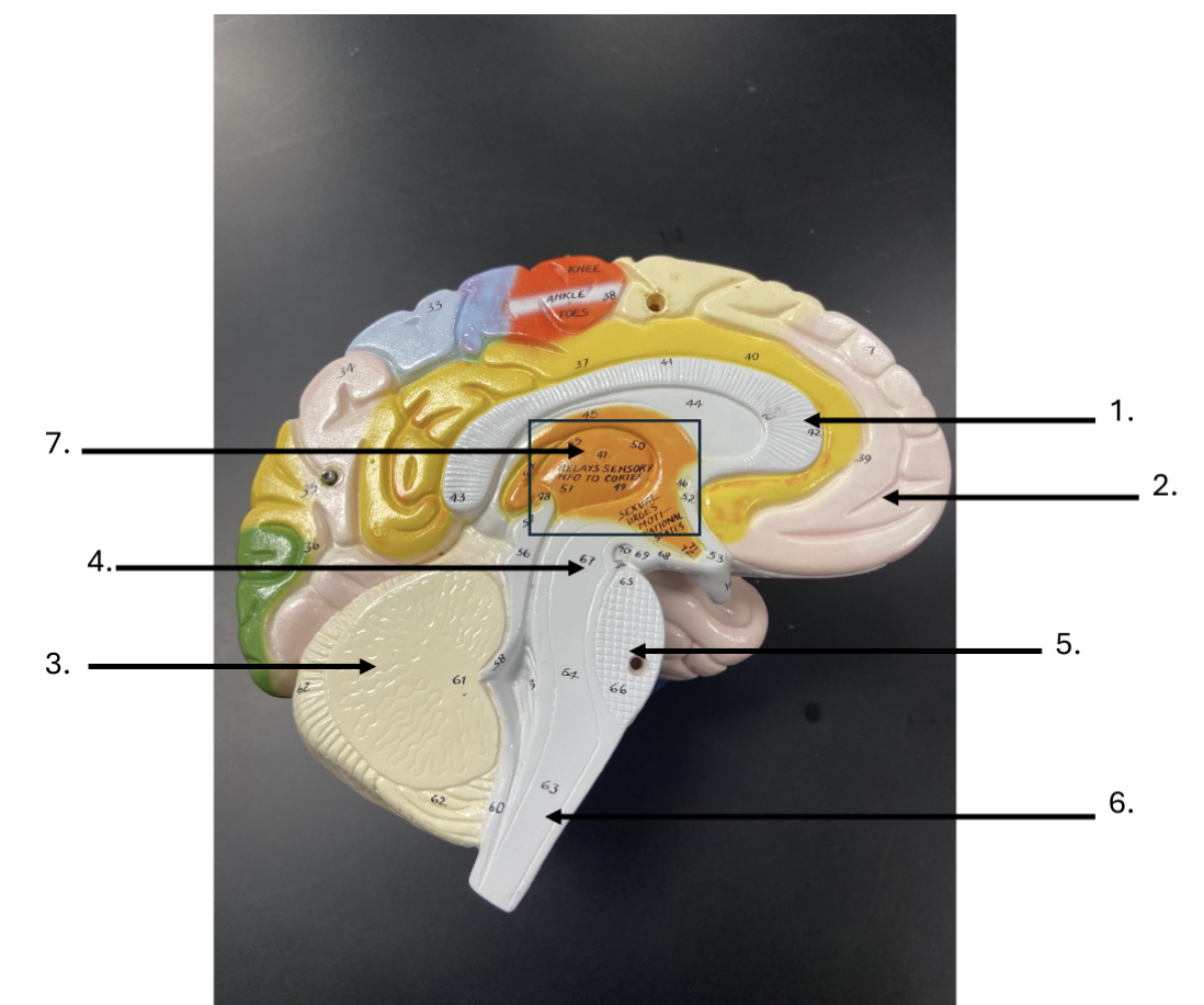
Within the area of the box are the thalamus and hypothalamus, what is the name of this generalized area indicated by #7?
diencephalon
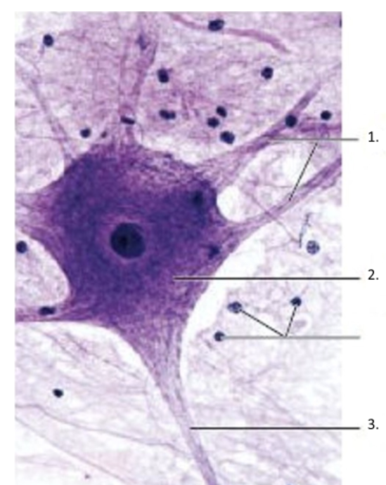
What is the structure indicated by #1 in the image provided below?
dendrites
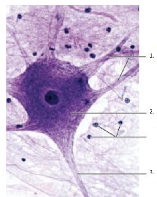
What is the structure indicated by #2 in the image provided below?
cell body
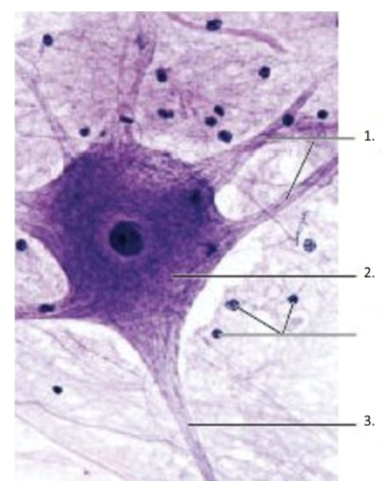
What is the name of the structure indicated by #3 in the image provided below?
axon
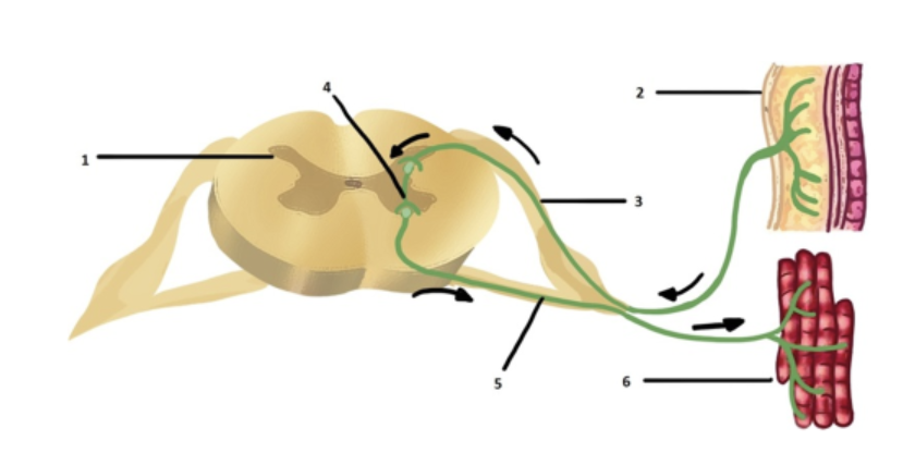
What is the structure indicated by #1 in the image provided below?
spinal cord
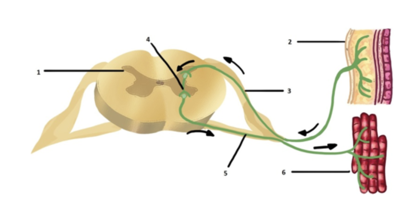
What type of neurons are represented by #2 in the image provided below?
sensory neurons
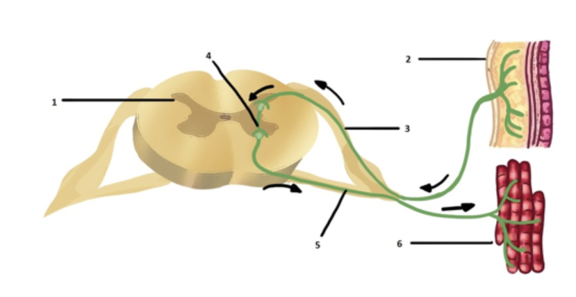
What is the name of the communication pathway indicated by #3 in the image provided below? Remember, A - AWAY from the reflex site
afferent pathway
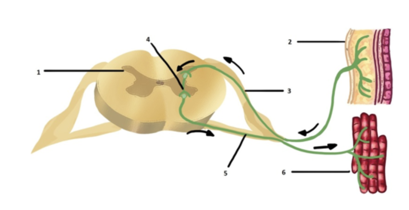
What is the name of the neurons indicated by #4 in the image provided below?
interneurons
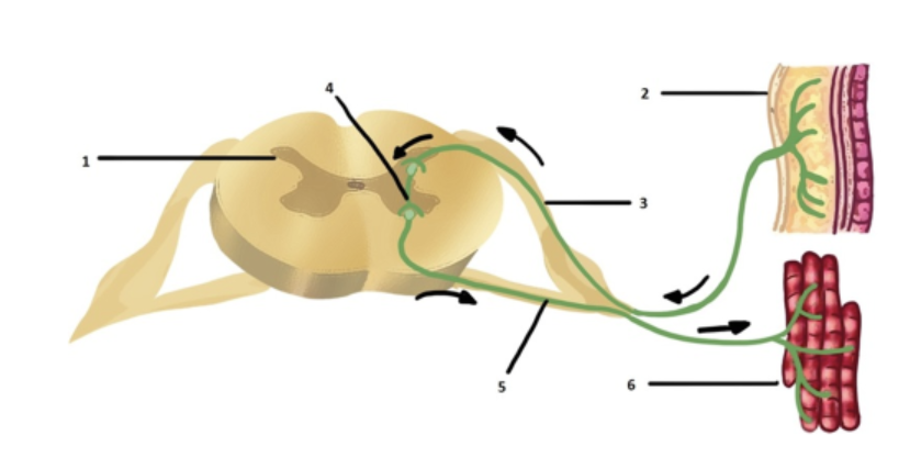
What is the name of the communication pathway indicated by #5 in the image provided below? *Remember, E -- EXCITE
efferent pathway

Provided the chart below where would #1 be located?
inner region

Based on the chart below, where would #2 be located?
outer region

Based on the chart below, where would #3 be located?
outer region

Based on the chart below, where would #4 be located?
inner region
General senses have simple neural pathways and include touch, temperature, pain, chemical, and pressure detection.
true
Special senses have complex pathways and contain receptors responsible for gustation, olfaction, vision, auditory, and equilibrium.
true
__________ receptors are sensors for changes in body temperature and have a wide distribution in the body, being found in the dermis, skeletal muscles, and hypothalamus.
Thermoreceptors
_______________, which are found in the medulla, in arteries near the heart, and in the heart, monitor changes in the concentrations of various chemicals present in body fluids.
Chemoreceptors
A _____________ is a touch-sensitive receptor in the dermal papillae.
Tactile corpuscles (Meissner's)
______________ pain means that the part of the body where pain is felt is different from the part of the body at which the painful stimulus is applied or is occurring.
Referred
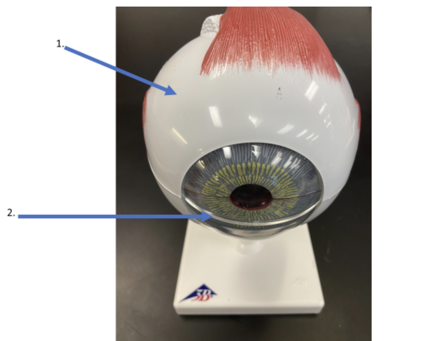
What is the structure indicated in #1?
sclera
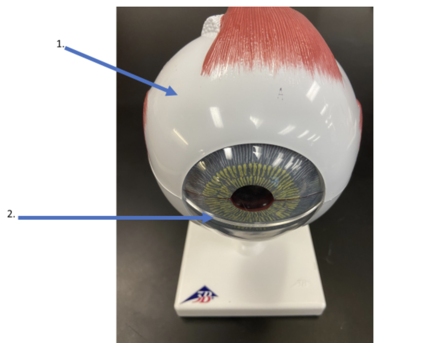
What is the structure indicated in #2?
cornea
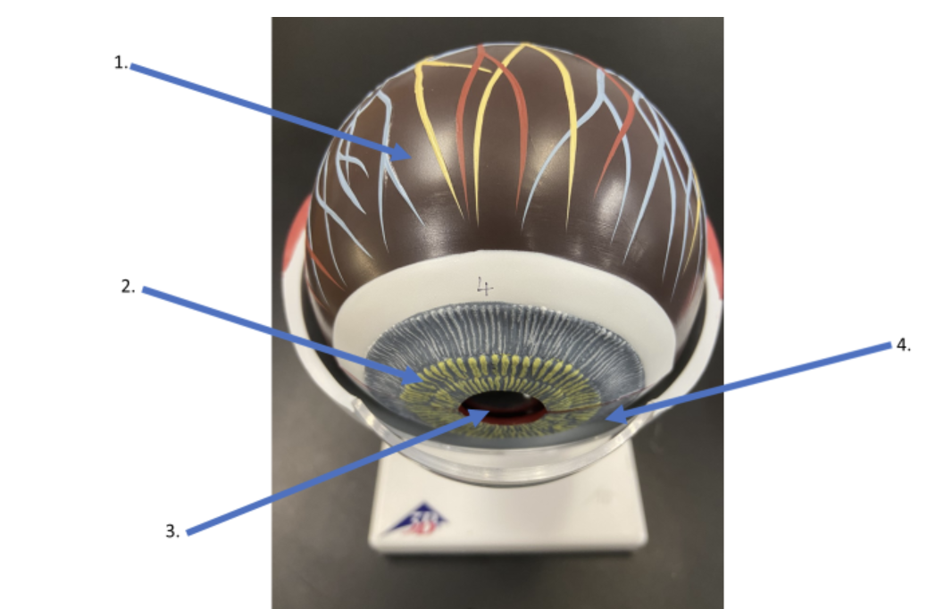
What is the layer indicated in #1?
choroid coat
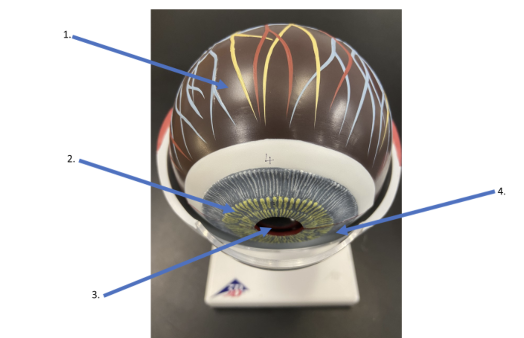
What is the structure indicated in #2?
iris
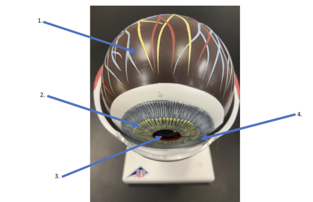
What is the structure indicated in #3?
pupil
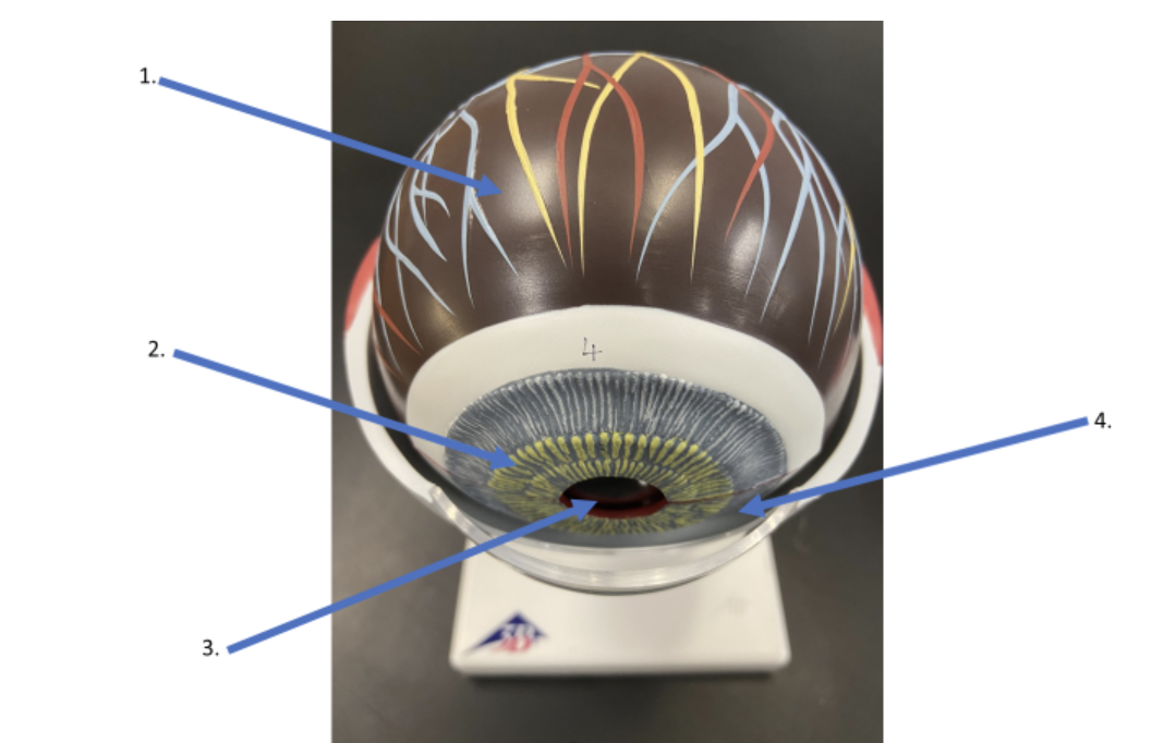
What is the fluid found in the anterior chamber as indicated by #4?
aqueous humor
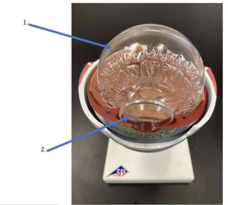
What is the name of the fluid that fills the posterior chamber of the eye as indicated by #1?
vitreous humor
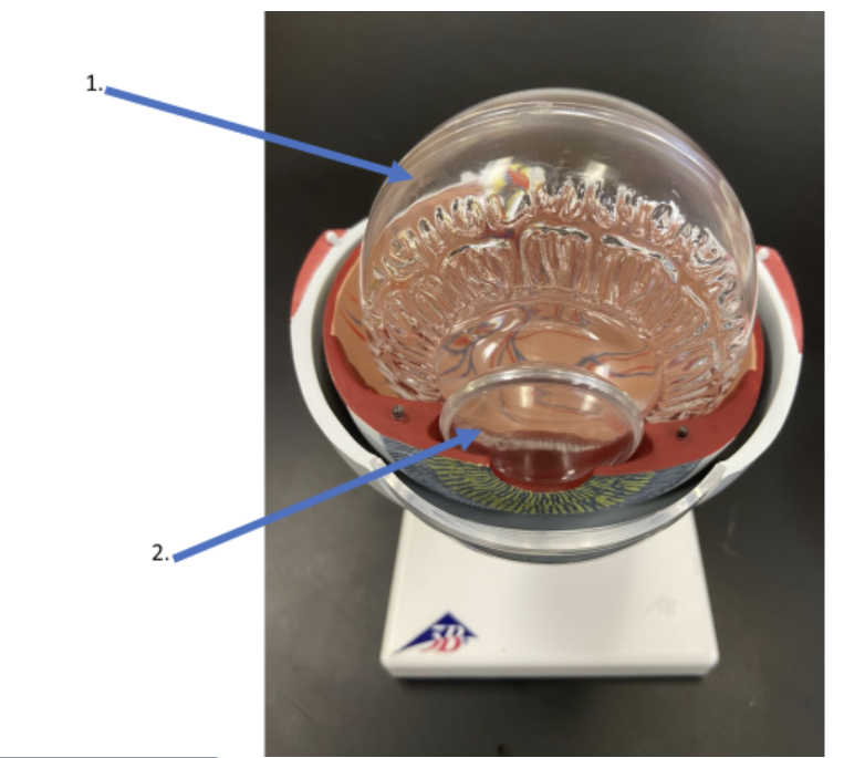
What is the name of the structure indicated by #2?
lens
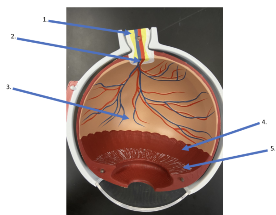
What is the name of the nerve indicated by #1?
optic nerve
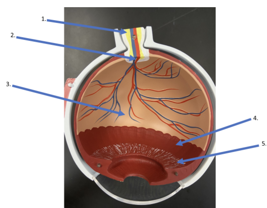
What is the name of this area where sharp vision will not take place as indicated by #2?
optic disc
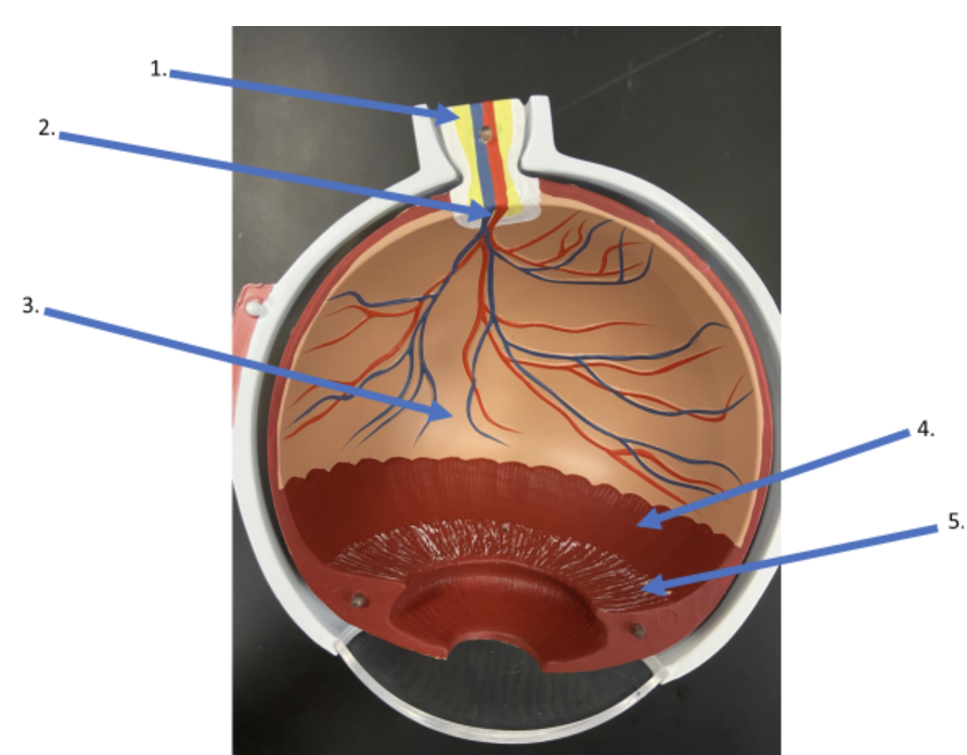
What is the name of the neural layer of the eye as indicated by #3?
retina
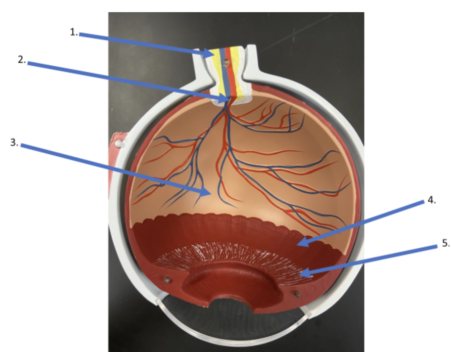
What is the name of the structure as indicated by #4?
ciliary body
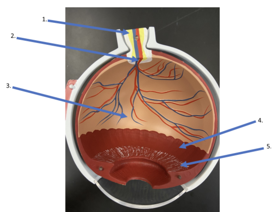
What is the name of the ligaments indicated by #5?
suspensory ligaments
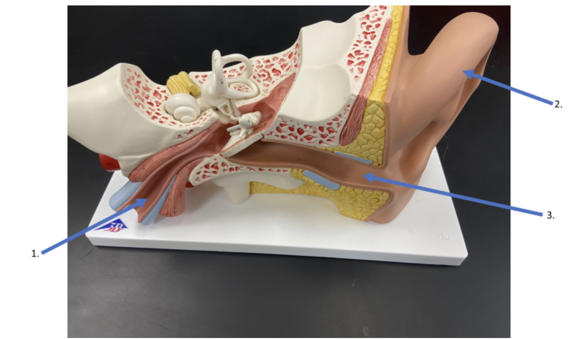
What is the name of the structure indicated by #1?
auditory tube
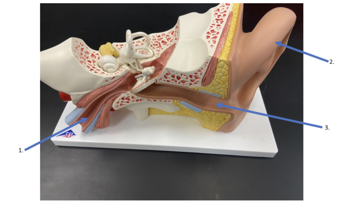
What is the name of the structure indicated by #2?
auricle
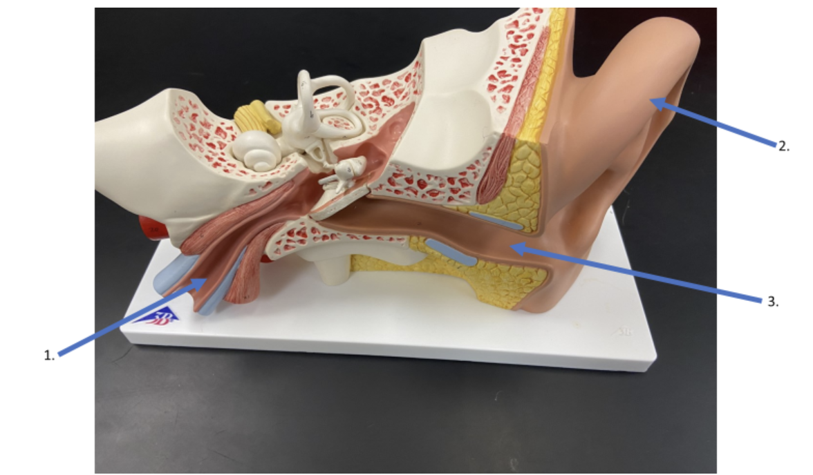
What is the name of the structure indicated by #3?
external acoustic meatus
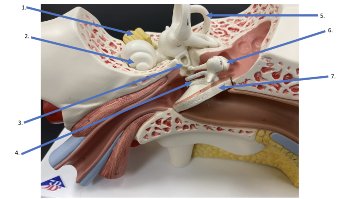
What is the name of the nerve indicated by #1?
vestibulocochlear nerve
(balance & hearing)
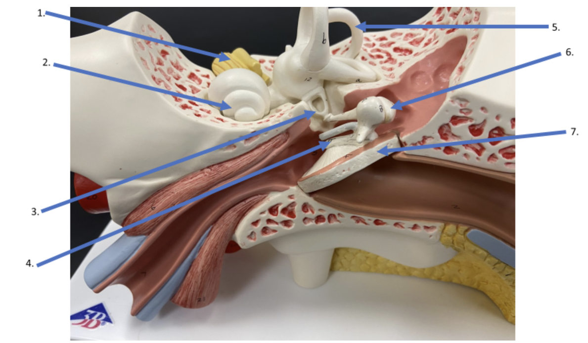
What is the name of the structure indicated by #2?
cochlea
(tonotopic frequency)
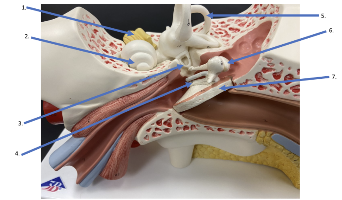
What is the name of the auditory ossicle indicated by #3?
stapes
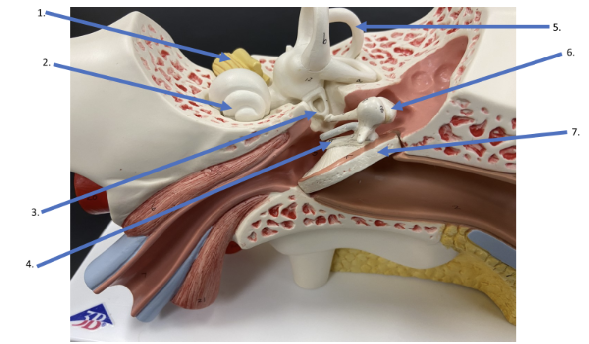
What is the name of the auditory ossicle indicated by #4?
malleus
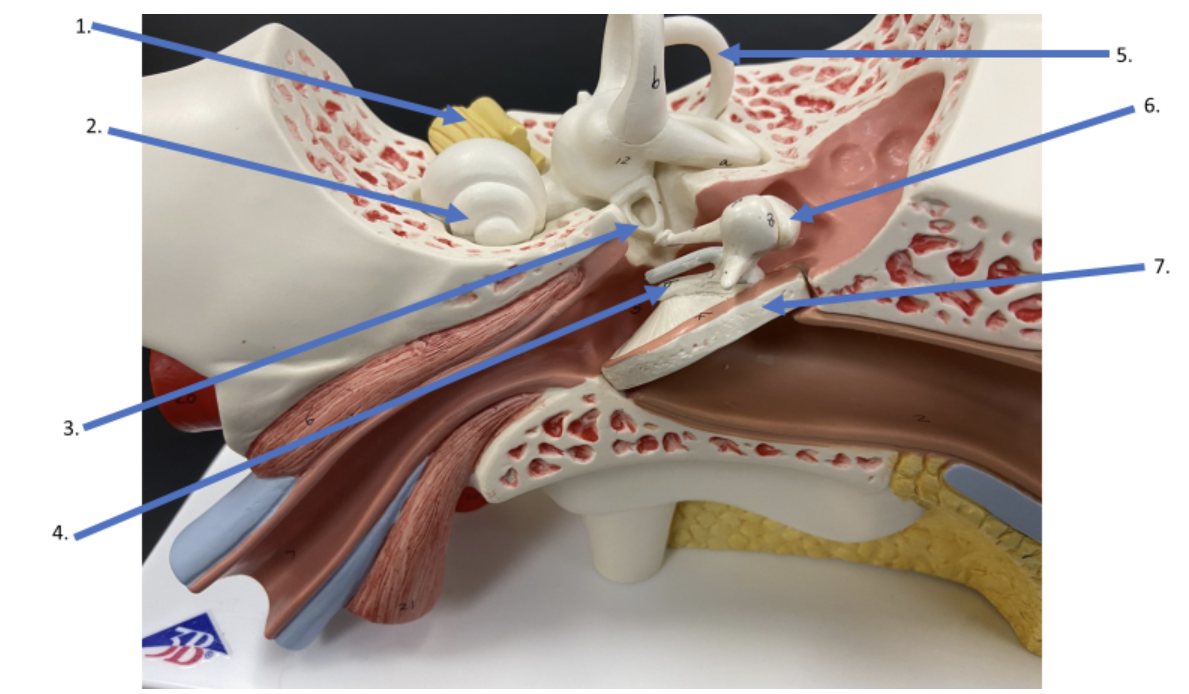
What is the name of the structure indicated by #5?
semicircular canals
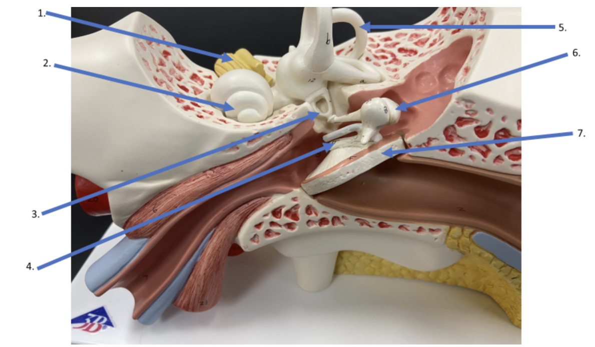
What is the name of the auditory ossicle identified by #6?
incus
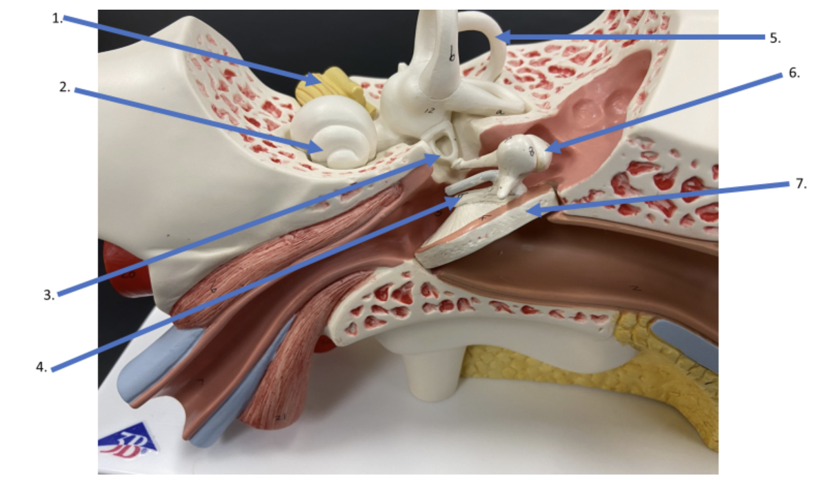
What is the name of the structure indicated by #7?
tympanic membrane