microscopes + cell fractionation + ultracentrifugation- in cell structure
1/27
There's no tags or description
Looks like no tags are added yet.
Name | Mastery | Learn | Test | Matching | Spaced |
|---|
No study sessions yet.
28 Terms
what are microscopes used for
to analyse cell components an observe organelles
define magnification
tells you how many times bigger the image produced by the microscope is than the real life object you’re viewing
define resolution
the ability to distinguish clearly between two objects
what are the two main types of microscopes
optical microscopes (light microscopes)
electron microscopes
describe optical (light) microscopes
they use light to form an image
using light limits the resolution of the image because it’s impossible to distinguish between two objects that are closer than half the wavelength of light to each other → hard to see smaller organnels such as ribosomes, ER
the wavelength of light is between 500-650 nm → optical microscopes have a resolution of around 200 nm → can see nuclei, mitochondria, chloroplasts
can observe live specimens (don’t use a vacuum) → look at the movement of chromosomes during mitosis and meiosis
describe electron microscopes
they use electrons to form an image
electron microscopes have a higher resolution than optical microscopes → can resolve between two objects that are extremely close together → more detailed image
wavelength of light- 500nm
wavelength of a beam of electrons- 0.5 nm
this means that electron microscopes can view smaller organelles such as ribosomes, ER
what are the two types of electron microscope
transmission electron microscopes (TEM’s)
scanning electron microscopes (SEM’s)
what are transmission electron microscopes (TEM’s)
they use electromagnets to focus a beam of electrons
the beam of electrons is transmitted through the specimen
denser parts of the specimen absorb more electrons (appears darker in image)
thinner parts of the specimen absorb less electrons (appear lighter in image)
this produces a contrast
what are the advantages of TEM’s
high resolution images
can see the internal structures of cells and organelles in detail
what are the disadvantages of TEM’s
they can only be used with vey thin specimens
they can’t be used to observe live specimens (vacuum inside the TEM, so all water must be removed → specimens must be dead)
they don’t produce a colour image (optical microscopes do)
what are scanning electron microscopes (SEM’s)
they scan a beam of electrons across the specimen
the electrons bounce off the surface of the specimen and the electrons are detected which forms an image
what are the advantages of SEM’s
they can be used on thick or 3D specimens (TEM’s can’t → specimen must be thin)
they produce 3D images that show the surface of specimens
what are the disadvantages of SEM’s
they give lower resolution images than TEMs
they can’t be used to observe live specimens (use a vacuum → no water)
they don’t produce a colour image (optical microscopes do)
why should you always start with the low power objective lens first when using an optical microscope
it’s easier to find what you’re looking for in the FOV as it’s less zoomed in
it helps to prevent damage to the lens or coverslip incase the stage has been raised too high
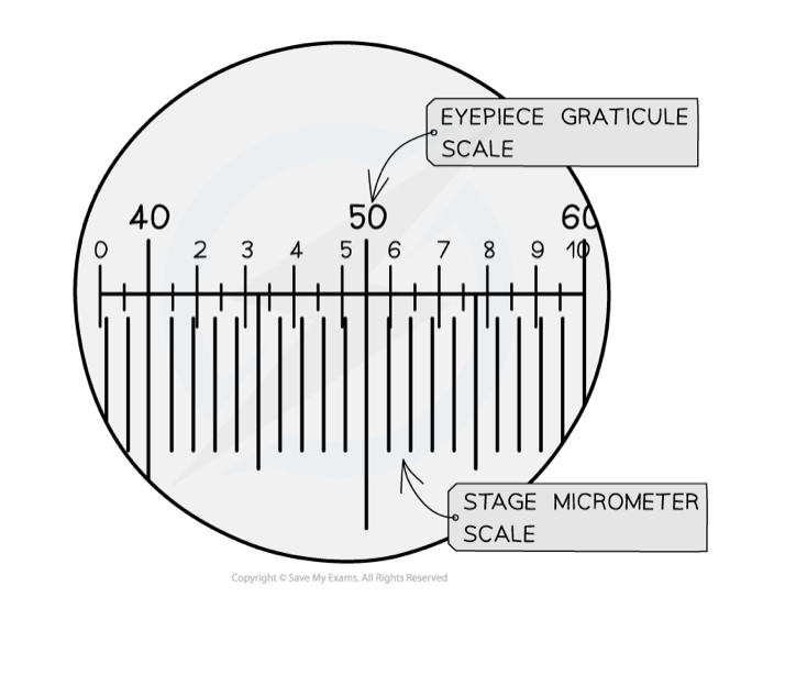
what is an eyepiece graticule used for
a graticule is a small disc that has an engraved scale
it can be placed into the eyepiece of a microscope to be used as a ruler in the FOV → to measure the length of a cell or organelle
a graticule has no fixed units so must be calibrated for the objective lens that’s in use
how is an eyepiece graticule calibrated
uses a scale engraved on a microscope slide (a stage micrometer)
by using the two scales together, the number of micrometers each graticule unit is worth can be worked out
after this is known, the graticule can be used as a ruler in the FOV
why is a stain used
it makes it easier to see the specimen under a microscope by making the specimen darker
how should the cover slip be applied
slowly and at angle to avoid any air bubbles getting trapped under the cover slip which could obstruct your view of the specimen
what are the two types of lens’ that optical (light) microscopes use
eyepiece lens- usually a magnification of x10
a series of objective lens’ each with a different magnification
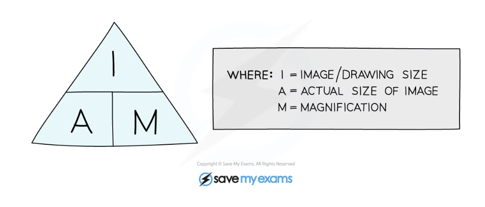
how do you calculate total magnification
total magnification = eyepiece lens magnification x objective lens magnification
how do you calculate magnification
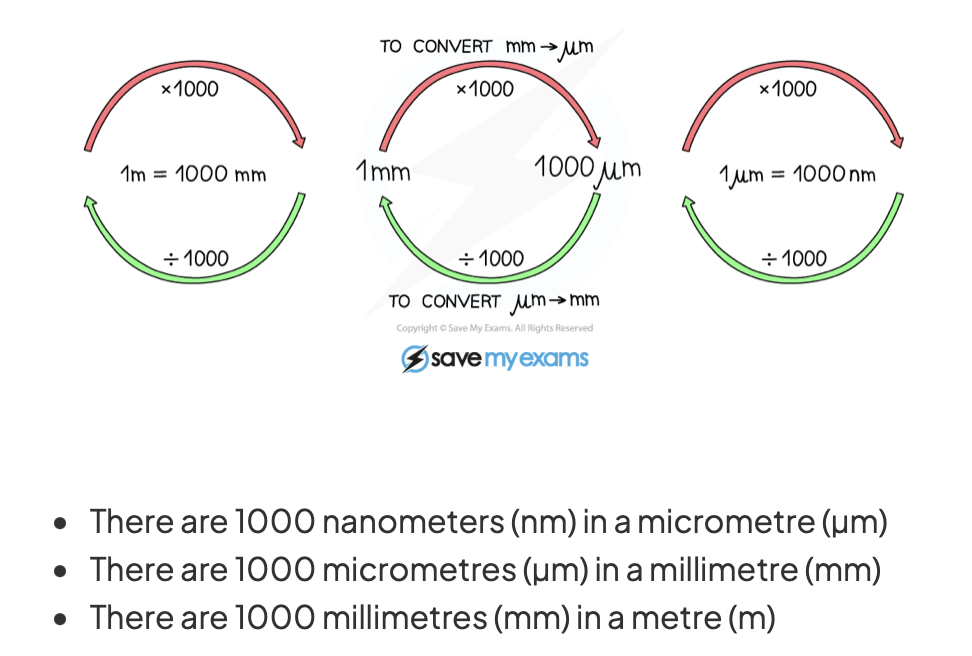
how do you convert between metres and nano meters
why may scientists want to look at one specific organelle
to study its structure under an electron microscope
to conduct research on it and learn more about its function
what different processes are involved in cell fractionation
homogenisation
filtration
ultracentrifugation
what happens in homogenisation
get a sample of tissue (containing the cells to be broken up)
place the sample of tissue in a cold, isotonic, buffered solution
cold- reduce the activity of enzymes
isotonic- the solution must have the same water potential as the cells being broken up to prevent water moving into the cells via osmosis and damaging them
buffered- to prevent organelle proteins, including enzymes from becoming denatured
the tissue containing solution is then homogenised using a homogeniser which grinds up the cells and breaks the plasma membrane of the cells and releases the organelles into a solution called the homogenate
what happens in filtration
the homogenate is filtered through a gauze
this is to separate out any large cell or tissue debris that wasn’t broken up by the homogeniser
the organelles are much smaller so they can pass through the gauze and aren’t filtered out
this forms the filtrate (contains a mixture of organelles)
what happens in ultracentrifugation
the filtrate is placed into a tube and the tube is placed into a centrifuge
The filtrate is first spun at a low speed
This causes the largest, heaviest organelles (such as the nuclei) to settle at the bottom of the tube, where they form a thick sediment known as a pellet
The rest of the organelles stay suspended in the solution above the pellet
This solution is known as the supernatant
The supernatant is drained off and placed into another tube, which is spun at a higher speed
Once again, this causes the heavier organelles (such as the mitochondria) to settle at the bottom of the tube, forming a new pellet and leaving a new supernatant
The new supernatant is drained off and placed into another tube, which is spun at an even higher speed
This process is repeated at increasing speeds until all the different types of organelle present are separated out (or just until the desired organelle is separated out)
Each new pellet formed contains a lighter organelle than the previous pellet
what is the order of mass of the organelles (from heaviest to lightest)
nuclei
chloroplasts
mitochondria
lysosomes
endoplasmic reticulum
ribosomes