Ch. 1 Periodontium: The tooth supporting structures
 Call Kai
 Learn
Learn Practice Test
 Spaced Repetition
 Match
Match Flashcards
 Knowt Play
1/30

There's no tags or description
Looks like no tags are added yet.
Name | Mastery | Learn | Test | Matching | Spaced | Call with Kai | Chat |
|---|
No analytics yet
Send a link to your students to track their progress
31 Terms
Periodontium
Functional system of tissues that surrounds teeth and attaches them to jawbone
Perio = around
Odontos = tooth
*aka- supporting tissues of teeth, attachment apparatus
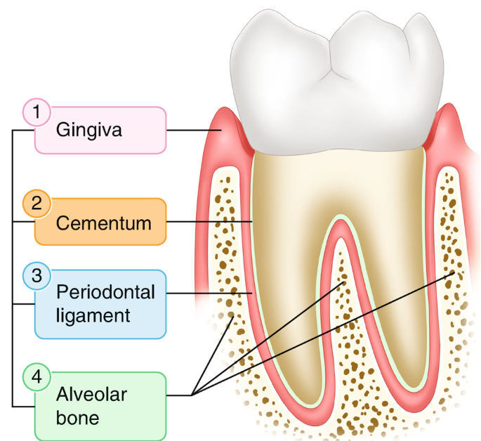
Tissues compromising the Periodontium
Gingiva
Cementum
Periodontal Ligament
Alveolar Bone
Function of the Gingiva
Provides a tissue seal around the cervical portion (neck) of the tooth
Covers the alveolar processes of the jaws
Holds the tissue against the tooth during mastication
Function of the Periodontal Ligament
Suspends and maintains the tooth in its socket
Function of Cementum
Anchors the ends of the periodontal ligament fibers to the tooth so that the tooth stays in its socket
Protects the dentin of the root
Function of the Alveolar Bone
Surrounds and Supports the roots of the tooth
Gingiva
Contour and shape of gingival margin approximates scalloped curvature of cementoenamel junction (CEJ)
Position may be age dependent
Health young- at or coronal to the CEJ
Healthy older- apical to CEJ
Attached by junctional epithelium
Composed of thin outer layer of epithelium and underlying layer of connective tissue
has 4 anatomical areas
function- protects underlying tooth-supporting structures of the periodontium from oral environment
Boundaries of the Gingiva
Gingival margin
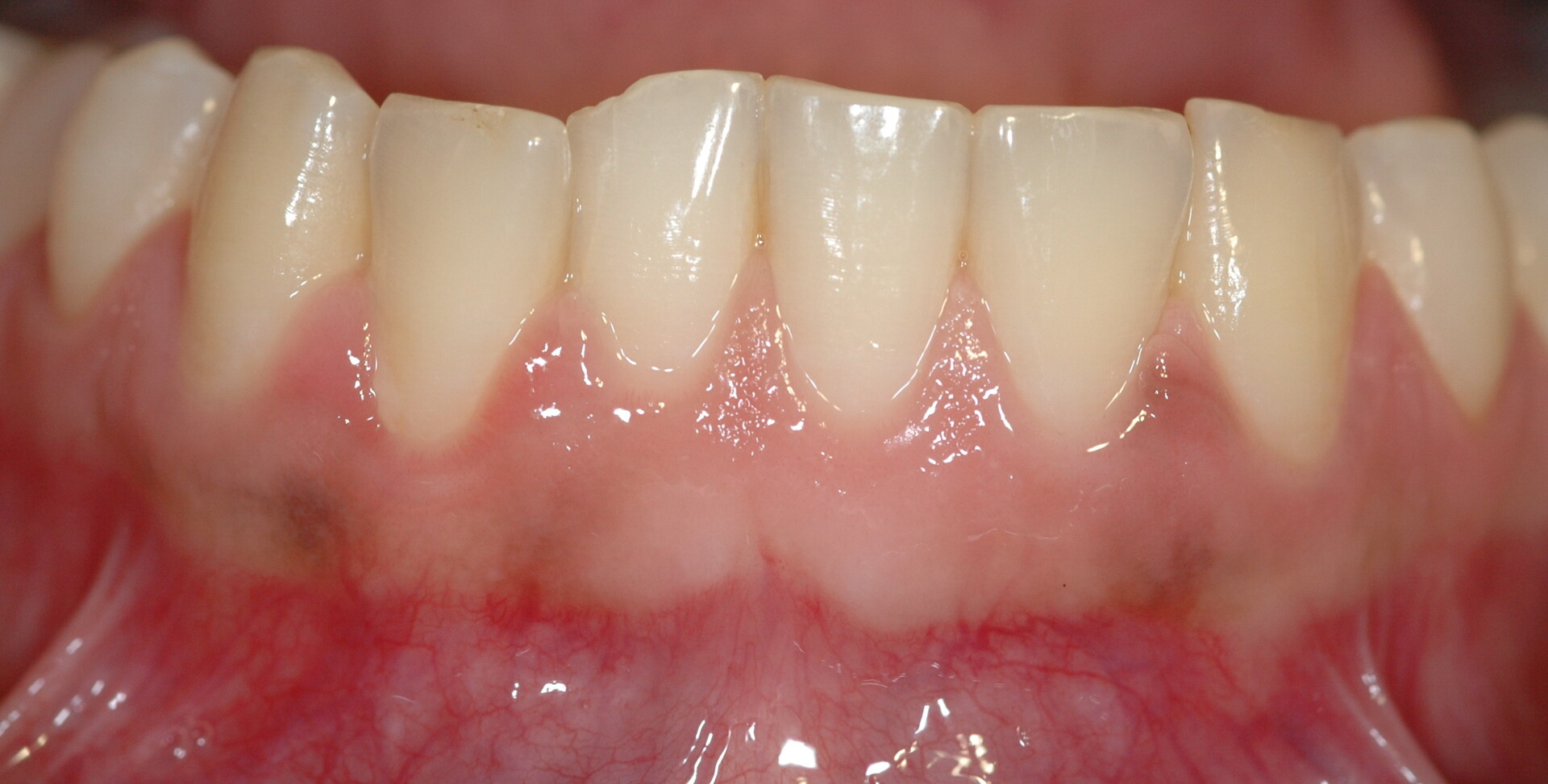
Alveolar mucosa: dark, red smooth shiny
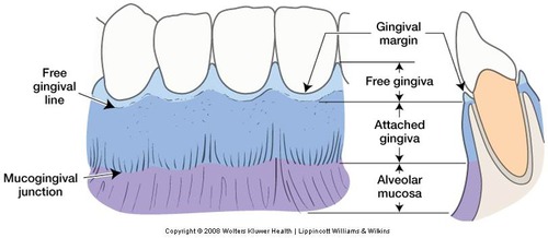
Free gingival groove - Gingiva Demarcations
shallow linear depression separating free and attached gingiva (may be hard to see with the naked eye)
Mucogingival junction - Gingival Demarcation
where the pink attached gingiva meets the red alveolar mucosa (clinically visible)
What are the two gingival demarcations?
Free gingival groove
Mucogingival Junction
Free gingiva
Unattached portion surrounding tooth in CEJ region
Also called unattached gingiva or marginal gingiva
Surrounds tooth in turtleneck manner
Attaches by junctional epithelium
Fits closely around tooth, but not directly attached
Forms soft tissue lateral wall of gingival sulcus
Free gingival margin- thin, rounded edge where free gingiva meets tooth follows scalloped, curved contours of underlying CEJ.
Attached gingiva
Continuous with free gingiva
Tightly bound to - underlying cementum on cervical-third of periosteum of alveolar bone
WIDEST in incisors and molar regions
NARROWEST in premolar regions
NOT measured on palate
Function : allows gingival tissue to withstand mechanical forces created during chewing, speaking and tooth brushing
prevents free gingiva from being pulled apically from tooth when tension is applies to alveolar mucosa ( ex. if you pull or tug on lip, the gingiva helps keep it in place)
Color of attached Gingiva
Pale or light coral pink when healthy
** May have physiologic pigmentation caused by increased melanin production
Texture of attached gingiva
May exhibit stippling on healthy attached and interdental gingiva
** NOT always present
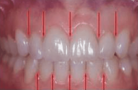
Interdental gingiva
Portion of gingiva that fills interdental embrasure between two adjacent teeth apical to contact area
Consist of facial papillae and lingual papillae
interdental col - depression apical to contact area of adjacent teeth that connects facial and lingual papillae
Function: Prevents food from packing between teeth when chewing
Gingival Sulcus
V-shaped shallow space between free gingiva and tooth surface
Encircles neck of tooth
1-3mm deep when clinically healthy (measured with periodontal probe)
Base interfaces with most coronal portion of junctional epithelium
Gingival Crevicular Fluid
also called Gingival sulcular fluid
seeps from underlying connective tissue
little to none in healthy sulcus
Flow increases in response to gingival stimulation (greatly increase with inflammation)
Which part of the gingiva is tightly bound to the underlying cementum and alveolar bone?
attached gingiva
What is the base of the gingival sulcus formed by?
most coronal junction of the coronal epithelium
Periodontal Ligament (PDL)
Network of soft connective tissue fibers that attach root of the tooth to bony walls of tooth socket
Primarily composed of dense fibrous connective tissue (fibers attach to root cementum and alveolar bone)
Serves to:
Connect tooth to alveolar process
support tooth in socket
Absorb mechanical loads placed on tooth
Functions of the Periodontal Ligament (5)
Supportive
Sensory
Nutritive
Formative
Remodeling
Supportive
suspends and maintains tooth in socket
Sensory
nerve endings relay sensory information
Nutritive
Blood vessels provide oxygen and nutrients
Formative
certain components build and maintain attachment apparatus and may aid periodontal regeneration
Remodeling
can remodel alveolar bone in response to pressure
Cementum
Thin layer of hard, mineralized tissue that covers surface of root
Overlies dentin
Softer than dentin or enamel
55% organic, 45% inorganic
More resistant to resorption than bone (makes orthodontic treatment possible)
undergoes continuous resorption and repair throughout lifetime of tooth
becomes thicker with age
two types: CELLULAR and ACELLULAR
Acellular Cementum
Forms BEFORE teeth are in occlusion
Devoid of cells
Covers cervical two-thirds of root
plays an important role in tooth support
Cellular Cementum
Forms AFTER teeth have reached occlusion
Composed of cementocytes
Covers apical one-third of root
Compensates for active eruption and normal tooth wear by continuous deposition of cementum