Chapter 11 slides lecture 1 narrated
 Call Kai
 Learn
Learn Practice Test
 Spaced Repetition
 Match
Match Flashcards
 Knowt Play
1/9

There's no tags or description
Looks like no tags are added yet.
Name | Mastery | Learn | Test | Matching | Spaced | Call with Kai | Chat |
|---|
No analytics yet
Send a link to your students to track their progress
10 Terms
Nervous system does three things
sensory input
- information gathered by sensory receptors about internal and external changes
- vision, taste, hear, touch, etc
- sensory information comes from peripheral receptors into CNS where there is integrationIntegration:
- interpretation of sensory input
- only takes place in CNS (spinal cord or brain)
- have input have to decide what to do, this is where the output or motor comes inMotor output:
- activation of effector organs (muscles and glands) produces a response
- ex: contract skeletal muscle, smooth muscle, cause glandular secretion, or nothing
How is the nervous system organized?
Central Nervous System (CNS)
- made up of neurons and supporting cells (glial cells)
- receives information from and sends information to the peripheral nervous system
- brain and spinal cord
- integrative and control centers, where conscious thought takes placePeripheral Nervous System (PNS)
- made up of cranial nerves (nerves that branch off brainstem) and spinal nerves (nerves that branch off the spinal cord)
- these nerves carry information away or to the CNS
- Sensory (afferent) division
- carries information toward the CNS
- Somatic (body):
- carries information from the body surface to the CNS
- Visceral (organ)
- carries information from the organs to the CNS
- Motor (efferent) division
- all about output
- carries information away from the CNS
- can carry information to skeletal muscle, to smooth muscle or to glands
- Somatic (body):
- voluntary motor system
- controls and how we chose to stimulate certain muscles to contract
- Autonomic:
- involuntary
- visceral mater: controls organ functions, glandular secretion, smooth muscle, cardiac muscle, glands, NOT SKELETAL MUSCLE
- Sympathetic division:
- associated with flight or fight
- mobilizes body systems during activity
- parasympathetic division:
- rest and digest
- aids in digesting food
- conserves energy
Histology of Nervous Tissue
There are two principal cell types in the nervous system
1. Neuroglia (glial cells)- supporting cells:
- makes neurons function better
- support the functions of the neurons
1. Astrocytes (CNS)
2. Microglia (CNS)
3. Ependymal cells (CNS)
4. Oligodendrocytes (CNS)
5. Satellite cells (PNS)
6. Schwann cells (PNS)
2. Neurons
- excitable cells that transmit information at synapses signals
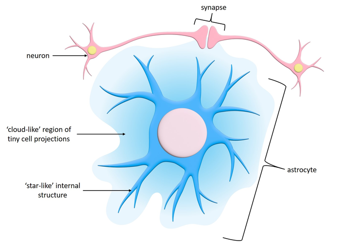
Astrocytes
they have soma, then lot of projections out like starburst
most abundant, versatile and highly branched glial cells
cling to neurons, synaptic endings, and capillaries
support and brace neurons
help determine capillary permeability
- the cells of the capillaries are normally bound together by tight junctions and astrocytes can regulate the tightness of these tight junctions
- astrocytes can make tight junctions tight or loose to let nutrients to cross or nothelp regulate access to nutrients for neurons
hold large portions of the CNS together
- hold blood vessels in place, neurons in place, etc because there is little connective tissueclean up and take care of the environment of the neurons
help regulate the whats in the interstitial fluid such as potassium which can be toxic if theres to much
Microglia
immune cell
can phagocytosis a macrophage
- this is how it “sounds the alarm”dendritic cell
helps clean injured neurons, dead neurons and removes them
small ovoid cells with thorny processes
migrate toward injured neurons

Ependymal cells
line the central cavities of the brain and spinal column
separate the CNS interstitial fluid from the cerebrospinal fluid
produce CSF at choroid plexuses (location within the ventricles)
may be ciliated
- cilia helps the CSF move through the canal like systemrange in shape from squamous to columnar
line all of the ventricles and the spaces associated with the ventricles in the CNS such as:
- cerebral aqueduct
- central canal
- spinal cordsome areas the ependymal cells have tight junctions, other areas tight junctions are present but not as tight

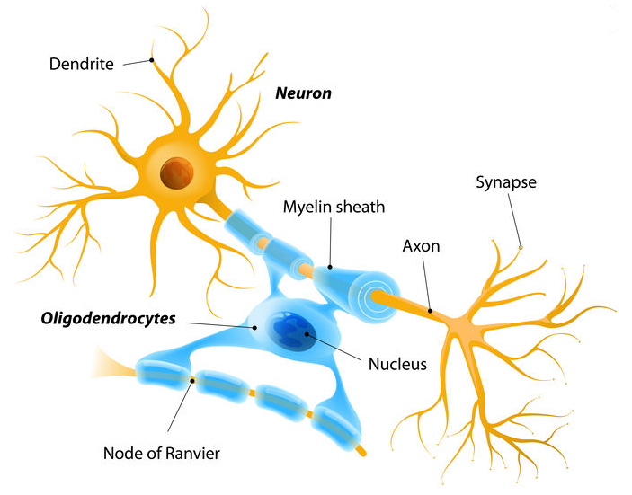
Oligodendrocytes
cells within the central nervous system that produce myelin
- myelin sheath is insulator, wraps around the axons of neurons and this helps to speed up the conduction of action potentials down the length of the axonbranched cells
processes wrap around multiple axons in CNS which is what forms the myelin sheath
multiple sclerosis (demyelination, lost of oligodendrocytes, in the CNS)
- oligodendrocytes die and are no longer present
Satellite cells
PNS
surround nueron cell bodies in the PNS
regulate environemnt and access to nutrients for neurons
help hold and bind things together
similar to astrocytes

Schwann cells (neurolemmocytes)
PNS
surround peripheral nerve fibers and form myelin sheaths
vital to regeneration of damaged peripheral nerve fibers
produce myelin similar to the way oligodendrocytes produce myelin
wrap around axons and help speed up the conduction of action potentials down the length of the axon
pathology: degenerative disease where schwann cells die: Lou Gehrig's disease or ALS
- ALS leads to death of neurons and loss of overall function

Neurons (nerve cells)
primary cell in the nervous system
long lived: 100 years or more
amitotic- few exceptions
expensive to maintain because they consume large amounts of energy
plasma membrane functions in:
- electrical signaling
- cell-to-cell interactions during development
