MODULE I - OVERVIEW OF CLINICAL HEME
 Call Kai
 Learn
Learn Practice Test
 Spaced Repetition
 Match
Match Flashcards
 Knowt Play
1/33

Earn XP
Description and Tags
WEEK 01 - plasma & formed elements
Name | Mastery | Learn | Test | Matching | Spaced | Call with Kai | Chat |
|---|
No analytics yet
Send a link to your students to track their progress
34 Terms
what is hematology?
the study of blood & blood forming organs
what are the two component of blood?
plasma: watery, straw colored liquid containing formed elements
formed elements: cellular components - erythrocytes, leukocytes, thrombocytes (cell fragments important for clotting)
define phlebotomy
the process of drawing blood from patients for diagnostic testing
describe the process of phlebtomoy.
set up with tourniquet, tubes, patient information, cotton, needle (gauge dependent on procedure & patient - smaller gauge = larger needle), ppe
tie the tourniquet 3-4 inches above the antecubital, make sure it is disinfected
uncap needle & inspect bevel for burrs
place thumb on forearm in line with chosen vein (median cubital is great for blood draws, bad for IVs - cephalic vein is great for IVs) & poll taut to anchor
advance needle into vein with bevel at 15-30° angle & insert quickly/smooth
insert tubes in order of draw (yellow, light blue, red, gold, green, purple, gray) using C method
on last tube, remove the tourniquet & remove needle simultaneously placing cotten
invert 2-3 times each tube
describe the order of draw - include additives
yellow - SPS
light blue - sodium citrate
red - clot activator
gold - SST
green - heparin
purple - EDTA (ethylenediaminetetra-acetic acid) - anticoagulant to preserve blood in liquid state (NO CLOTTING)
EDTA chelates (actively binds) calcium to keep from clotting
grey - sodium fluoride
what is a waste tube?
in a winged butterfly needle there is air inside. light blue (sodium citrate) tubes require an exact ratio of blood to additive. attach a tube with no additive to remove air
what elements make up whole blood?
55% plasma:
91% water
7% proteins (hormones, albumin, clotting proteins, antibodies)
2% solutes (sodium, calcium, chloride, etc)
45% formed elements
>99% erythrocytes
<1% leukocytes
<1% platelets
describe erythrocytes (rbc)
small, non-nucleated cells made of hemoglobin & water used to transport oxygen & remove CO2 waste
biconcave
central pallor makes up 1/3 of the cell size & shape, allows for proper gas exchange
live & die in 120 days
derived from a reticulocyte
describe thrombocytes (platelets)
small, acellular (not a cell, cell fragment), circulating nuclear membrane bound structures derived from megakaryocyte
bioconvex shape
become plump in the presence of EDTA anticoagulant
important for blood coagulation, inflammation & allergic reactions
name the types & subtypes of leukocytes (wbcs)
granulocytes: leukocytes containing cytoplasmic granules that contain microbe-killing toxins
neutrophils
basophils
eosinophils
agranulocytes: agranular, not containing cytoplasmic granules
lymphocytes
monocytes
protect us against foreign cells, organisms, & materials
what are neutrophils? what do they look like?
granulocyte - cytoplasmic granules
“neutral liking” - neutral rxn to stain, show more purplish granules that do not stick out as much
polymorphonuclear cells (PMN) - nucleus is never the same
circulating phagocytic cell, mostly in circulatory system
important in innate immunity

what are eosinophils? what do they look like?
granulocyte
“eosin stain” - pinkish
circulating phagocytic cells
important in innate immunity from worm parasites (helminths)
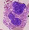
what are basophils? what do they look like?
granulocyte
“big blue granules”
circulating non-phagocytic cells
important in innate immunity
allergic responses

what are monocytes? what do they look like?
agranulocytes
mononuclear phagocytic wbc that circulates in the blood for ~8hrs before moving to the tissues where it matures into a macrophage
important for innate & acquired immunity

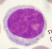
what are lymphocytes? what do they look like?
agranulocyte
small ring of cytoplasm
small, motile mononuclear, nonphagocytic cells that circulate between blood & lymph.
make up majority of lymphatic system
important for acquired immunity
T cells & B cells: adaptive immunity modulate specific immune response & produce antibodies
NK cells “natural killer”: innate immunity, tumor surveillance
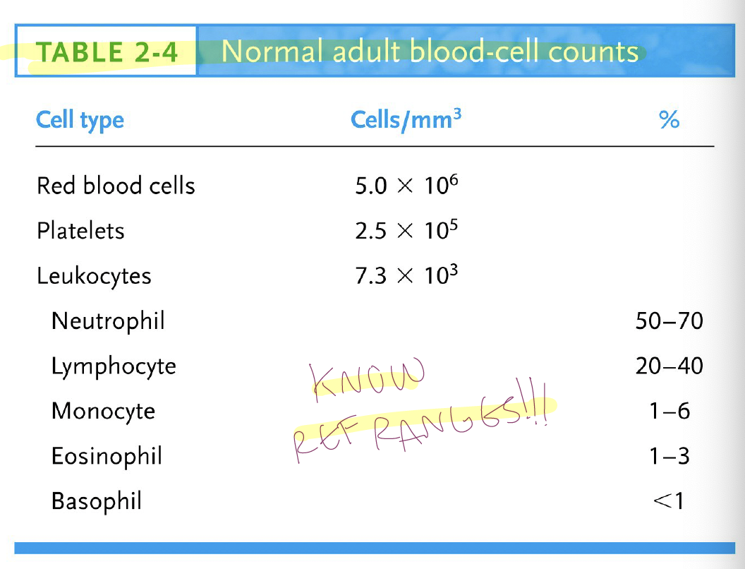
what are normal adult blood-cell counts?
what is a complete blood count? what tube is it collected in?
calculation of rbs, wbcs, & plts in the blood
purple top (EDTA)
what is a hemacytometer?
a quantitative method for determining the concentration of rbcs, wbcs, & plt in diluted blood
what are manual cbcs essential for?
quality control checks
low or abnormal counts
body fluid counts (CSF, synovial, serous fluids)
instrument backup or verification
what is the principle of a cbc?
a known dilution of blood is placed in a counting chamber of a known volume, and cells are counted under a microscope
describe the hemacytometer slide
thick glass slide with 2 counting chambers etched with grid pattern (improved neubauer ruling)
coverslip placed over chamber with a depth of 0.2mm
what is the improved neubauer ruling?
describes the grid layout on a hemacytometer
each ruled area = 3 mm x 3mm (9mm²)
central square: 1mm² divided into 25 large squares each with 16 small squares - RBCs & PLTS @ 40x
four corner squares: each 1mm² with 16 small squares - WBCs @ 10x
each large square represents a volume of 0.1mm³ or 0.1uL
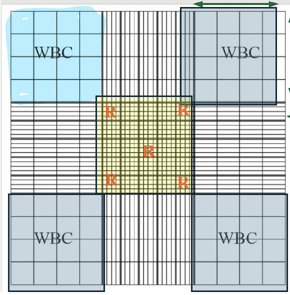
where on the grid of a hemacytometer do you not count cells?
cells touching bottom & right lines
what are the 3 rules to counting RBCs, WBCs, and PLTs? explain.
different dilution factor
RBCs: 1:200
PLTs: 1:100
WBCs: 1:20
different squares
RBCs: 4 corners & middle of central square
PLTs: all of central square
WBCs: 4 corners
different magnification
RBCs & PLTs: 40x
WBC: 10x
what 2 equations can we use for hematcytometer counting?
(# cells counted x dilution factor)/(area counted mm² x depth 0.1)
(# cells counted x dilution factor x depth 10)/area counted mm²
result: # cells/mm³
how do we convert mm³ to standard units (US)
# cells/uL
RBCs = # cells x 10^6 cell/uL
PLTs & WBCs = # cells x 10³ cells/uL
how do we convert mm³ to international units (SI)?
# cells/L
RBCs: # cells x 10^12 cells/L
PLTs & WBCs: # of cells x 10^9 cells/L
what is the normal range for rbcs in SI units (x10^12/L)
male: 4.60 - 6.00
female: 4.00 - 5.40
what is the normal range for plts in SI units (x10^9/L)?
male or female: 0.5 - 1.5
what is the normal range for wbcs in SI units (x10^9/L)
male or female: 4.5 - 11.5
what types of sources of error are there in manual cell counts?
pre-analytical
analytical
post-analytical
describe pre-analytical sources of error
describe analytical sources of error
describe post-analytical sources of error