BIO 201 Urinary and Reproductive Systems
 Call Kai
 Learn
Learn Practice Test
 Spaced Repetition
 Match
Match Flashcards
 Knowt Play
1/159

There's no tags or description
Looks like no tags are added yet.
Name | Mastery | Learn | Test | Matching | Spaced | Call with Kai | Chat |
|---|
No analytics yet
Send a link to your students to track their progress
160 Terms
Urinary System
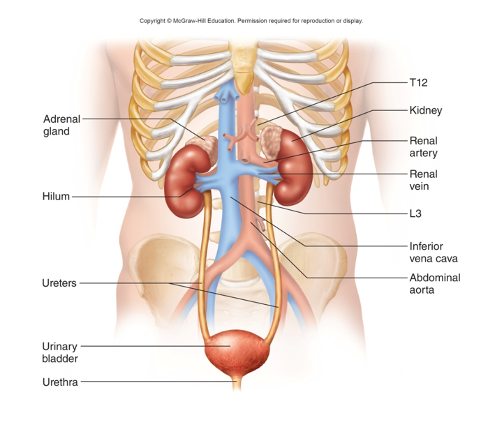
Organs of excretion: composed of a pair of kidneys and the urinary tract.
Bean-Like
Left kidney is positioned between T12 - L3
Right kidney is slightly inferior
11th and 12th ribs provide some protection for both kidneys
Adrenal glands sit on top of the kidneys
Urinary Tract
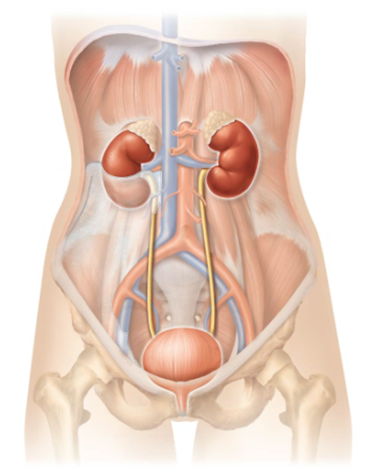
Composed of:
Pair of ureters
Urinary bladder
Single urethra
Pathway:
Urine exits kidneys → ureters → urinary bladder (urine is stored) → urethra → exits the body
Urinary System Functions
Removal of metabolic wastes
Regulate fluid & electrolyte balance
Regulate acid-base balance
Maintenance of blood pressure
Regulation of erythropoiesis
Performing other metabolic functions
Removal of Metabolic Wastes
A function of the urinary system.
Waste products in the blood are eliminated via urine
Ex. urea, uric acid, creatinine, salts, etc
Regulate Fluid & Electrolyte Balance
A function of the urinary system.
Regulate osmolarity
Osmolarity - number of particles in a solution
Either conserve or eliminate water & electrolytes (Na+, K+, Ca2+)
Regulate Acid-Base Balance
A function of the urinary system.
Assists in keeping blood neutral
Long term regulation of blood pH by conserving or eliminating hydrogen (H+) & bicarbonate (HCO3-) ions
Maintenance of Blood Pressure
Function of the urinary system.
Directly influenced by controlling blood volume and sodium excretion
Secrete an enzyme (renin) that influences blood volume and peripheral resistance via the RAAS (renin-angiotensin-aldosterone-system)
Regulation of Erythropoiesis
Function of the urinary system
Regulation of red blood cell production
Release hormone erythropoietin (stimulates RBC production)
Other Metabolic Functions of the Urinary System
Detoxifying substances in the blood
Activating vitamin D via calcitriol production (promotes bone growth)
Making new glucose (gluconeogenesis)
External Layers of the Kidneys
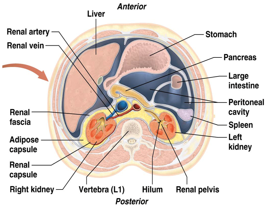
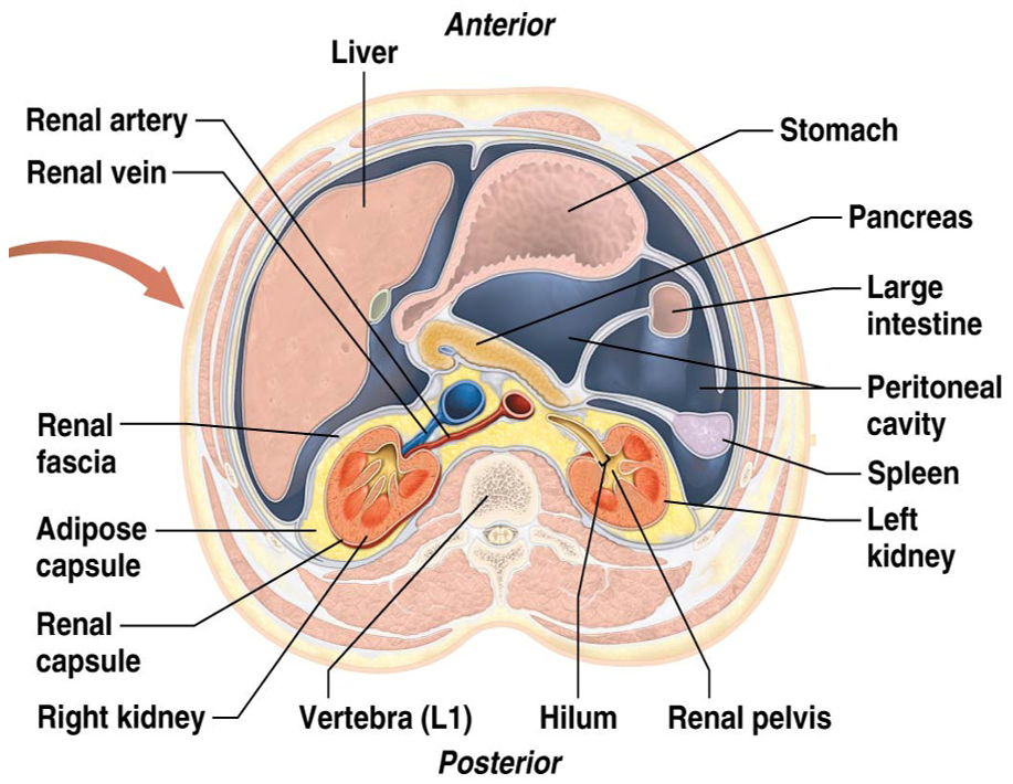
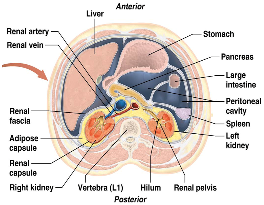
Each kidney held in place and protected by three layers of connective tissue, from superficial to deep:
Renal Fascia
Adipose Capsule
Renal Capsule
Renal Fascia
Most superficial external layer of the kidneys
Made of dense connective tissue
Anchors each kidney to the peritoneum & muscles
Adipose Capsule
Middle external layer of the kidneys.
Thickest layer
Wedges each kidney in place
Shields them from physical shock
Renal Capsule
Deepest external layer of the kidneys.
Thin layer of dense irregular connective tissue
Covers exterior of each kidney
Protects kidneys from infection and physical trauma
Hilum
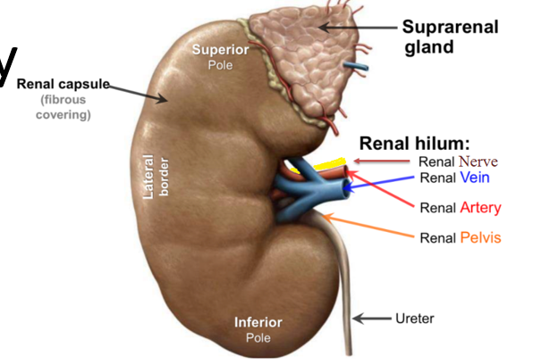
Opening on the medial surface of the kidneys where the renal artery, vein, nerves, and ureters enter and exit.
Renal Sinus
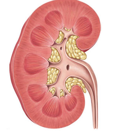
Cavity lined by the renal capsule.
Anchors the ureter, blood vessels, & nerves in place
Filled with urine-draining structures and adipose tissue
Internal Anatomy of the Kidneys
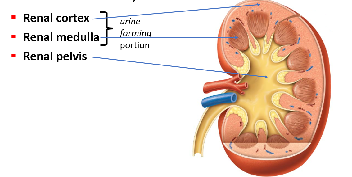
Three distinct regions of the renal sinus.
Renal Cortex
Renal Medulla
Renal Pelvis
Renal Medulla
Region of the renal sinus within the kidneys.
Houses cone-shaped renal pyramids
Darker in color with fewer blood vessels
Nephrons
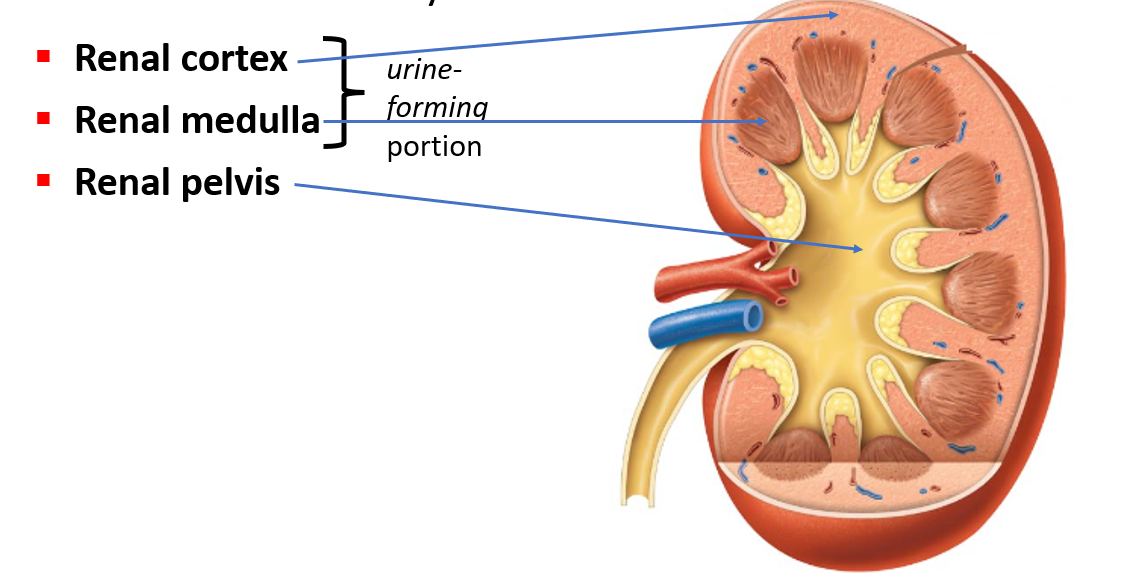
Functional unit of the kidneys.
Over one million filtering apparatuses
Found within the cortex and medulla
Two main components:
Globe-shaped renal corpuscle
Long Renal Tubule
Renal Pyramids
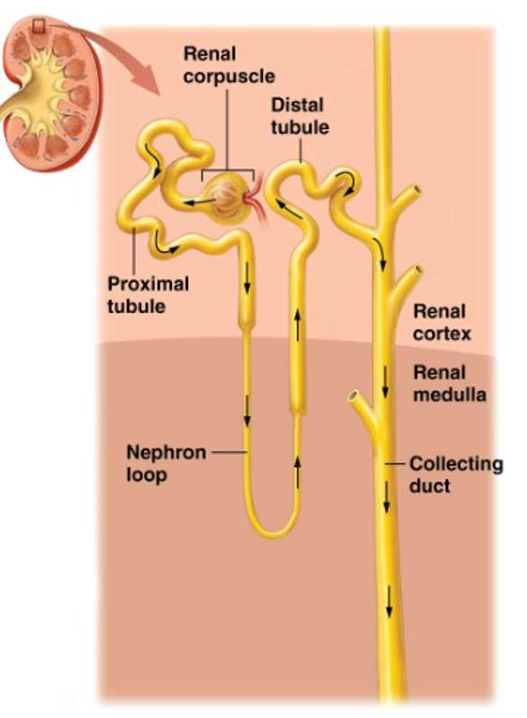
Housed in the medulla of the kidney.
Smooth muscle within walls of calyces and renal pelvis propel urine towards the ureter
Tapers into a slender papilla → minor calyx → major calyx → renal pelvis → ureter
Blood Supply to Kidneys
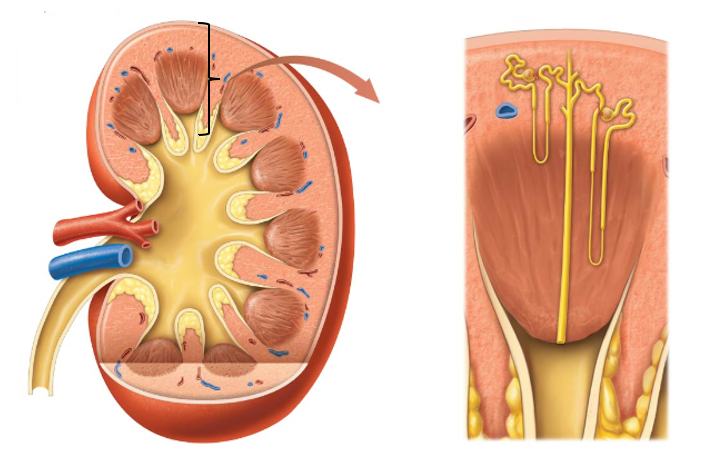
Blood travels from the abdominal aorta → left and right renal arteries → kidneys
Kidneys receive about ¼ of total cardiac output (25%)
~1200 ml of blood per minute
Renal arteries fan out into ever-smaller vessels
Blood flows from the largest to the smallest arteries
1. Renal Artery →
2. Segmental Artery →
3. Interlobar Artery →
4. Arcuate Artery →
5. Interlobular Artery →
6. Afferent Arterioles →
7. Glomerulus →
8. Efferent Arterioles →
9. Peritubular Capillaries (Second Capillary Bed) →
10. Interlobular Veins →
11. Arcuate Vein →
12. Interlobar Vein →
13. Renal Vein →
Inferior Vena Cava
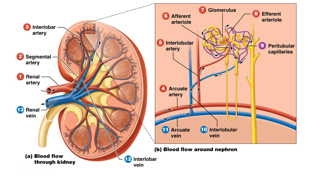
Capillary Bed System of the Kidneys
Unusual system where arterioles both feed and drain capillaries.
Peritubular Capillaries
Form a network or plexus surrounding the renal tubule of each nephron.
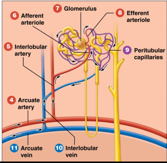
Renal Corpuscle
Round-shaped structures found on nephrons.
Responsible for filtering blood
Composed of:
Glomerulus
Glomerular Capsule (Bowman’s Capsule)
Glomerulus
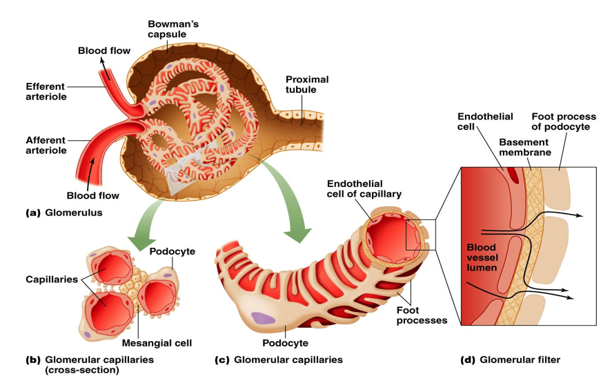
Group of looping fenestrated capillaries; extremely “leaky” or permeable
Housed within the renal corpuscle
Surrounded by Bowman’s capsule
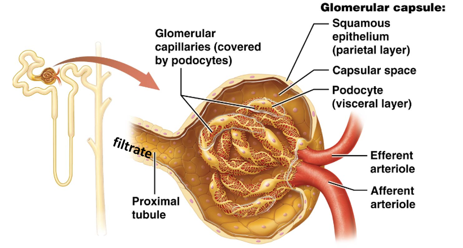
Glomerular Capsule (Bowman’s Capsule)
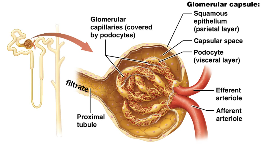
Double-layered sheath of epithelial tissue surrounding the glomerulus.
Consists of outer parietal layer and inner visceral layer
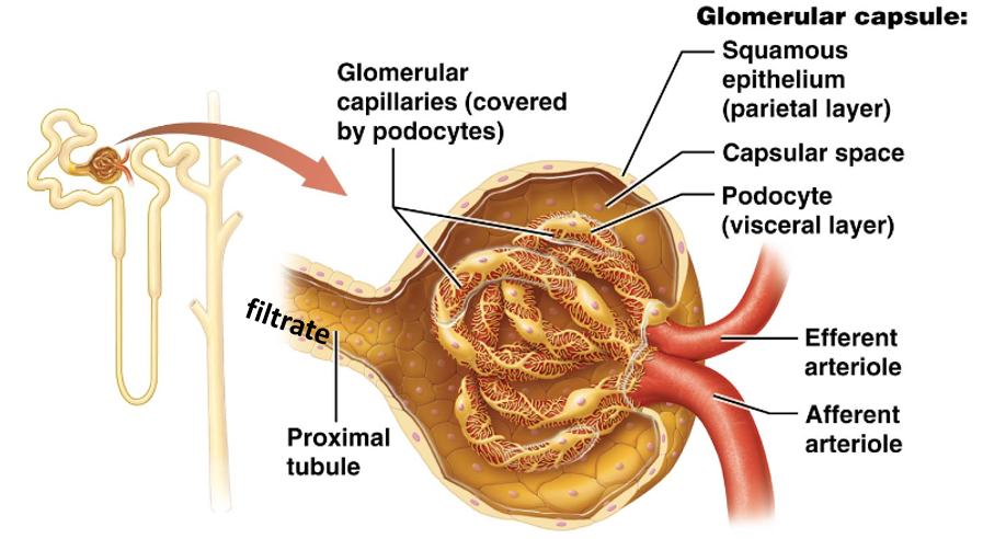
Podocytes
Have special extensions called foot processes, or pedicels.
Surround glomerular capillaries, forming filtration slits.
Renal Tubule
The pipes of the nephron that collect and modify the filtrate (“proto-urine”).
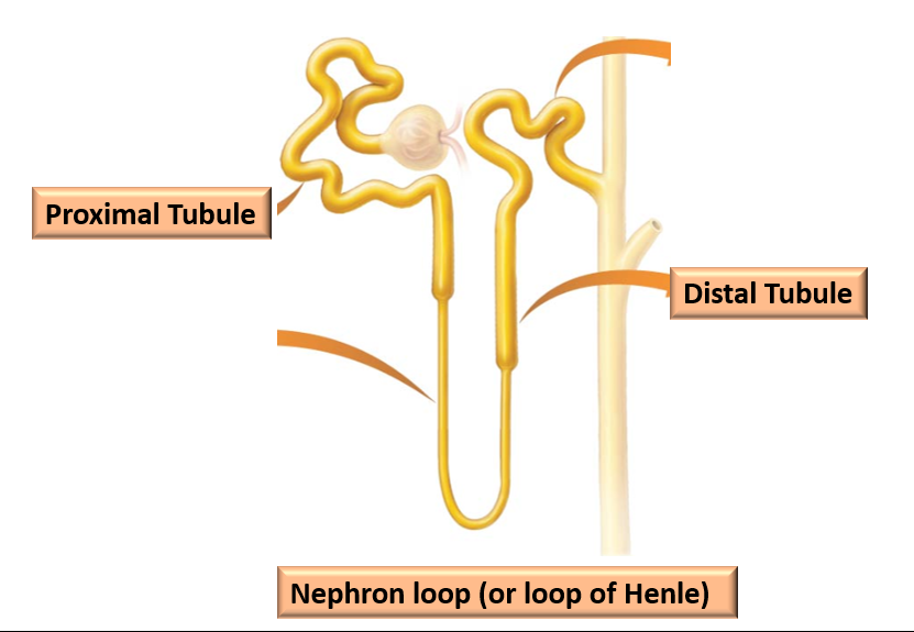
Three structurally and functionally distinct regions:
Proximal Tubule
Nephron Loop
Distal Tubule
Proximal Tubule
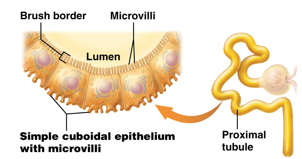
First and longest segment of the renal tubule.
Walls made of simple cuboidal epithelial cells with prominent microvilli
Project into the tubule lumen to form the brush border
Greatly increases surface area of this region
Nephron Loop (Loop of Henle)
Middle segment that filtrate enters after the proximal tubule.
Only part of renal tubule to dip into the renal medulla
Consists of a descending and an ascending limb
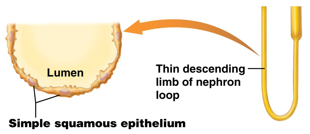
Descending Limb
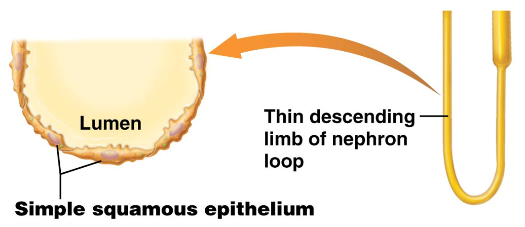
Thin limb of nephron loop that travels towards the renal medulla.
Made of simple squamous epithelial cells
Ascending Limb
Once descending limb turns 180 degrees.
Some nephrons have a thin ascending limb at the bend because the region is made of thin simple squamous epithelial cells
Majority is made of simple cuboidal epithelial cells, and referred to as the thick ascending limb
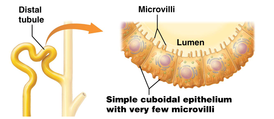
Distal Tubule
The last segment of the renal tubule that filtrate passes through.
Made of simple cuboidal epithelium without a brush border and very few microvili
Filtrate will be known as proper urine after passing through this part
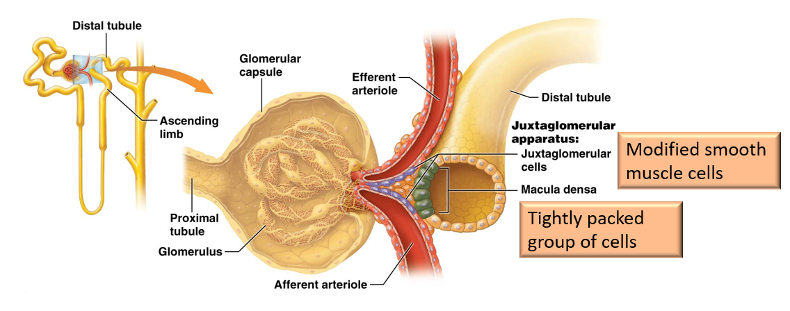
Juxtaglomerular Apparatus (JGA)
Composed of both macula densa and juxtaglomerular (JG) cells.
Found at transition point between ascending limb of nephron loop and distal tubule
Closely associated with the afferent/efferent arterioles
Regulates blood pressure and glomerular filatration rate
Collecting Tubule
Where urine in nephrons drains into.
Multiple of these empty into larger collecting ducts
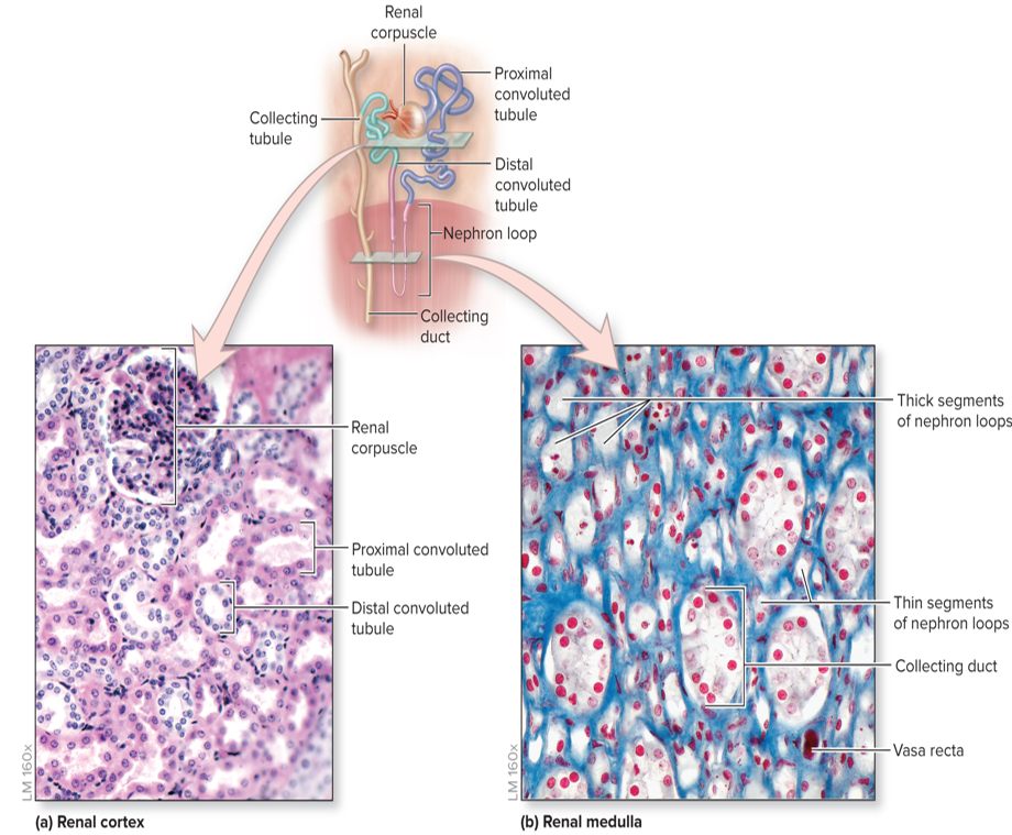
Collecting Ducts
Numerous collecting tubules drain urine into these.
Multiple of these structures drain into the renal papilla
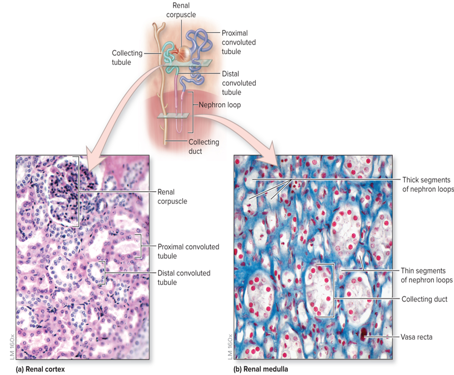
Urine Drainage
Renal Corpuscle →
Proximal Convoluted Tubule →
Nephron Loop (Loop of Henle) →
Distal Convoluted Tubule →
Collecting Tubule →
Collecting Duct →
Renal Papilla →
Minor Calyx →
Major Calyx →
Renal Pelvis →
Ureter →
Urinary Bladder →
Excreted through the Urethra
Types of Nephrons
Cortical Nephrons
Juxtamedullary Nephrons
Classified based on:
Relative position of renal corpuscle in the cortex
Length of the nephron loop
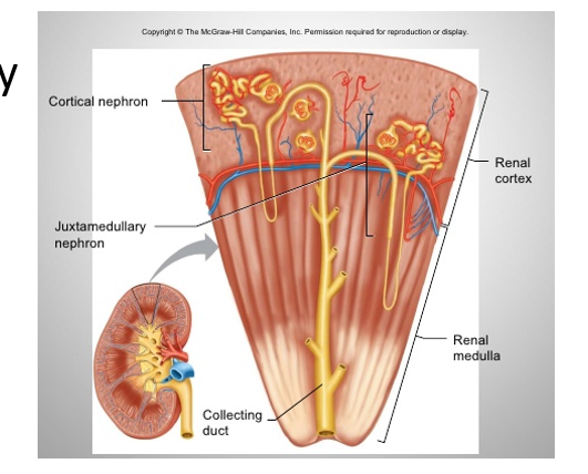
Cortical Nephrons
Oriented with renal corpuscles near the outer surface of the renal cortex
Short nephron loop barely penetrates the renal medulla
85% of nephrons
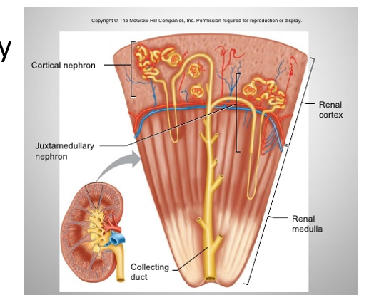
Juxtamedullary Nephrons
Renal corpuscles located near the border between the renal cortex and medulla
Long nephron loop extends deep into the renal medulla
Help establish salt concentration gradient in interstitial space
Allows for regulation of urine concentration by antidiuretic hormone (ADH)
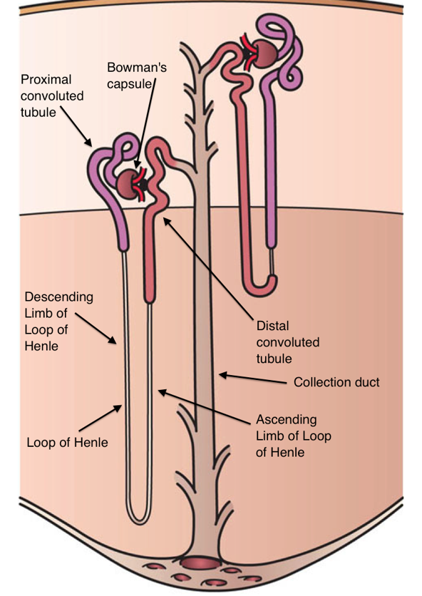
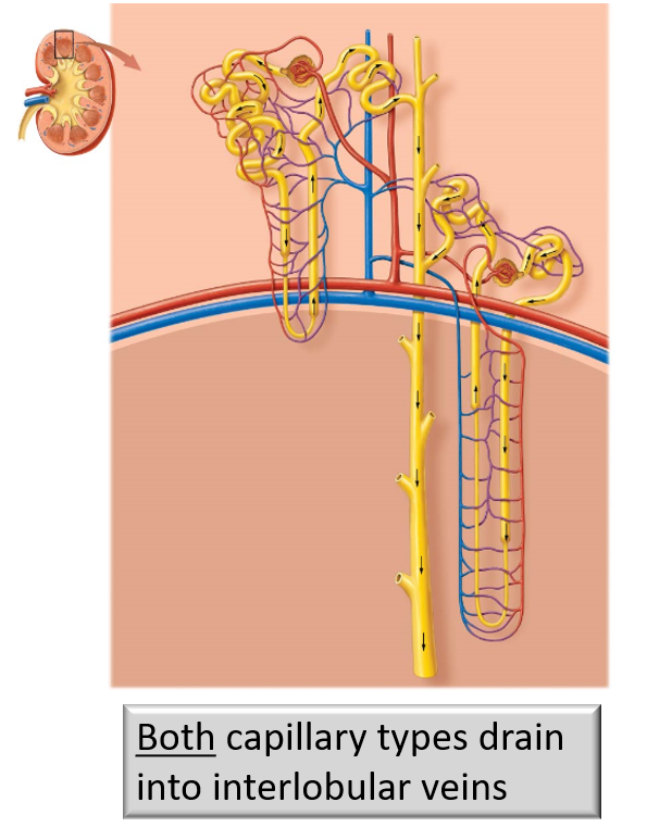
Capillary Types
Cortical Portion of Nephron Tubules
Surrounded by peritubular capillary branches
Nephron Loop
Surrounded by a ladder-like network of capillaries called the vasa recta
Unique capillary structural arrangement allows juxtamedullary nephrons to control volume and concentration of urine
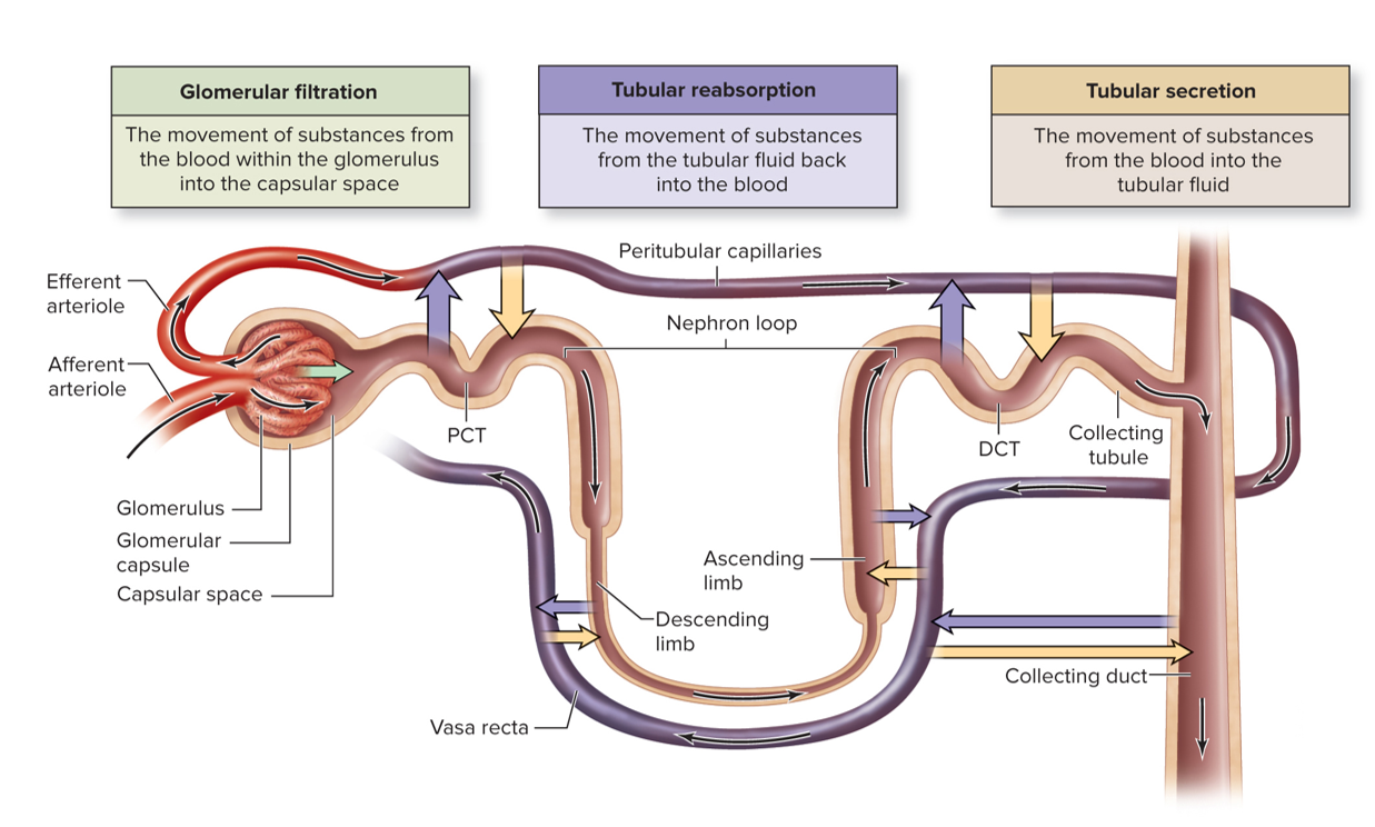
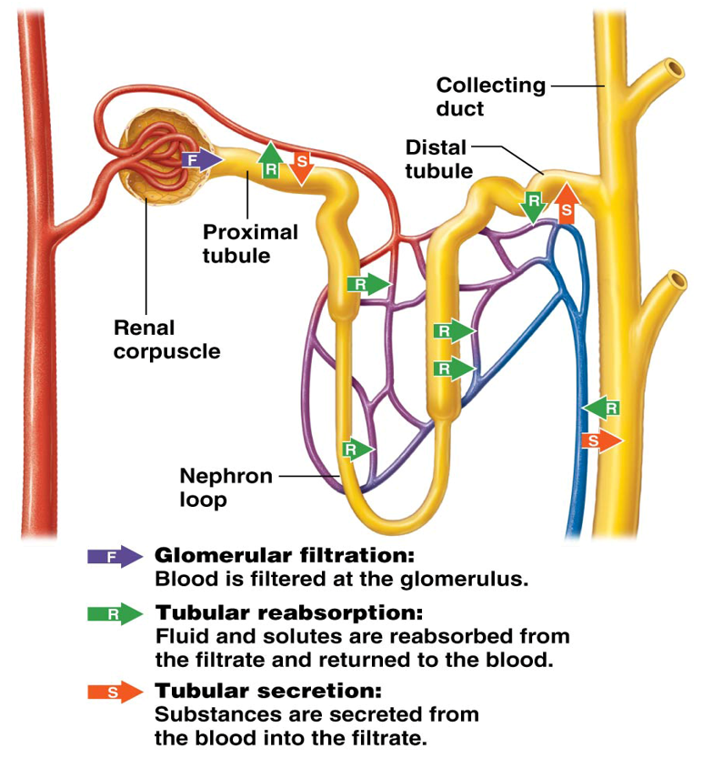
Physiological Proccesses Overview of the Kidneys
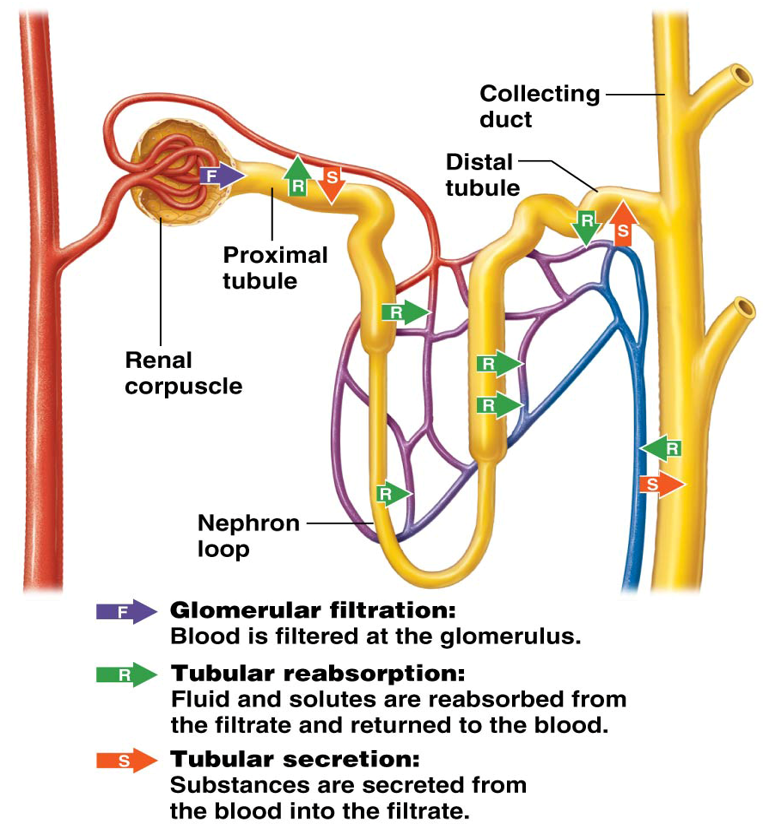
Glomerular Filtration
Tubular Reabsorption
Tubular Secretion
Glomerular Filtration
Initial process of nephrons used to filter blood
Circulating blood → glomerular capsule → proximal tubule
The glomerular capillary filters blood content based on size
Large cells and proteins cannot enter
Smaller substances can enter from blood into the glomerular capsule, and become components of the filtrate
Water
Acids and Bases (H+ and HCO3- Ions)
Electrolytes (Na+ and K+ Ions)
Organic Molecules
Metabolic Wastes
Filtration rate is controlled by pressure
Tubular Reabsorption
Substances are reabsorbed/reclaimed from filtrate back into the blood (in peritubular capillaries)
Water, Glucose, Amino Acids, and Electrolytes
The nephron can reabsorb the majority of filtered water and solutes from the proximal tubule and nephron loop
Tubular Secretion
Peritubular capillaries add substances to the filtrate in the tubule for excretion from the body.
Occurs along the entire tubule
Helps maintain
Electrolyte Homeostasis
Acid-Base Homeostasis
Removes toxins from the blood that did not enter tubular fluid by filtration
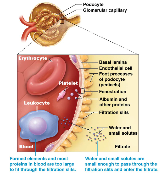
Gomerular Filtration Membrane
Consists of three layers that collectively create a selectively permeable barrier
Fenestrated (leaky) glomerular capillary endothelial cells
Prevent formed elements from passing through
Basal Lamina (extracellular contact between endothelial cells and podocytes)
Prevent most proteins from passing through
Podocytes
Prevent smaller molecules from passing through
Nitrogenous Wastes
Group of small substances that are readily filtered
Urea and ammonium ions (NH4+) from protein metabolism
Creatinine - prodiced by enzyme creatine kinase in muscle
Uric Acid - product of nucleic acid metabolism
Filtration Fraction
Percentage of plasma that becomes filtrate in capsular space
Average about 20% - 1/5th of the plasma that enters the glomerulus becomes filtrate
Glomerular Filtration Rate (GFR)
Amount of filtrate formed by both kidneys in one minute
125 ml/min or equivalent of filtering all 3 liters of blood plasma about 60 times per day
Fluid Movement Driven By:
Pressure Gradient
Difference between blood pressure in the glomerulus vs. fluid pressure of filtrate in the glomerular capsule
Two Forces Generate Filtration Pressures:
Hydrostatic Pressure
Colloid Osmotic Pressure
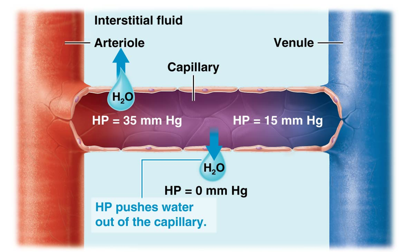
Hydrostatic Pressure (HP)
The force of a fluid pressing on the wall of a container
Equals to the blood pressure → tends to push water out of capillary into the interstitial space
Colloid Osmotic Pressure (COP)
Pressure generated by solutes, primarily albumin, in plasma.
Osmotic gradient pulls water into capillaries by osmosis
The side with more solutes will pull water towards it
Blood plasma contains a high concentration of proteins
Net Filtration Pressure (NFP)
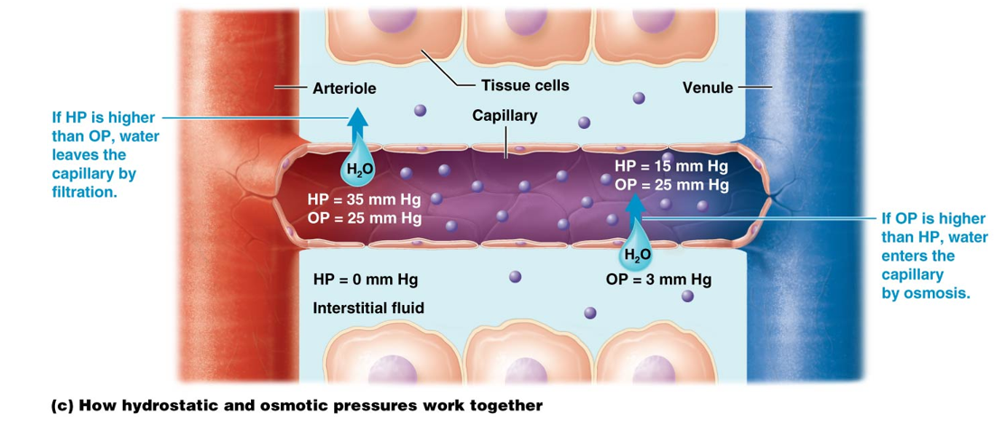
The interaction between hydrostatic and colloid osmotic pressures determines this and the direction in which water will flow.
Water moves out of the capillary when HP is higher than the COP
Water moves into the capillary when COP is higher than HP
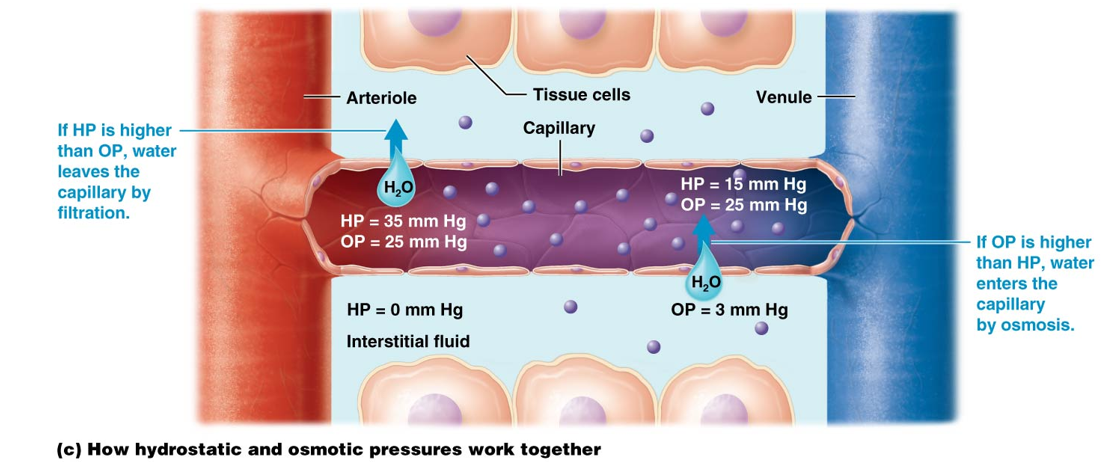
Net Filtration Pressure (NFP) at Glomerulus
Overall pressure that drives fluid out of the blood and into the glomerular capsule. Determined by three forces:
Glomerular Hydrostatic Pressure (GHP)
Tendency for fluid to be pushed out of the glomerular blood vessels
Glomerular Colloid Osmotic Pressure (GCOP)
Tendency for fluid to be sucked into the capsule
Capsular Hydrostatic Pressure (CHP)
Tendency for fluid in the capsule to be pushed back into the blood vessels
Affects of Glomerular Filtration Rate
Internal Factors:
Autoregulation
External Factors:
Hormonal
Neuronal
Autoregulation
Internal kidney mechanisms that work together to maintain GFR within a normal range.
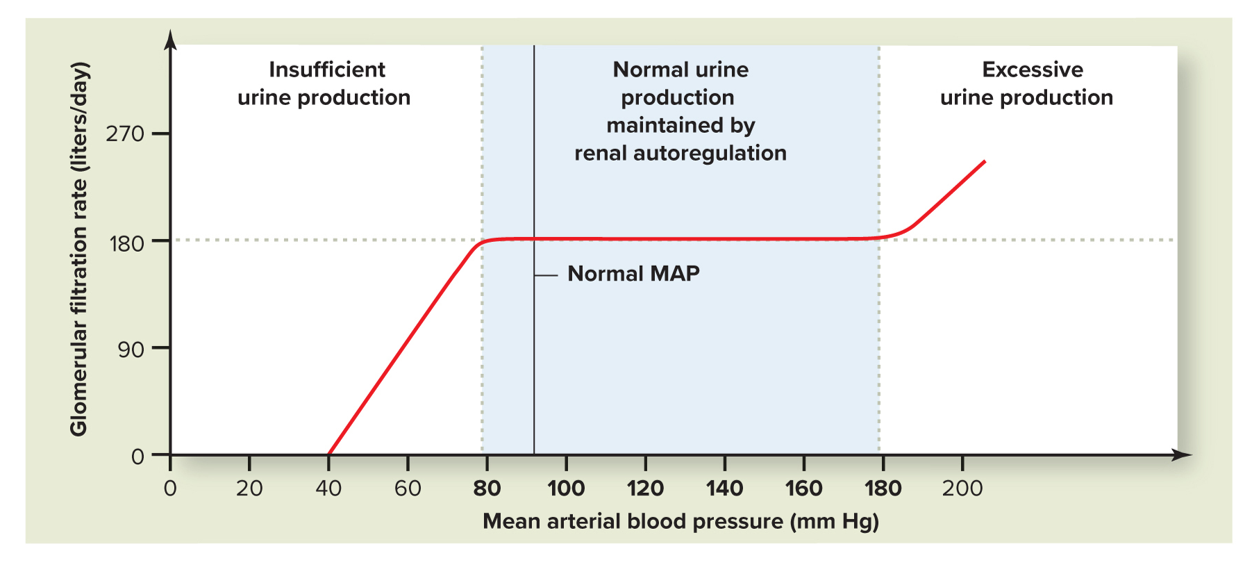
GFR is fairly constant despite normal blood pressure fluctuations
Consists of two negative feedback processes:
Myogenic Mechanism
Tubuloglomerular Feedback
Myogenic Mechanism
One of the negative feedback processes of autoregulation of GFR.
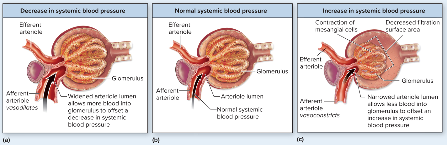
Contraction or relaxation of the smooth muscle of the afferent arteriole in response to stretch
Increased systemic blood pressure → stretches afferent arteriole → increases GFR → smooth muscle responds by constricting the afferent arteriole → reduces blood flow through the glomerulus → returns GFR back to normal
Decreased systemic blood pressure → stretches afferent arteriole less → reducing GFR → smooth muscle responds by relaxing → increasing blood flow through the glomerulus → causing an increase in GFR back toward normal range
Works best for systemic blood pressure changes between 80 and 180 mm Hg to rapidly restore GFR to normal
Tubuloglomerular Feedback (NaCl)
Another autoregulation mechanism of GFR.
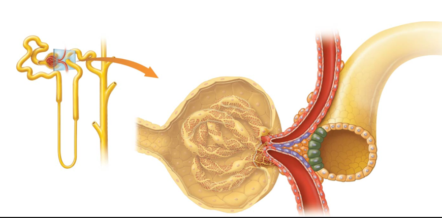
Involves the juxtaglomerular apparatus
Part of the negative feedback loop that controls pressure in the glomerulus in response to the NaCl (sodium chloride) concentration of filtrate
Tubuloglomerular Feedback Response to Increased GFR
As the glomerular filtration rate (GFR) increases → volume of the filtrate flowing through the renal tubule increases → more Na+ and Cl- ions are absorbed into the macula densa
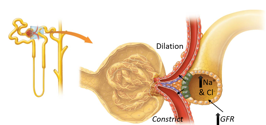
Macula Densa
Cells release chemicals that tell the afferent arteriole to constrict
Also signals juxtaglomerular cells to reduce the release of the hormone renin → reduction of angiotensin II → dilation of the efferent arteriole → a decrease in GFR towards normal
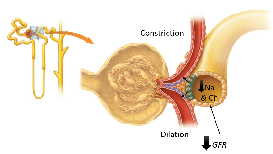
Tuboglomerular Feedback Response to Decreased GFR
Decreases in GFR → reduce Na+ and Cl- ions absorbed by macula densa → triggers dilation of afferent arteriole and constriction of efferent arteriole → increases glomerular hydrostatic pressure to restore GFR
Hormonal Control of GFR
Hormonal effects on GFR are part of a larger system that involves regulation of systemic blood pressure and includes:
Angiotensin II
Natriuretic Peptides
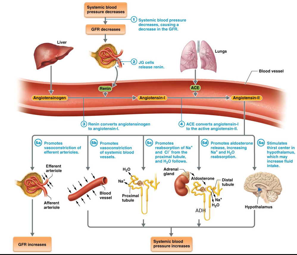
Renin-Angiotensin-Aldosterone System (RAAS)
A system that primarily functions to stabilize overall systemic blood pressure
By doing so, it also stabilizes GFR
Responds to three conditions:
1. Stimulation by the sympathetic nervous system
2. Low glomerular hydrostatic pressure
3. Stimulation from the macula densa
Atrial Natriuretic Peptide (ANP)
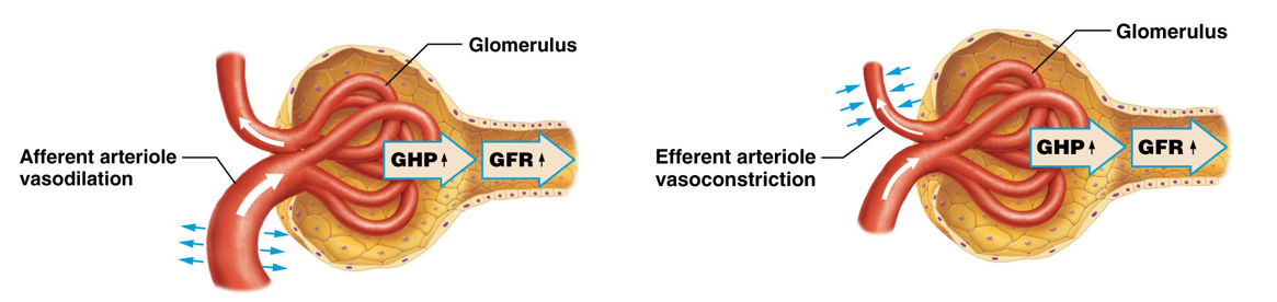
A hormone released by heart cells in the atria in response to increasing fluid volume → lowers blood volume and blood pressure to reduce the workload of the heart
ANP increases → dilates afferent arterioles and constricts efferent arterioles → increases glomerular hydrostatic pressure → blood volume reduction by favoring fluid loss from the kidneys → reduces systemic blood pressure
Neural Regulation of GFR
Primarily involves the sympathetic division of the NS and the hormone norepinephrine (NE)
Works with a larger system to control systemic blood pressure
Increased sympathetic activity → increases release of NE → constriction of afferent arterioles like most systemic blood vessels → increases systemic blood pressure
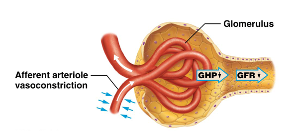
Control of GFR
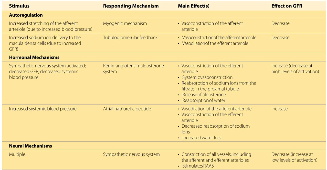
Autoregulation
Myogenic Mechanism
Decreases GFR
Tuboglomerular Feedback
Decreases GFR
Hormonal
Renin-Angiotensin-Aldosterone System (RAAS)
Primarily increases GFR
Decreases GFR at high levels of activation
Atrial Natriuretic Peptide
Increases GFR
Neural
Sympathetic Nervous System
Primarily decreases GFR
Increases at low levels of activation
Reabsorption in the Proximal Tubule
Large % of electrolytes: sodium, potassium, chloride, sulfate, and phosphate ions
Ensures electrolyte homeostasis
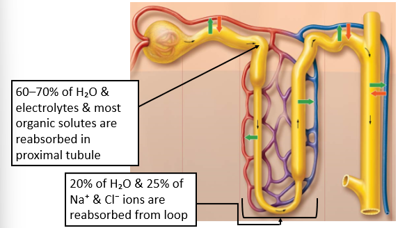
Almost 100% of nutrients, including glucose, amino acids, & other organic substances (ex., water-soluble vitamins, small proteins, and lactic acid)
Vital for nutrition purposes
90% of bicarbonate ions
Ensures acid-base homeostasis
About 65% of filtered water
Ensures the body's fluid homeostasis
Secretion in the Proximal Tubule
Hydrogen Ions (H+)
Ammonium Ions (NH4+)
Creatinine (Metabolic Byproduct)
Uric Acid (how humans get rid of most nitrogenous waste)
First half of tubule, most uric acid is reabsorbed
Nearly all of it is secreted back into the filtrate in the second half of the tubule
Small amounts of Urea
Alternative means of getting rid of nitrogenous waste
Drugs
Including Penicillin and Morphine
Gout (Clinical Connections)
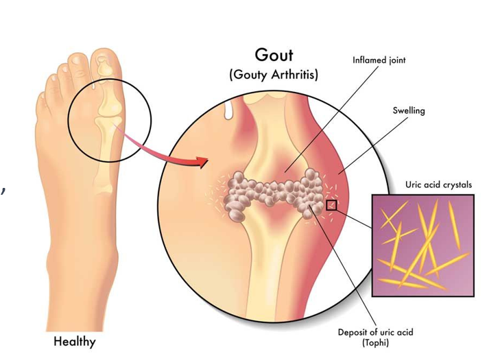
A type of arthritis that is caused by the deposition of uric acid crystals in the joints
Causes inflammation, pain, and immobility
Kidneys play a crucial role in regulating the level of uric acid in the blood
Risk Factors include
Obesity
High BP
Diabetes
Diet high in purines
Diet rich in red meat and organ meat
Treatment
Medications
Lifestyle changes to reduce inflammation and prevent future attacks
Left Untreated
Joint damage
Kidney damage
Development of tophi (swollen joints)
Reabsorption in the Nephron Loop
20% of H2O
25% of Na+ & Cl- ions
Filtrate throughout the Nephron
Proximal Tubule:
Filtrate concentration is = interstitial fluid concentration
Nephron Loop:
Water reabsorbed → filtrate becomes more concentrated
Distal Tubule:
NaCl secreted into tubule cells → draws water into filtrate via osmosis → filtrate becomes less concentrated
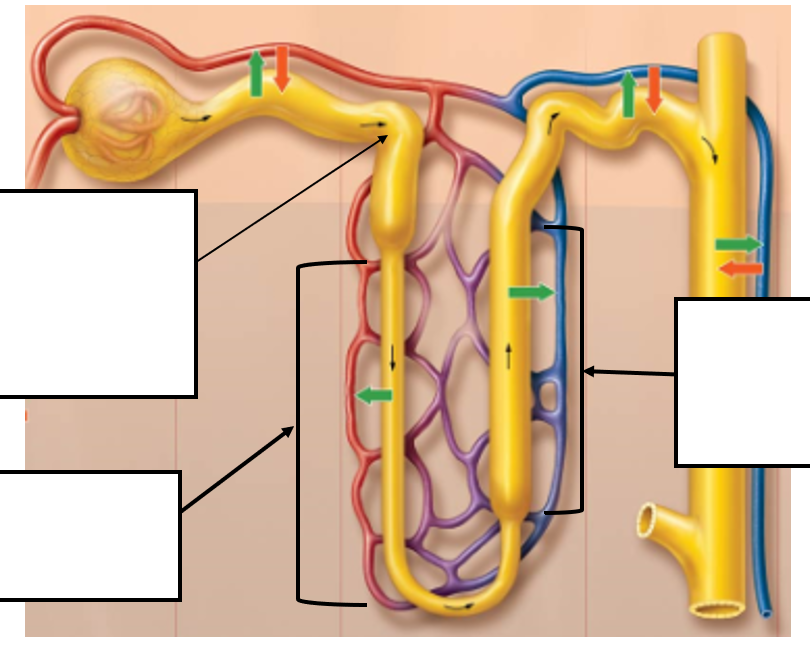
Reabsorption in the Distal Tubule and Collecting System
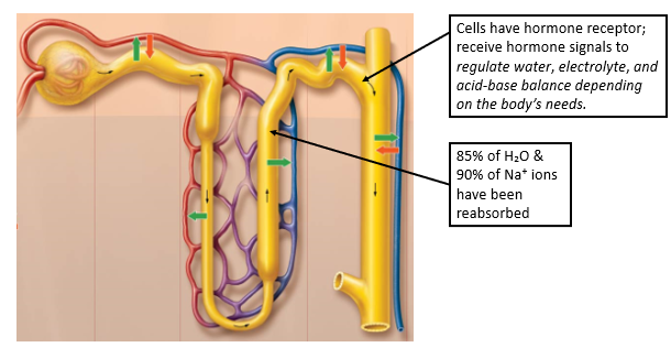
Cells with hormone receptors receive hormone signals to regulate water, electrolyte, and acid-base balance depending on the body’s needs
85% of H2O is reabsorbed
90% of Na+ ions have been reabsorbed
Renal Failure
When renal function is greatly diminished or absent.
Often from chronic disease affecting the glomerulus or small blood vessels
Dialysis
A treatment that can help remove waste products and excess fluid from the body when the kidneys are unable to do so on their own.
Kidney Transplant
In some cases, it may be a treatment option for renal failure.
Involves surgically placing a healthy kidney from a donor into the recipient’s body to replace the failed kidney
Ureters
Part of the urinary tract.
Contain smooth muscle cells that contract rhythmically (peristalsis) to propel urine toward the urinary bladder
Begin at L2 → Travel behind the peritoneum → Empty into the urinary bladder
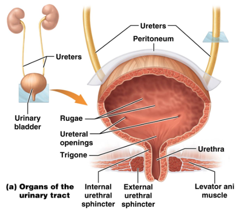
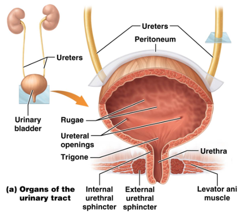
Urinary Bladder
A hollow, distensible organ found on the pelvic cavity floor that stores urine.
Held in place by the parietal peritoneum
Collapses when empty
Pear-shaped when full
Holds 700-800 ml of urine in males and slightly less in females
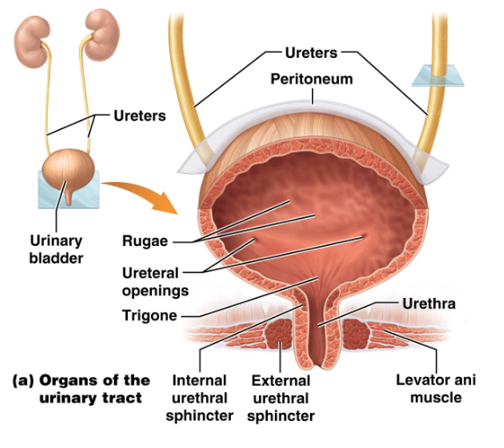
Trigone
Triangular region on the bladder floor made of
Two ureter openings with mucosal flaps
Internal urethral orifice
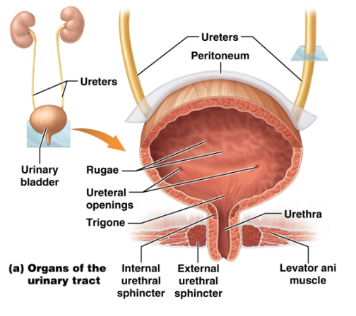
Urethra
Final segment of the urinary tract.
Histology is similar to the ureters, with the following exceptions:
The opening is surrounded by an internal urethral sphincter
The external urethral sphincter is formed by skeletal muscle
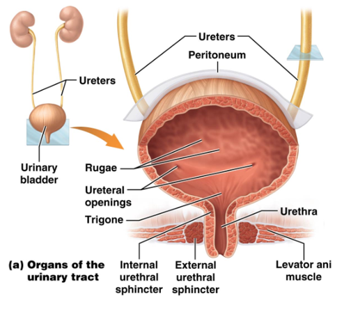
Internal Urethral Sphincter
Surrounds the opening of the urethra.
Only opens when urine is passing through
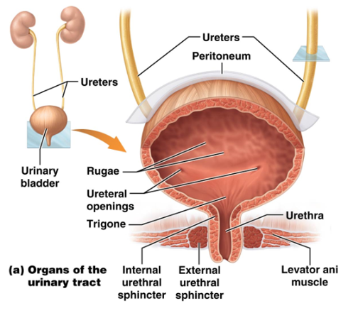
External Urethral Sphincter
Allows for voluntary control of urination.
Formed by skeletal muscle
Micturition
Also known as urination or voiding.
Discharge of urine from the urinary bladder to the outside of the body.
Time and training make this a voluntary process
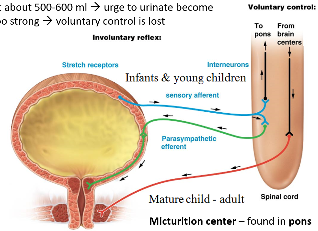
Micturition Reflex
Reflex arc mediated by the parasympathetic nervous system when urine fills the bladder and stretches the walls.
At about 500-600 ml → urge to urinate becomes too strong → voluntary control is lost
Micturition center is found in the pons
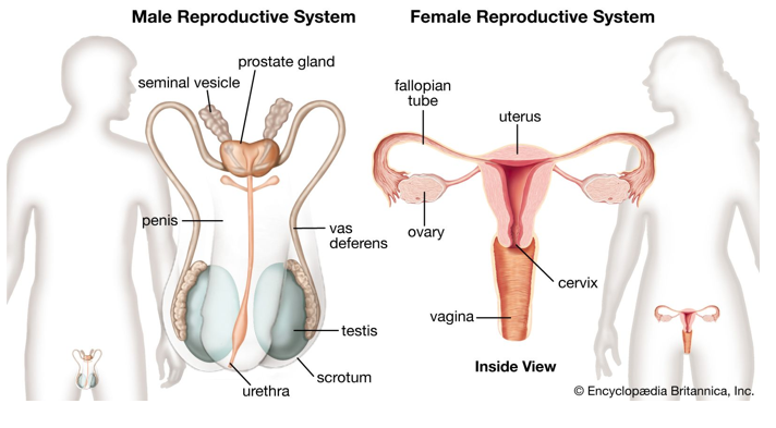
Major Functions of the Male Reproductive System
Produce / Maintain Sperm
Transport sperm and protective fluid (semen)
Dischrage sperm within the female reproductive tract during sex
Produce male sex hormones
Major Functions of the Female Reproductive System
Produce / Maintain Eggs
Transport egg cell
Protect and nourish the offspring until birth
Produce female sex hormones
Similarities Between Male & Female Reproductive Systems
Both have gonads (primary sex organs)
Secrete sex hormones (testosterone & estrogen)
Produce gametes (sex cells) through meiosis
Male → Sperm
Female → Ova / Egg
Have accessory reproductive organs
Contribute to the functioning of the reproductive system
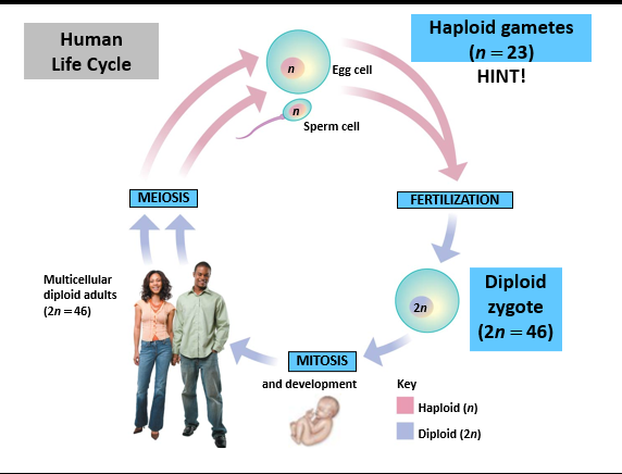
Human Life Cycle
Sperm (haploid n=23) fertilizes the Egg (haploid n=23)
Produces a diploid zygote (2n = 46)
Diploid
Containing 46 chromosomes (2 pairs, 2n)
Haploid
Containing 23 chromosomes (1 pair, n)
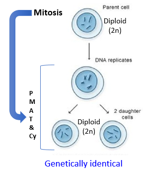
Mitosis
Cell division of somatic cells (or body cells)
Produces two genetically identical diploid daughter cells
For purposes of tissue growth & repair
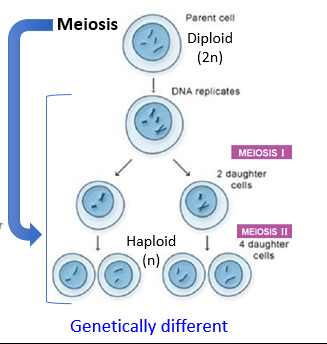
Meiosis
Cell division of germ cells (sex cells)
Produces 4 genetically different haploid daughter cells
For purposes of reproduction (including recombination)
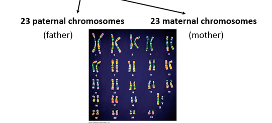
Human Somatic Cells
All have a nucleus with 46 chromosomes (23 pairs) = diploid (2n)
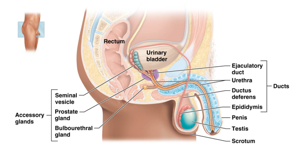
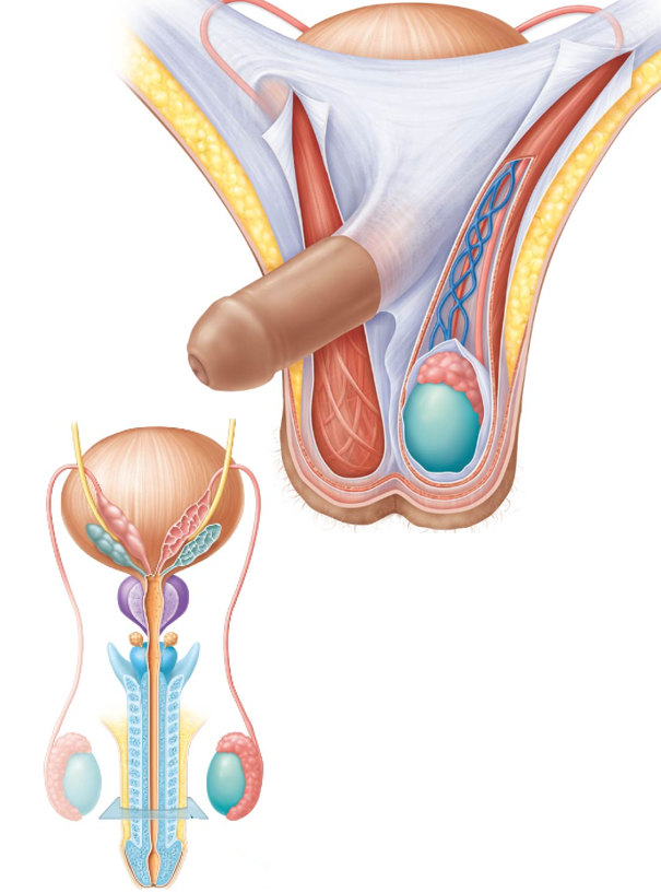
Male Reproductive System
Consists of the penis, testis, and scrotum
Accessory Glands include
Seminal Vesicle
Prostate Gland
Bulbourethral Gland
Ducts
Ejaculatory Duct
Urethra
Ductus Deferens
Epididymis
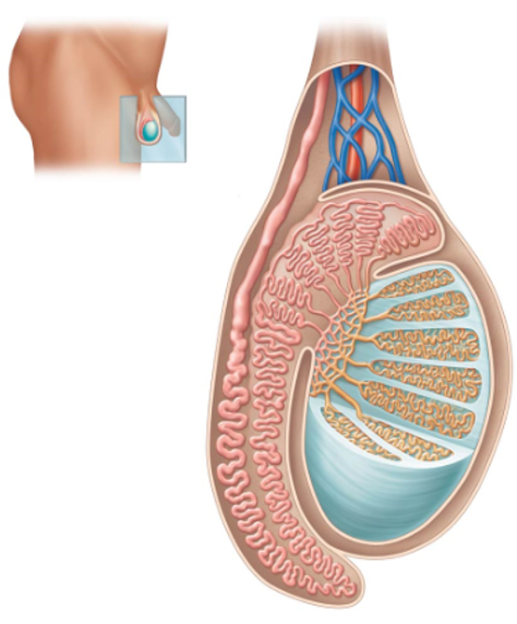
Testes (Testicles)
Ovoid structures located outside of the abdominopelvic cavity in a saclike structure called the scrotum.
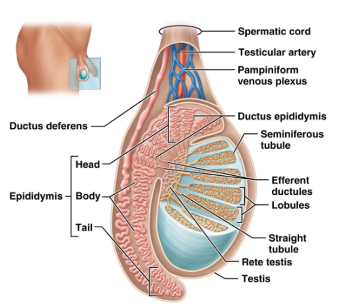
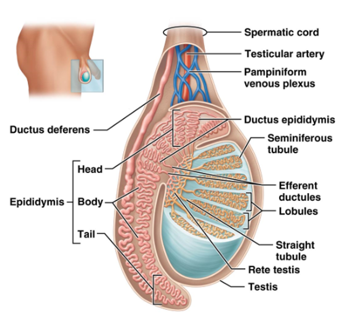
Contain numerous lobules that contain tightly coiled loops called seminiferous tubules
Innervated by the autonomic nervous system, but also contains a large number of pain and temperature receptors
Functions Include
Sperm Production
Secretion of androgen hormones, primarily testosterone
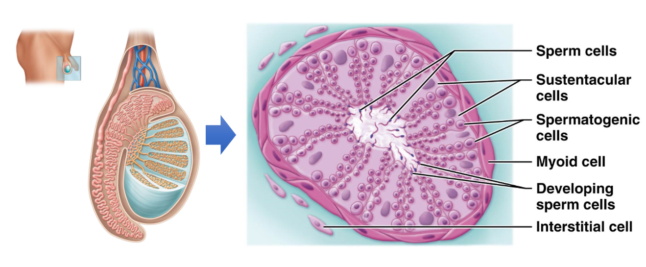
Seminiferous Tubules
Tightly coiled loops within lobules of the testes.
Contain two cell types:
Spermatogenic Cells
Sperm-Forming Cells
Sustentacular Cells (Sertoli Cells)
Support sperm production
Produce testicular fluid
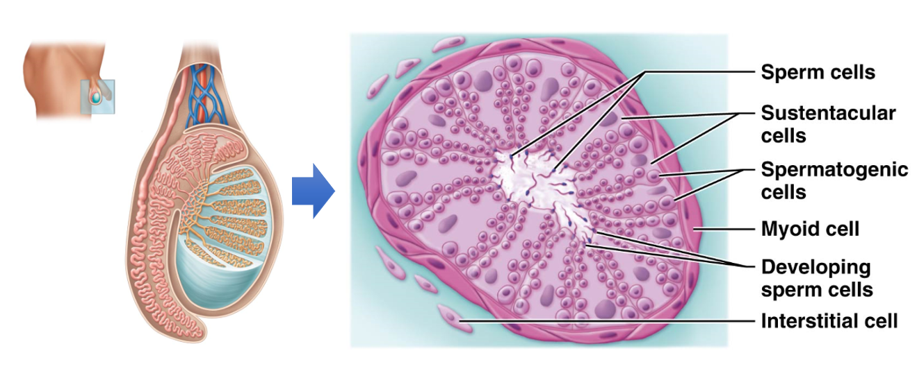
Interstitial Cells (Leydig Cells)
Cells found between the seminiferous tubules
Produce and secrete androgens, primarily testosterone, into the surrounding interstitial fluid
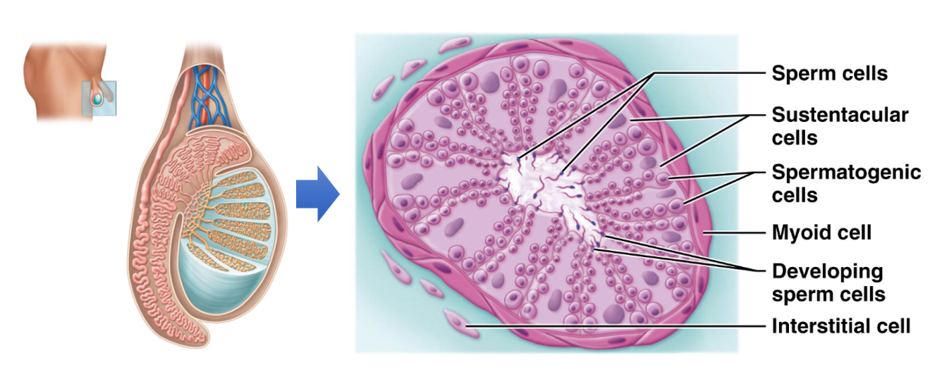
Myoid Cells
Muscle-like cells that surround seminiferous tubules
Contract to push sperm and testicular fluid through the tubules
Epididymis
Where sperm is stored after production.
Contains a head, body, and tail
Sperm exits through the Ductus (vas) Deferans
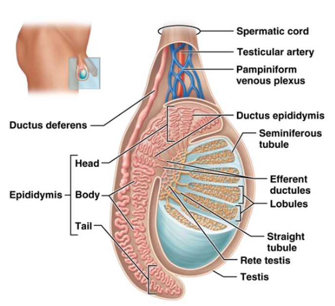
Ductus Deferens (Vas Deferens)
Duct through which sperm cells exit the epididymis/testes.
Begins at the tail of the epididymis
Enters the abdominopelvic cavity through a fibrous tunnel (inguinal canal) → loops over the ureter on the posterior side of the bladder → joins the seminal vesicles → enters the prostatic urethra
Testicular Artery
Branch from the abdominal aorta that supplies blood to the testes.
Pampiniform Venous Plexus
Drains blood from the testes into the testicular veins
Helps regulate the temperature of the testes for proper sperm production
Semen
Contains both lubricating and nourishing fluids for sperm.
Produced by the seminal vesicles, prostate, and bulbourethral glands
Alkaline (neutralizes the acidic pH of the female reproductive tract)
pH also helps make the sperm fully motile and allows them to begin the process of capacitation