TV Disease & Congenital Defects
1/23
There's no tags or description
Looks like no tags are added yet.
Name | Mastery | Learn | Test | Matching | Spaced | Call with Kai |
|---|
No analytics yet
Send a link to your students to track their progress
24 Terms
What is the most common cause of tricuspid stenosis?
Rheumatic disease
What is rheumatic tricuspid stenosis?
Narrowing of TV that is characterized by thickening of leaflet tips and restricted mobility with diastolic doming due to inflammation
What is carcinoid tricuspid stenosis?
Narrowing of TV that is characterized as entire leaflet thickening due to carcinoid tumor in GI tract or pancreas
What are symptoms of tricuspid stenosis?
Dyspnea or SOB
Palpitations
IJV distension due to backup of blood flow in SVC
What is the sonographic appearance of tricuspid stenosis?
Increased PHT
Mean gradient > 2 mmHg
TVA < 1 cm²
RA and IVC dilation
Decreased RV size
Increased E wave velocity > 1 m/sec
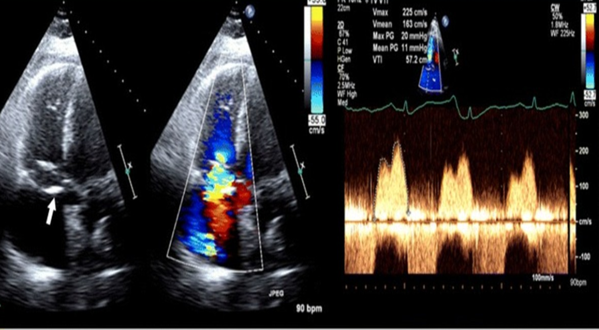
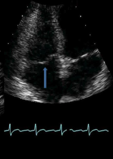
Identify this image.
TS
How is vena contracta performed for TR?
Performed in PLAX or PSAX
Zoom on valve
DECREASE COLOR SCALE TO 50-60 cm/sec
Measure smallest jet diameter
How is PISA performed for TR?
Performed in A4C
Zoom on valve
DECREASE COLOR BASELINE 30-40 cm/sec (aliasing velocity)
Measure color shell above leaflet tips

What is the most common form of physiologic regurgitation?
TR
What is primary or degenerative TR?
TR due to defect in valve or apparatus
What is the most common cause of primary TR?
Myxomatous degeneration
What is the sonographic appearance of primary or degenerative TR?
Eccentric color jet
Associations: Congenital anomalies, TVP, leaflet thickening, endocarditis
What is secondary or functional TR?
TR due to complication of another abnormality
What is the most common cause of secondary or functional TR?
Pulmonary HTN
What is the sonographic appearance of secondary or functional TR?
Central color jet
Associations: Dilated RA or RV, CHF, pulmonary HTN, AFIB
What is the sonographic appearance of severe TR?
Jet area: > 10 cm²
Vena contracta: > 7 mm
PISA: > 9 mm
Systolic flow reversal in HV
Dilated RA and IVC
Triangular spectral contour
* Increased severity upon inspiration
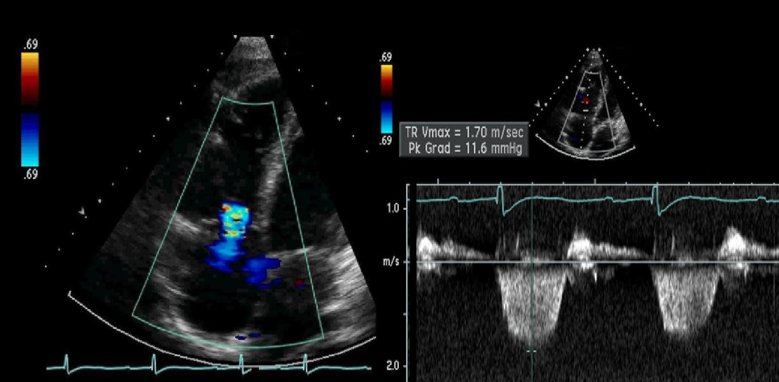
Identify this image.
TR
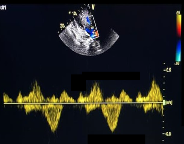
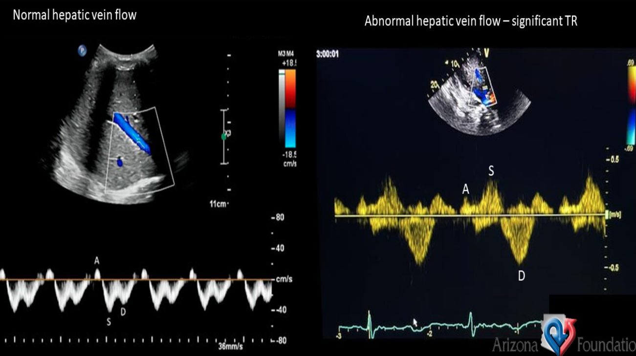
Identify this image.
Abnormal HV flow due to severe TR
What is tricuspid valve prolapse (TVP)?
When anterior or septal TV leaflet is displaced into RA during systole
What congenital conditions are associated with TVP?
Marfan syndrome
Ostium secundum defect
Ebstein anomaly
Identify this image.
TVP
What is a flail tricuspid leaflet?
When TV leaflet is dyskinetic due to ruptured papillary muscle or chest trauma
What is tricuspid atresia?
Absence of TV
What is the sonographic appearance of tricuspid atresia?
Echogenic plate-like structure between RA and RV
Small RV
Dilated RA
VSD and PFO present