Neuroscience Chapter 2
 Call Kai
 Learn
Learn Practice Test
 Spaced Repetition
 Match
Match Flashcards
 Knowt Play
1/53

There's no tags or description
Looks like no tags are added yet.
Name | Mastery | Learn | Test | Matching | Spaced | Call with Kai | Chat |
|---|
No analytics yet
Send a link to your students to track their progress
54 Terms
Neurons (nerve cells)
these communicate information between other neurons, glands, muscles and organs in the body
Glial cells
provide support for information processing neurons
Dendrites (input zone)
cellular extensions that receive information
Cell body (integration zone)
integrates and processes information
Axons (conduction zone)
carry information away from the cell body
Axon terminals (output zone)
transmit signals across synapses
most common type of neuron
multipolar (one axon many dendrites)
Motor neurons
stimulate muscles or glands
Sensory neurons
respond to environmental stimuli, such as light, odor, or touch
Interneurons
receive input from and send input to other neurons
Golgi stain shows
cell body and processes, but only stains a very small percentage of cells
Nissl stain shows
cell bodies only. Stains all cells
can be used to visualize cells that contain specific proteins, enzymes, etc
Immunocytochemistry
Tract tracing
chemicals are carried down axons to show where axons travel in the nervous system
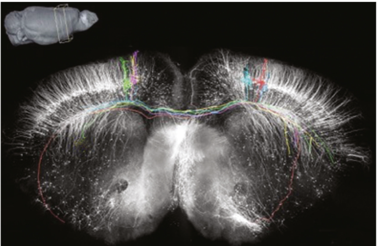
Axonal transport
The movement of materials within an axon via motor proteins
Anterograde transport
The movement of substances from the cell body to the axon terminal
Retrograde transport
The movement of substances from the axon terminal to
the cell body
function of glial cells
support and enhance neurons
Astrocytes
many processes receive neuronal input and monitor activity
Microglial cells
small cells remove debris from injured cells
Myelination
glial cells wrap axons with a fatty sheath, myelin, to insulate and speed conduction
Multiple sclerosis
a demyelinating disease that causes an interruption in information flow down the axon
Schwann cells
form myelin sheath for cells outside the brain and spinal cord, such as in peripheral nerves
Oligodendrocytes
form myelin sheath in brain and spinal cord (but not in the peripheral nervous system)
Nodes of Ranvier
gaps between sections of myelin where the axon is exposed
Central nervous system (CNS)
the brain and spinal cord
Peripheral nervous system (PNS)
all parts of the nervous system found outside the skull and spinal column (ie, everything outside of the central NS)
Motor nerves
transmit information from the CNS to muscles, organs, and glands. A.K.A. Efferent Nerves
Efferent Nerves
transmit information from the CNS to muscles, organs, and glands
Sensory nerves
convey information from the body to the CNS. A.K.A. Afferent Nerves
Afferent Nerves
convey information from body to CNS
Somatic nervous system
connects brain and major muscles and sensory systems (voluntary)
Autonomic nervous system
Nerves that primarily control the viscera (organs) -- bodily functions you don’t normally have voluntary control over
Sympathetic nervous system
prepares the body for action
Parasympathetic nervous system
relax, rest and recover
Enteric nervous system
digestion/gut motility
two cerebral hemispheres of brain
cerebral cortex and folds
Cerebral cortex
folded outermost layer of the cerebral hemispheres, comprised mostly of neuron cell bodies, dendrites, and axons
Folds (gyri and sulci)
increase amount of cortex that can fit into the skull, and are grouped together into lobes
function of brain folds (gyri and sulci)
increase surface area
Gray matter
Contains more cell bodies and dendrites, which lack myelin
White matter
Consists mostly of axons with white myelin sheaths.
Basal ganglia (nuclei)
important in motor control; reciprocally connected with the cortex
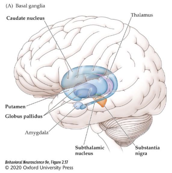
Limbic system
includes structures important for learning and memory, cognitive functions, emotional regulation, sense of smell

the two distinct branches that fuse to form each spinal nerve
dorsal and ventral roots
Dorsal root
carries sensory information from the body to the spinal cord
Ventral root
carries motor information from the spinal cord to the muscles
Computerized axial tomography (CAT or CT)—
a measure of X-ray absorption at several positions around the head; maps tissue density
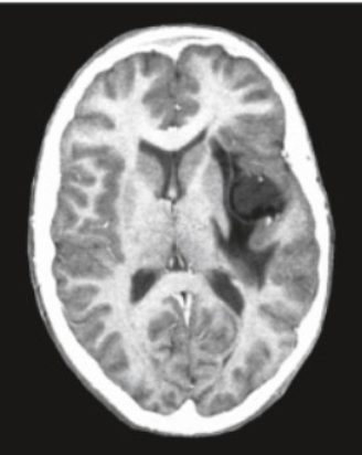
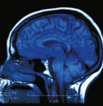
Magnetic resonance imaging (MRI)
produces high-resolution images using radio frequency energy
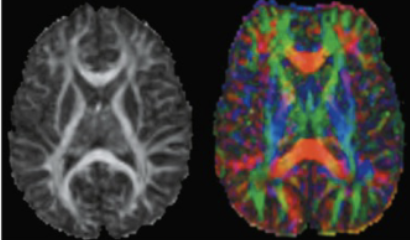
Diffusion tensor imaging (DTI)
uses MRI technology to study white matter tracts; based on fractional anisotropy (FA)
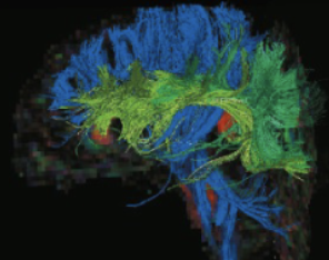
DTI tractography
uses mathematical manipulations to produce structural images of axonal fiber pathways
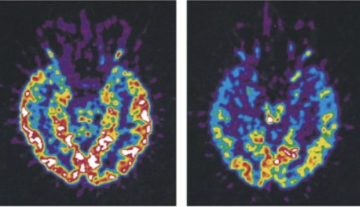
Positron emission tomography (PET)
produces images of brain activity: identifies brain regions that contribute to specific functions
Functional MRI (fMRI)
detects small changes in brain metabolism, such as oxygen use, in active brain areas
can show how networks of brain structures collaborate
fMRI