Orthopedic Neoplasm (CMPP)
 Call Kai
 Learn
Learn Practice Test
 Spaced Repetition
 Match
Match Flashcards
 Knowt Play
1/174

There's no tags or description
Looks like no tags are added yet.
Name | Mastery | Learn | Test | Matching | Spaced | Call with Kai | Chat |
|---|
No analytics yet
Send a link to your students to track their progress
175 Terms
Most malignancy of the bones are usually from
Metastasis
What defines the treatment of bone neoplasm
Type of Tumor
Primary or Metastatic
Extent of Tumor
Who do we refer bone neoplasms to
Orthopedic Surg ± Oncology
Whatt imaging may be needed in work-up orthopedic neoplasms
XR
CT / MRI
Bone Scan
What is the most reliable indicator of malignancy risk
Hx of Prior Malignancy
What are some red flags for bone malignancy
Pain, unrelieved by rest, nocturnal
Weight loss
Night sweats
Fatigue
Pathologic fracture
Swelling or palpable deformity
what lab may be elevated for orthopedic neoplasms
Alk Phos w/o GGT
What cancers can be test with AFP
Liver
Testicular
Liver Tumor Marker
AFP
Testicular Tumor Marker
AFP
b-hCG
LDH
Prostate tumor markr
PSA
Ovarian Tumor Marker
CA-125
Pancreatic Tumor Marker
CA 19-9
Breast Tumor Markers
CA 15-3
CA 27
CA 29
Colorectal Tumor Marker
CEA
Lung Tumor Markers
CEa
CYFRA 21-1
NSE
Stomach Tumor Markers
CA 72-4
CEA
Bladder Tumor Markers
NMP22
BTA
What information on bone neoplasms can gained from XR
Location
Border
periosteal reaction
matrix
How can we describe location of bone neoplasms
Specfic bone
Epihysis / Metaphysis / Diaphysis
How can we describe borders of bone neoplasms
Well-defined with or without sclerosis (geographic)
Moth eaten
Permeative
How can we describe matrix of bone neoplasms
Lytic - empty hole
Blastic - increased bone
Calcifications
Ground glass
Arcs and rings
How can we describe periosteal reaction of bone neoplasms
Onion skinning
Starburst
What tumor commonly presents with onion skinning periostealr reaction
Ewing Sarcoma
When is CT used for bone neoplasms
setting of metastatic disease
MRI can best assess what aspects of bone neoplams
Tumor Size
Local IO and extraosseous extent
identify both hypervascular and necrotic areas within tumor
What benefit can technetium bone scans provide
evaluate entire skeleton for the presence of multiple lesions
What can cause false positives on technetium bone scan
previous trauma
What cancers commonly spread to bone
Breast
Lung
Thryoid
Kidney
Prostate
Most Common Primary Bone Tumor
MM
Follwed by osteosarcoma
What are the types of benign bone tumor
Osteoid Forming
Chondrioid Forming
Fibrous
Cystic
Giant Cell Tumors
Hemangioma
Types of Osteoid Forming Benign Bone Tumors
Osteoid Osteoma
Osteoblastoma
Osteofibrous Dysplasia
Types of Chondrioid Forming Benign Bone Tumors
Enchondroma
Osteochondroma
Periosteal Chondroma
Chondroblastoma
Chondromyxoid Fibroma
Types of Fibrous Benign Bone Tumors
Fibrous Cortical Defect
Nonossifying Fibroma
Fibrous Dysplasia
Types of Cystic Benign Bone Tumors
Simple Bone Cyst
Aneurysmal Bone Cyst
Epidermoid Cyst
Osteoid Osteoma
Benign bone-forming tumor characterized by a small radiolucent nidus (<1 to 1.5 cm)
Who most commonly gets osteoid osteomas
Teenage males
Risk Factors for Osteoid Osteoma
None
Where is the most common site for osteoid osteoma
Promixal Femur
Presentation of Osteoid Osteoma
Progressively increasing pain worse at night
Limp
Decreased ROM
Swelling and Tenderness
Pain quickly relieved by NSAIDs
Nidus
The source of infection / disease
Why does pain of osteoid osteoma go away with NSAIDs
The tumor makes prostaglandins
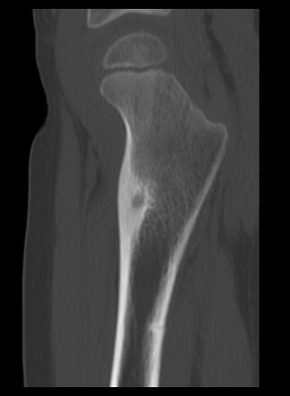
Osteoid Osteoma
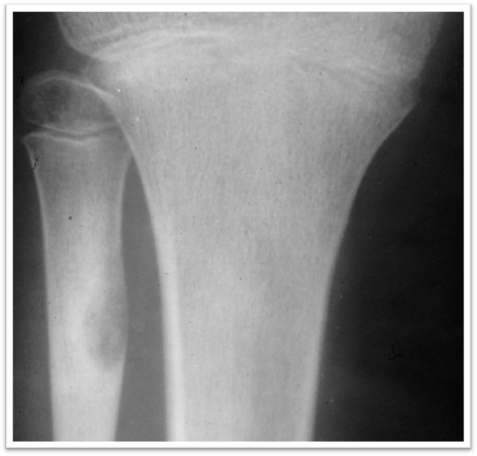
Osteoid Osteoma
How do Osteoid Osteomas appear on XR
small, round lucency (nidus) with a sclerotic margin
Preferred Moduality for dx of Osteoid Osteoma
CT
Why don’t we use MRI for osteoid osteomas
Cab be obscured by soft tissue involvement
Treatment Options for Osteoid Osteoma
Percutaneous radio frequency ablation
Curettage and bone grafting:
En bloc resection
Percutaneous radio frequency ablation
Uses radio frequencies passed beneath the skin through a needle to kill the tumor cells by heating them to a high temperature
Curettage and bone grafting:
tumor is scraped out of the bone
Remaining cavity is packed with donor bone tissue (allograft) or bone chips taken from another bone (autograft)
En bloc resection (Bone)
Surgical removal of bone containing the tumor
Internal fixation with pins may restore structural integrity of the bone
Enchondroma
Benign cartilage-forming tumors that develop in the medulla (marrow cavity) of long bones
Enchondromatosis
a syndrome of multiple Enchondroma
Forms of Enchondromatosis
Ollier disease
Maffucci syndrome – can also have vascular malformations
Who is most likely to present with a Enchondroma
Teens
Who is most likely to present with encondromatosis
children <10 years
What is the most common sites for enchondroma
tumor of the HAND (#1)
Also likes humerus and femur
Presentations of Enchondroma
Many will be asymptomatic until fracture
deformity due to widening of the bone
limb-length discrepancy
enchondromatosis in skull – headache and cranial nerve palsy
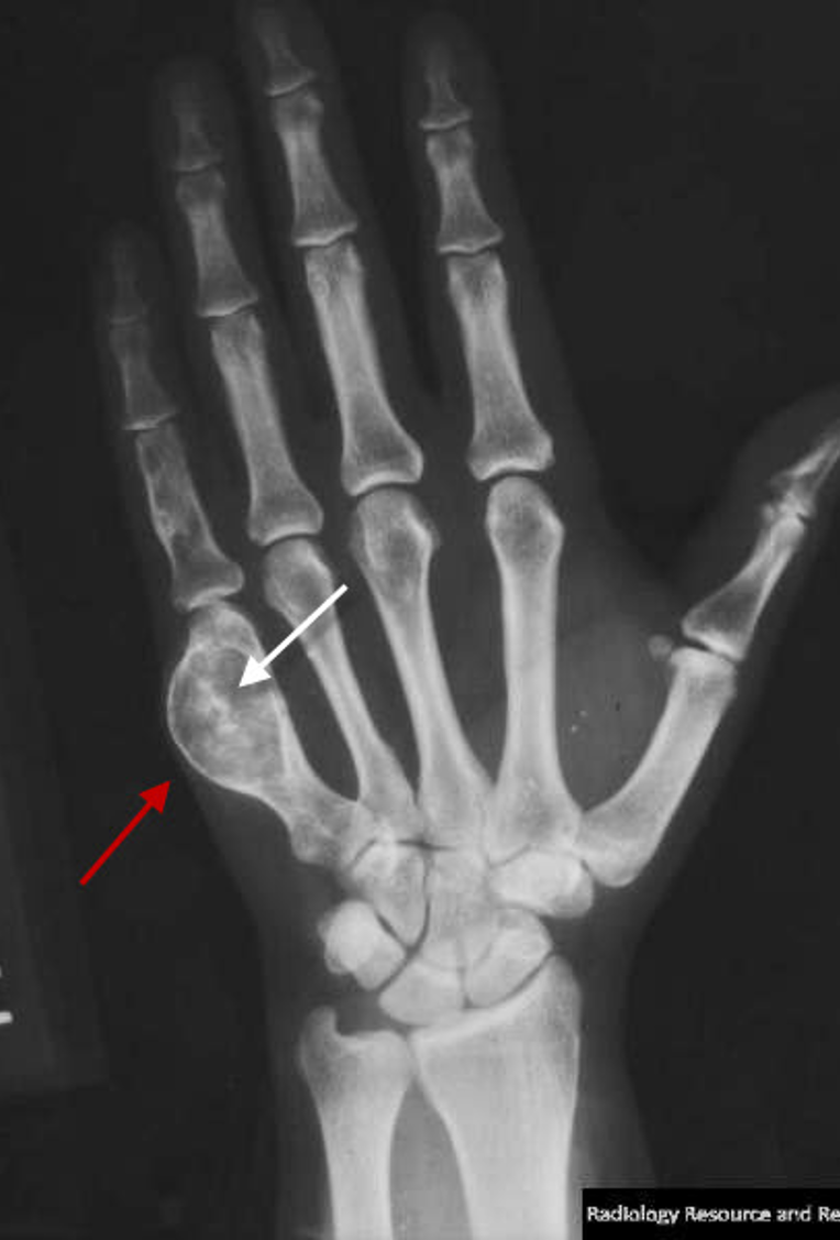
Enchondroma
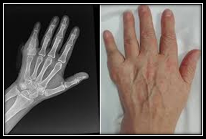
Enchondroma
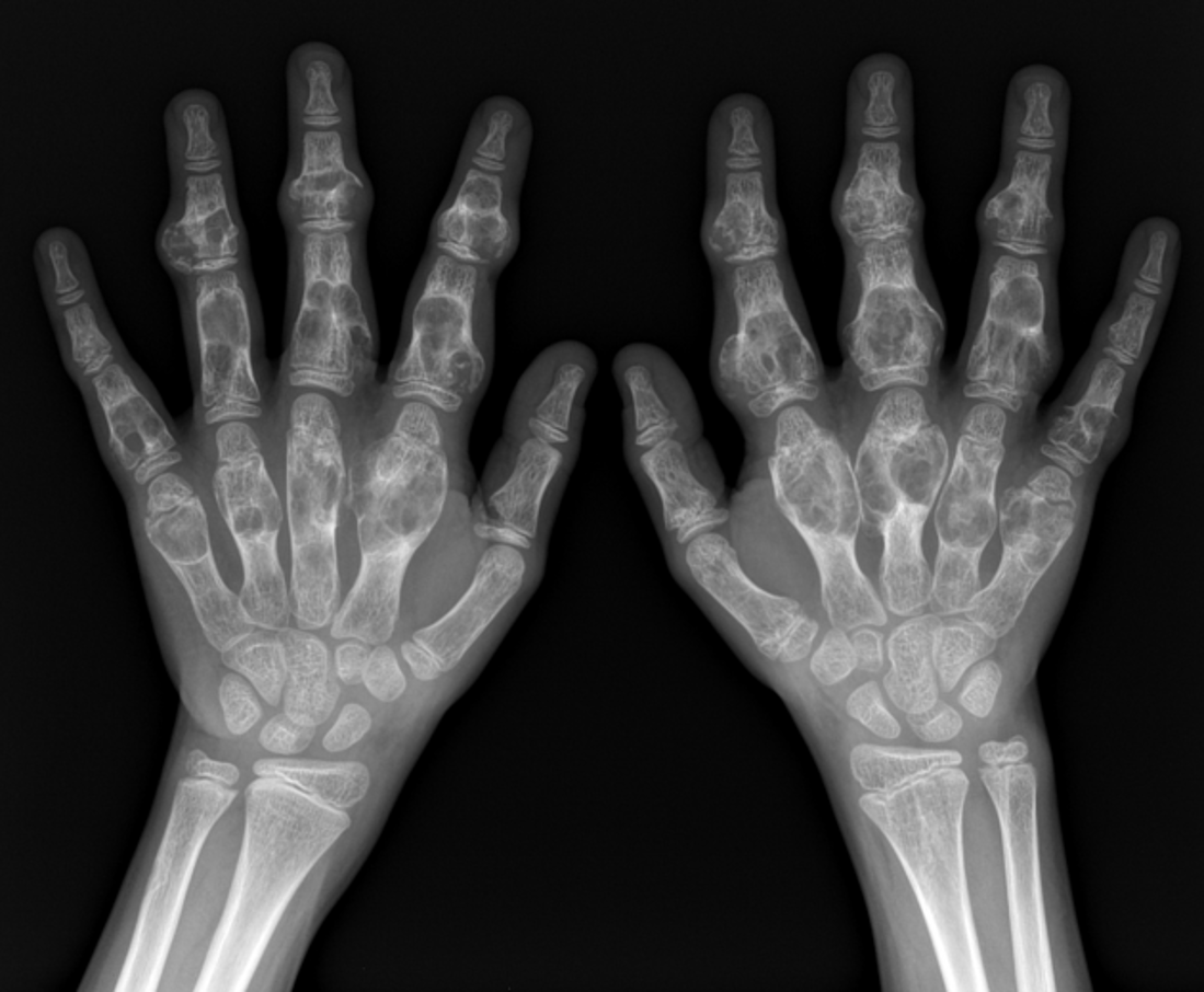
enchondromatosis
Preferred Test for Enchondramas
XR
How do enchondromas appear on XR
Lesion(s) often oval, well-circumscribed
Sclerotic or non-sclerotic margins
Central lucent lesion – may include matrix calcifications
Endosteal scalloping
What are flags that would indicate a bone tumor is not an enchondroma
gross bone destruction
periosteal reaction
soft tissue mass
Endosteal scalloping
resorption of the inner surface of bone cortex;
appears as indentations or erosions on the normally smooth margin of the bone's inner layer
What bone tumor presents with endosteal scalloping
Enchondroma
Treatment for Asymptomatic and Small Enchondrma
Observation
Who gets actual treatments for enchondroma
Long bones due to risk of fracture with weight-bearing bone
>25 mm in diameter
Involve >50 percent of the diameter of the cortex
Treatment for Symptomatic Enchondramsas
Curettage and Graft
Untreated enchondrams can transform into
Chondrosarcoma
What are signs that enchondrams have undergone malignany transformation into chondrosarcoma
worsening pain
Usually after skeletal maturity
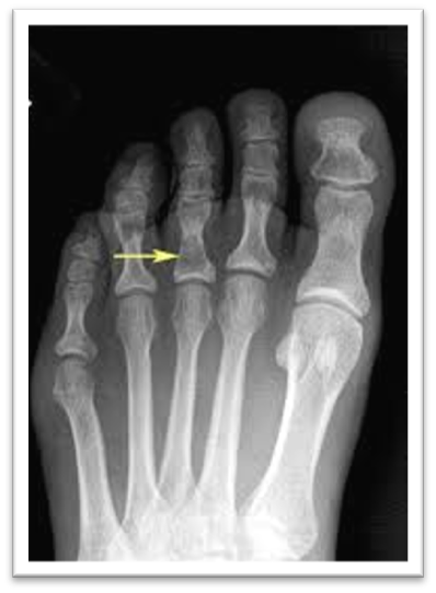
Enchondroma
Osteochondroma
Cartilage-capped bony spur arising on the external surface of a bone
Marrow cavity of the spur is continuous with the cavity of the underlying bone
Cap of cartilage is the source of growth
Where do osteochondrmas appear from
escaped physeal cells
What conditions do you se osteochondrmas with
Hereditary multiple osteochondromas (HMO)
Hereditary multiple exostoses (HME)
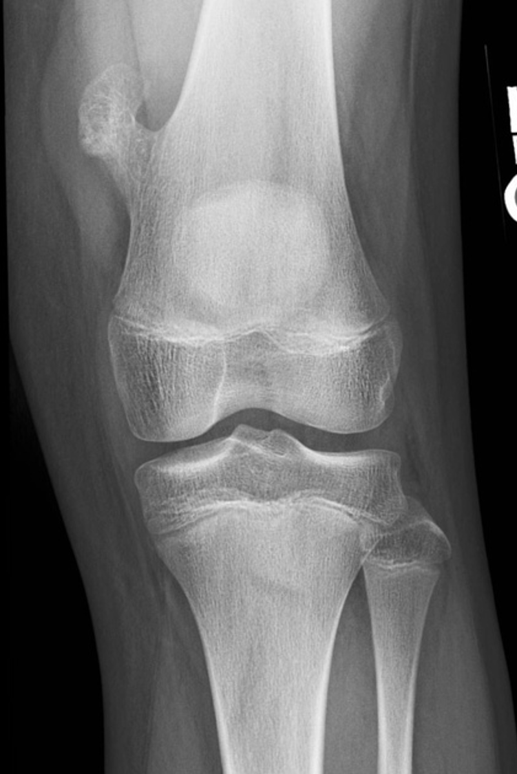
Osteochondroma
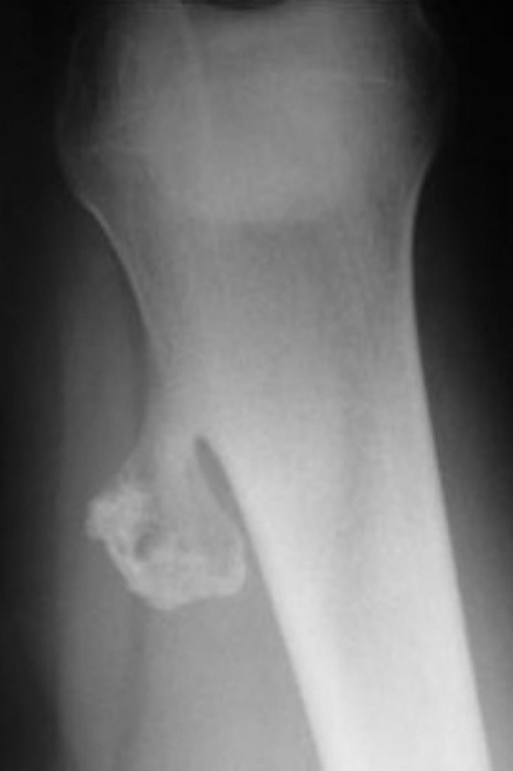
Osteochondroma
Presentation of Osteochondroma
painless mass – often found near a joint or on the axial skeleton
painful when associated with local trauma or with pathologic fracture
decreased range of motion
deformity
short stature,
limb length discrepancy,
varus/valgus deformities
Test of Choice for Osteochondroma
XR
Indications for MRi for Osteochondroma
Concern for adjacent soft tissue impingement
New focal pain at site, not apparent by X-ray
Concern for chondrosarcomatous (malignant) transformation
Indications for Excising Osteochondromas
Local irritation or deformity
Concern for malignant transformation
Simple Bone Cyst
Fluid-filled lesion with a fibrous lining
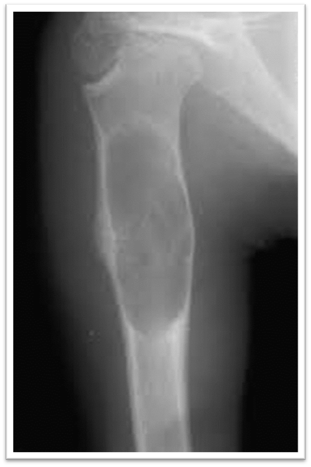
Simple Bone Cyst
Peak Incidence for Simple Bone Cysts
0-20 years
Most Common Sites for Bone Cysts
Proximal humerus
femur
Preesentation of Bone Cysts
localized pain,
decreased function/failure to use extremity
limp
Pathologic fracture
Test of Choice for Bone Cysts
XR
How do bone cysts appear on XR
Well-marginated cystic lesion of metaphysis or metadiaphysis without reactive sclerosis
"Fallen leaf" sign
"Fallen leaf" sign
Fragment of bone falls to the bottom of the cyst
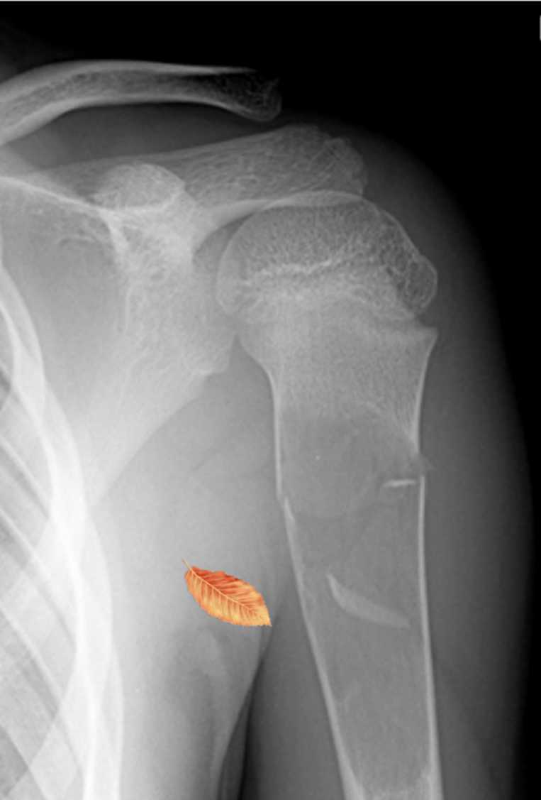
Bone Cyst
Treatment for Simple Bone Cysts
Observation with Serial XR q4-6months
Activity Restriction due to Fracture Risk
Aspiration and Methylprednisolone Injection (Large)
Surgery (Rare)
Giant Cell Tumors
Non-malignant but locally aggressive osteolytic skeletal neoplasm
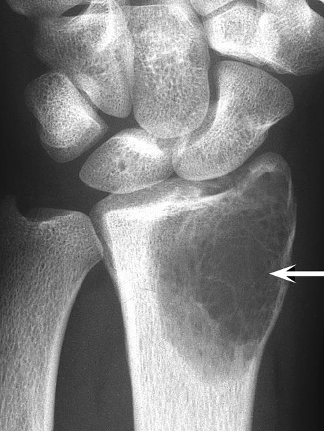
Giant Cell Tumors
Peak Incidence with Giant Cell Tumors
20-40 y/o
What condition are Giant Cell Tumors seen in
Paget’s Disease
Where do giant cell tumors form
meta-epiphyseal region of the long bones, usually around the knee
what is highly expreessed in giant cell tumors
RANK ligand
Presentation of Giant Cell Tumor
Pain
Swelling
Limitation of ROM
Pathologic Fracturee
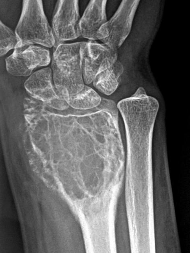
Giant Cell Tumor
How do Giant Cell Tumors present on XR
expansile, eccentric, lytic lesion in epiphysis and adjacent metaphysis
Absence of matrix calcification and periosteal reaction