EXAM 1- John O'Reilly-ZOO 251
 Call Kai
 Learn
Learn Practice Test
 Spaced Repetition
 Match
Match Flashcards
 Knowt Play
1/168

There's no tags or description
Looks like no tags are added yet.
Name | Mastery | Learn | Test | Matching | Spaced | Call with Kai | Chat |
|---|
No analytics yet
Send a link to your students to track their progress
169 Terms
layers of pericardium
parietal pericardium, pericardial cavity, visceral pericardium, pericardial fluid

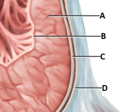
epicardium
visceral pericardium (C)
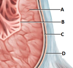
Myocardium
heart muscle (A)
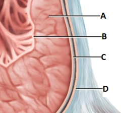
endocardium
atria and ventricles (B)
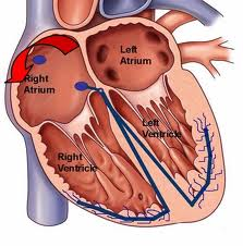
Chambers of the heart
right atrium, right ventricle, left atrium, left ventricle
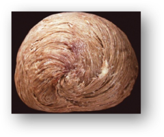
myocardial vortex
Formed by a spiraling pattern of cardiac muscle cells
antrioventricular valves
valves between the atria and ventricles
Right AV valve
tricuspid

Left AV
bicuspid/mitral

semilunar valves
valves between ventricles and pulmonary arteries & aorta
semilunar valves
pulmonary and aortic
left coronary artery (LCA)
anterior interventricular branch (left anterior descending) & circumflex branch
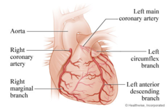
right cornary artery (RCA)
right marginal branch & posterior interventricular branch
which are the major branches of your left coronary artery (LCA)?
(a) anterior interventricular (LAD) branch & circumflex branch
(b) right marginal branch & posterior interventricular branch
(c) both a and b
(d) neither a nor b
(a) anterior interventricular (LAD) branch & circumflex branch
the valves in your heart that are found between your atria and ventricles are called?
(a) tricuspid
(b) mitral
(c) bicuspid
(d) both a and b
(e) all of the above
(e) all of the above
Blood flow of the heart
1. Superior Vena Cava/Inferior
2. R. atrium
3. tricuspid valve
4. R. ventricle
5. pulmonary valve
6. pulmonary artery to the lungs
7. Pulmonary veins from the lungs
8. L. atrium
9. bicuspid valve
10. L. ventricle
11. aortic valve
12. body

Cardiac Physiology
electrical and mechanical pacemaker potential and sinoatrial node
pacemaker potential
slow Na+ inflow (if funny current) through HCN channels

Cardiac Action Potential
1) Rapid depolarization due to Na+ influx
2)*Plateau Stage due to slow Ca+ influx (responsible for the refractory period)*
3) Repolarization due to K+ efflux - RMP reestablished

Conduction pathway of the heart
1. SA node
2. AV node
3. Bundle of His
4. Bundle branches
5. Purkinje fibers

sinoatrial (SA) node
pacemaker of the heart

internodal pathway
fast
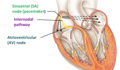
Atroventricular Node
slowest
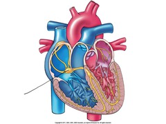
Bundle of His
fast

bundle branches
faster

Purkinje fibers
fastest

cardiocytes
striated, intercalated discs, gap junctions

intercalated discs
connections between cardiac muscle cells
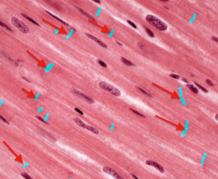
gap junctions
electrically coupled cardiocytes
Diastole
Relaxation of the heart
Systole
Contraction of the heart
filling
-diastole
-no change in pressure
-increase in volume
isovolumetric contraction
-systole
-no change in volume
-increase in pressure

ejection
-systole
-increase in pressure
-decrease in volume

isovolumetric relaxation
-diastole
-no change in volume
-decrease in pressure

P wave
atrial depolarization

QRS complex
ventricular depolarization and atrial repolarization

T wave
ventricular repolarization

PR interval
AV node delay & atrial contraction

ST segment
time during which ventricles are contracting and emptying

TP interval
ventricles relaxing and filling

what happens if PR interval longer than 0.2s?
then AV conduction block
what happens if the ST interval is too short or long?
then ventricular ischemia or hypoxia
what happens if the QT interval is too long?
then tachyarrhythmias
cardiac output
stroke volume x heart rate

increased heart rate
tachycardia
decreased heart rate
bradycardia
autonomic nervous system
sympathetic and parasympathetic
sympathetic
increases heart rate
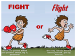
Parasympathetic
decreases heart rate

Stroke volume is directly proportional to what?
-End Diastolic Volume (EDV, aka, preload)
-strength of ventricle contraction
Stroke volume is inversely proportional to what?
-Peripheral Resistance (after load)
sympathetic
norepinephrine
Parasympathetic
acetylcholine
sympathetic
depolarization
Parasympathetic
hyperpolarization
Baroreceptors
carotid sinus (IX) & aortic arch (X)

Chemoreceptors
aortic bodies (vagus X) & carotid bodies (glossopharyngeal IX)

Baroceptors
pressure
Chemoreceptors
chemicals
Arteries
Blood vessels that carry blood away from the heart

Veins
Blood vessels that carry blood back to the heart

Capillaries
nutrient and waste exchanges with tissues
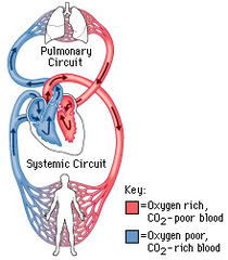
pulmonary circulation
flow of blood from the heart to the lungs and back to the heart

systemic circulation
circulation that supplies blood to all the body except to the lungs
lymphatic system
extracellular fluid uptake & return to circulatory system

layers of artery wall
tunica interna, tunica media, tunica externa (deep to superficial)
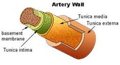
tunica interna (intima)
-endothelium
-connective tissue

tunica media
-smooth muscle
-collagen
-elastin

tunica externa
connective tissue

Three types of capillaries
continuous, fenestrated, sinusoidal
continuous capillaries
Least permeable (muscle, peripheral nerves)
fenestrated capillaries
medium permeability (kidney, intestines, endocrine glands)
sinusoidal capillaries
most permeable (liver, bone marrow, spleen)
one way valves
prevent backflow of blood
blood reservoir
veins
pressure reservoir
arteries
pulmonary artery
from heart to lungs (oxygen-poor; low O2)
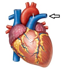
pulmonary vein
from lungs to heart (oxygen-rich; high O2)

brachiocephalic trunk
shoulder & head

R/L common carotid artery
head
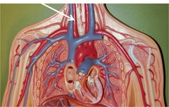
R/L subclavian artery
shoulder

ascending aorta
superior

descending aorta
inferior

internal carotid artery
brain

external carotid artery
head

vertebral artery
spinal cord and brain

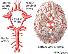
basilar artery
base of the brain

Circle of Willis
-basilar a.
-internal carotid a.
-anterior & posterior communicating a.
-anterior & posterior cerebral a.
internal jugular vein
brain

Poiseulle's law
shows the relationship among blood flow, pressure, and resistance
blood flow = difference between P1 and P2/resistance
or Q = delta P/R
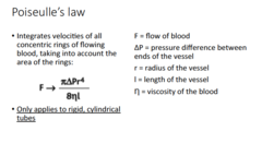
Flow is directly proportional to what?
pressure gradient
systolic pressure
peak pressure exerted in arteries when blood is pumped into them during ventricular systole
diastolic pressure
lowest pressure exerted in the arteries when blood is draining into the vessels during ventricular diastole
pulse pressure
difference between systolic and diastolic pressure
mean arterial pressure
average blood pressure throughout the cardiac cycle
mean arterial pressure
2/3 diastolic + 1/3 systolic
blood vessel diameter
decreases from arteries to capillaries, then increases in veins
total x-section area
increases from arteries to capillaries then decreases in veins
Blood pressure
decreases from arteries to capillaries to veins