Di Imaging 2: Exam #1
 Call Kai
 Learn
Learn Practice Test
 Spaced Repetition
 Match
Match Flashcards
 Knowt Play
1/160

There's no tags or description
Looks like no tags are added yet.
Name | Mastery | Learn | Test | Matching | Spaced | Call with Kai | Chat |
|---|
No analytics yet
Send a link to your students to track their progress
161 Terms
CT
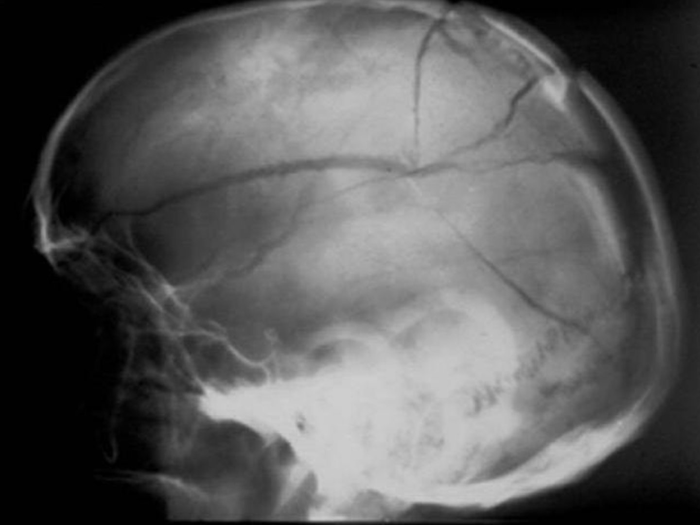
What imaging is best for determining a skull fracture?
Linear Skull Fracture (it's most common)
What is the diagnosis?
True
T/F: A tripod fracture involves the seperation of all 3 major attachements of the Zygomatic bone to the rest of the face
Le Fort Fractures
What are types of facial fractures involving the maxillary bone and surrounding structures?
subdural hematoma
What is the diagnosis?
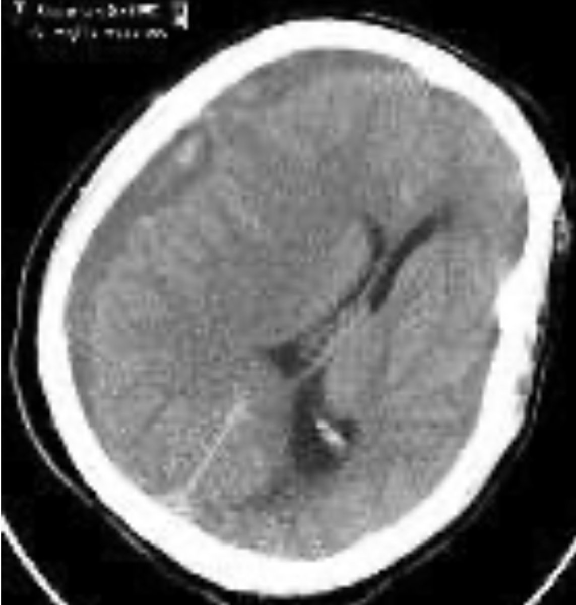
Convex
A subdural hematoma is usually ______ in shape
venous
A subdural hematoma is due to ________ blood
Epidural Hematoma
What is the diagnosis?
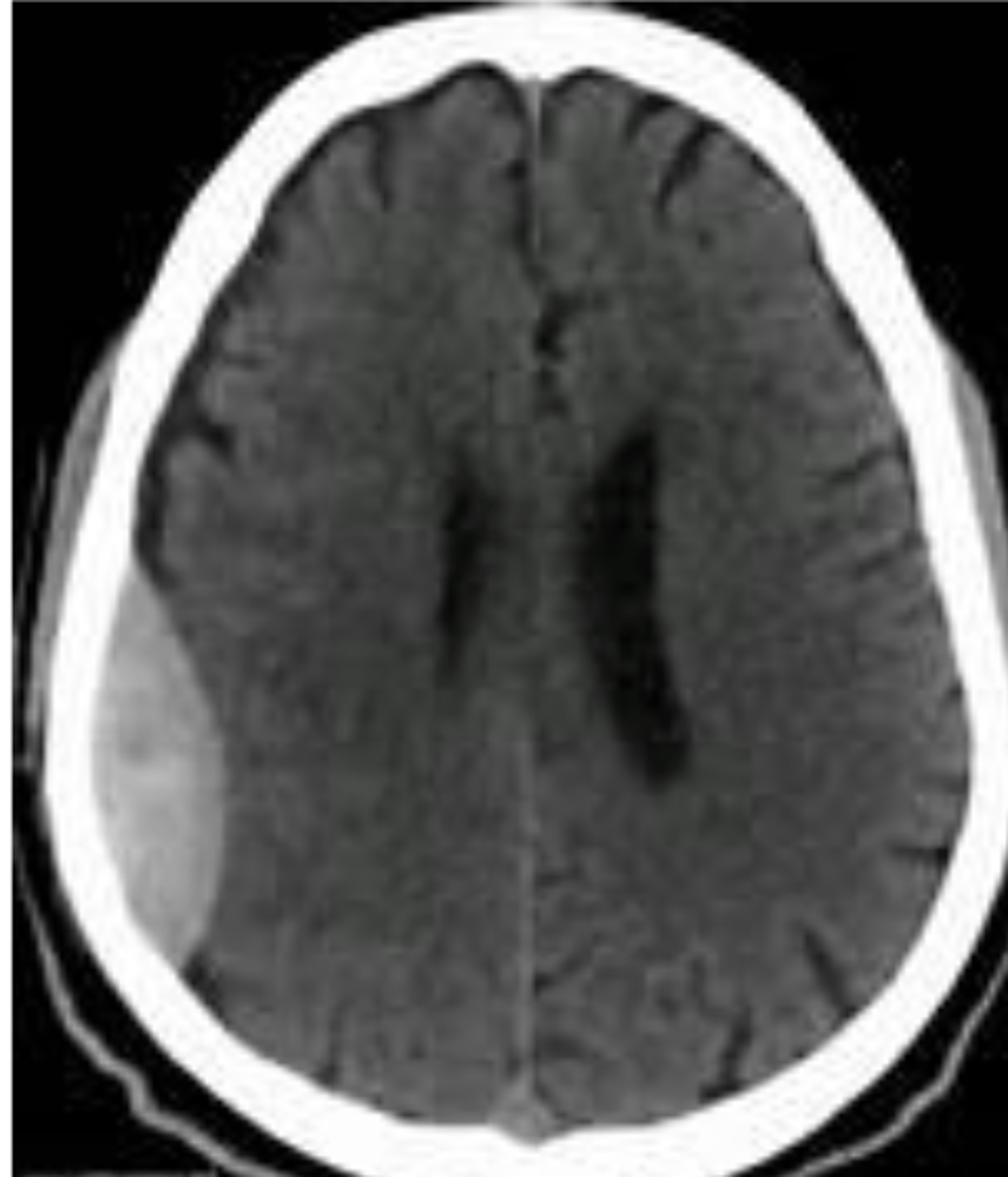
Concave
A epidural hematoma is usually ______ in shape
arterial
An epidural hematoma is due to ________ blood
Direct Trauma
What is the most common mechanism of a Blowout fracture?
- Compression Fracture
- Bilateral interfacetal dislocation
- Flexion teardrop fracture
- Clay shoveler's fracture
- Anterior subluxation
- Dens fx
Name the fractures we have discussed that happen due to a Hyperflexion mechanism?
- Unilateral interfacetal dislocation & fractrue
Name the fractures we have discussed that happen due to a Hyperflexion and rotation mechanism?
- avulsion of the anterior tubercle of C1
- hyperextension fx-dislocation
- hyperextension dislocation
- posterior arch fx of C1
- extension teardrop fx
- Hangman's fx
- lamina fx
- dens fx
Name the fractures we have discussed that happen due to a Hyperextension mechanism?
- Jefferson's Fracture of C1
- Bursting Fracture of lower cervicals
Name the fractures we have discussed that happen due to a Vertical Compression mechanism?
Posterior Arch Fracture
What is the mc fracture of atlas?
C1-2, C5-7, T12-L1
What are the most common locations for Fractures/Dislocations?
1. Lateral
2. APOM
3. AP Cervical
4. Lt & Rt. Oblique
5&6 Flexion & Extension
7. Swimmers
List the Order of the Davis Series
If more than one column is disrupted or if the middle column is disrupted
Using the 3 column method, describe how a segment would be considered unstable?
Children: 14mm
Adults: 22mm
What should the Retrotracheal space in children and adults not exceed?
7mm
What should the Retropharyngeal space not exceed?
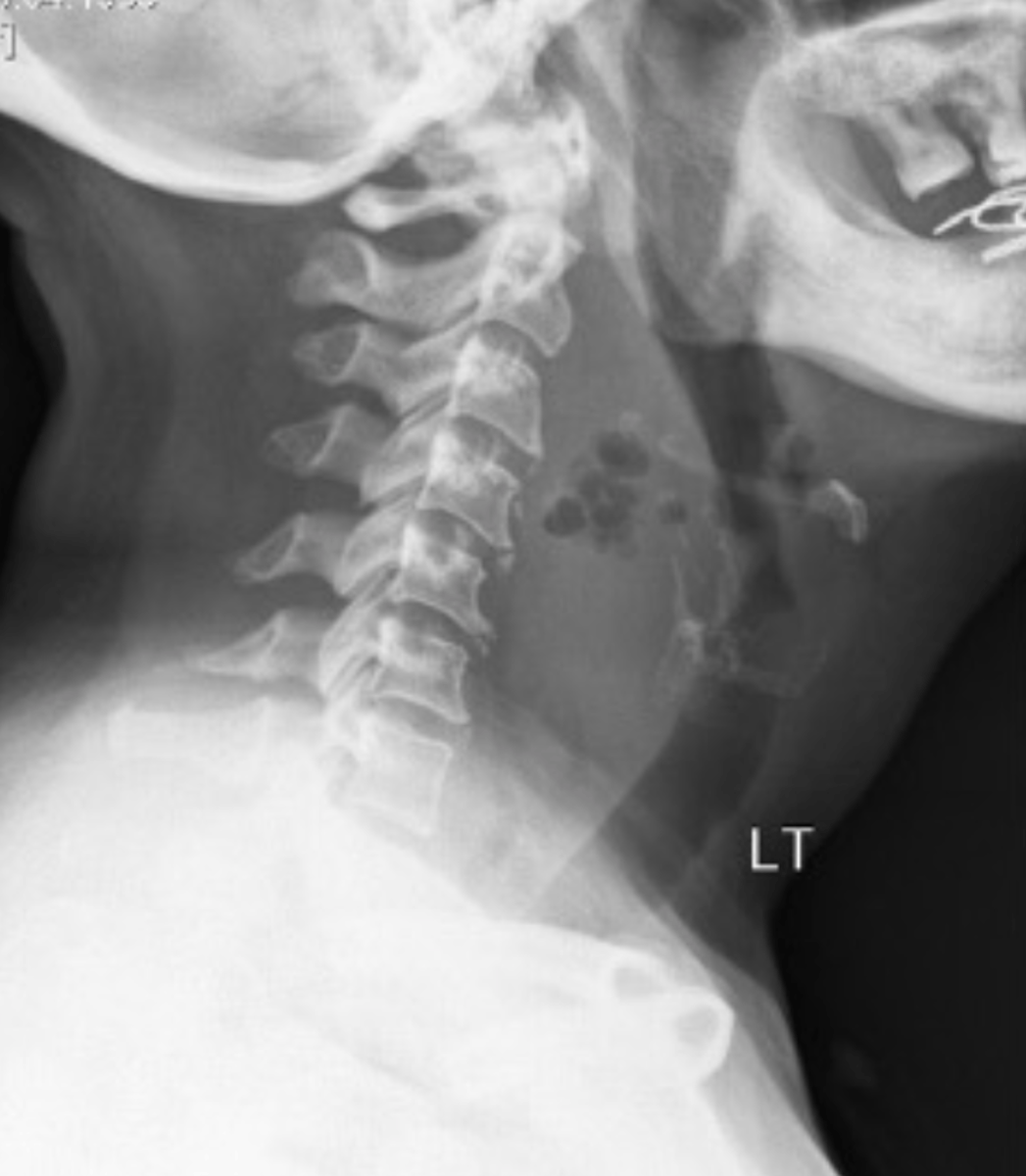
Increased Retrotracheal Space
What is your finding or diagnosis? (honestly I'm not sure which this is classified as)
Children: <5mm
Adults <3mm
What is the maximum ADI in children & adults
~3-6 months
How long do spinal fractures take to heal?
C1 Posterior arch fracture
What is the diagnosis?
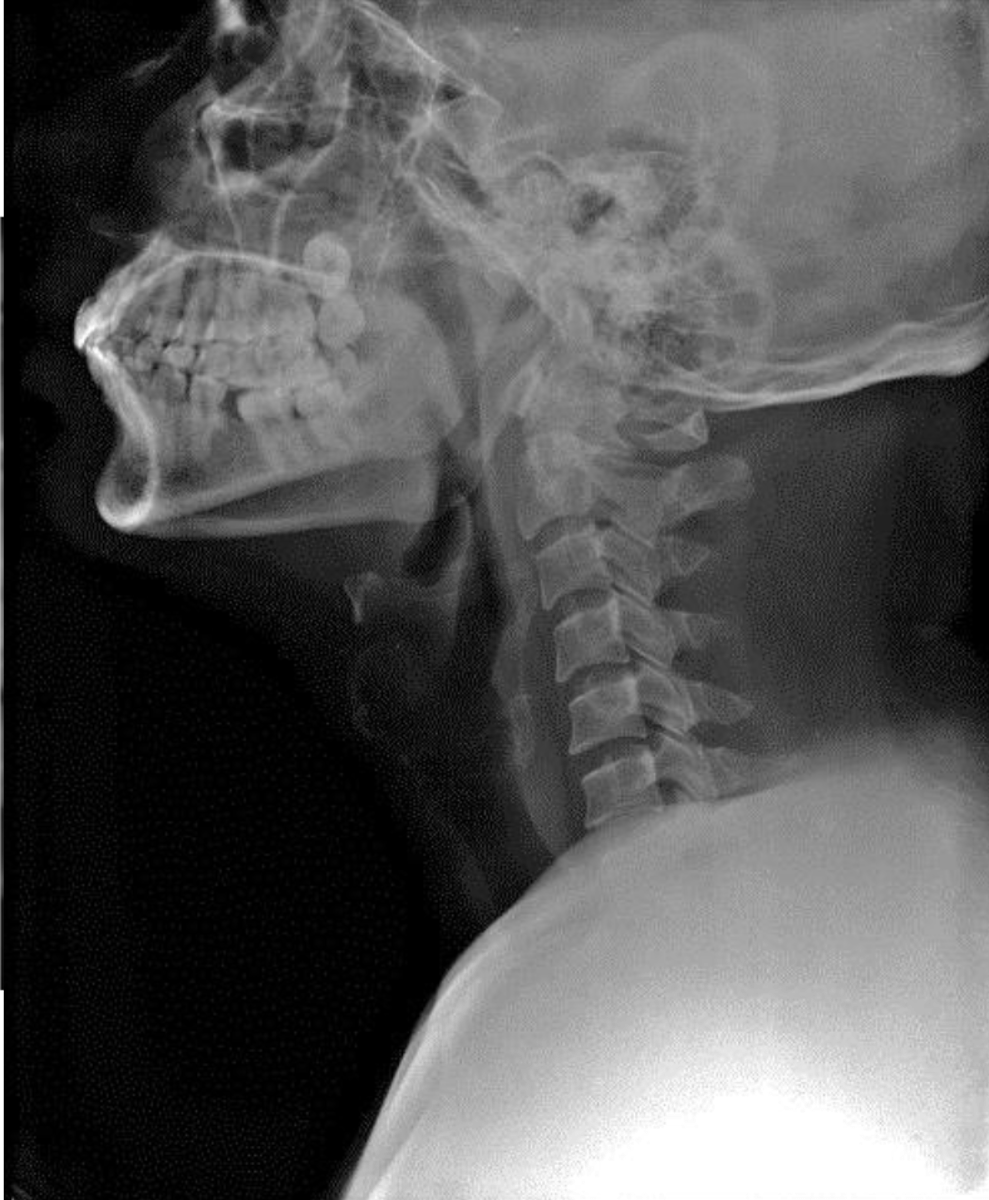
Posterior arch fracture and increased ADI
What are the findings?
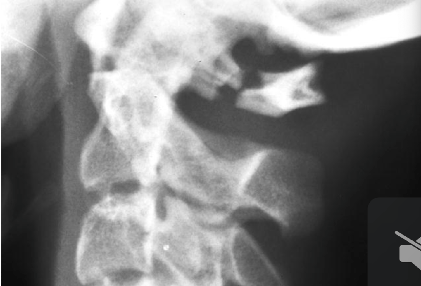
-bilateral fracture of anterior & posterior arches
- lateral masses are offset
- transverse ligament tor >7mm
What is a Jefferson's Fracture?
AP Open Mouth
What x-ray view is best for determining a Jefferson's Fracture?
Jefferson's Fracture
What is the potential diagnosis for this male with an occipital headache after head butting someone playing football?
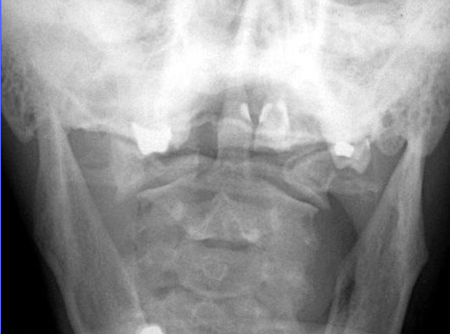
Increased atlanto-dental overhang sign
What is the finding for a Jefferson's Fracture?
hyperextension
An avulsion fracture of the anterior tubercle results from ________ of the neck against contraction of the long flexors
Rheumatoid Arthritis
Atlantoaxial dislocations usually associated with ___________
3mm
What is the maximum upper limit for an ADI in adults?
5mm
What is the maximum upper limit for an ADI in children?
-normal variant
-trauma
-Down's syndrome
-major upper cervical anomalies
-inflammatory arthropathies
What are some etiological factors of a increased ADI?
oblique fracture of the tip (4%)
What is a type 1 odontoid fracture?
Fracture at the base of the dens (66%) (aka High Dens Fracture)
What is a type 2 odontoid fracture?
Fracture into the body of C2 (30%)
What is a type 3 odontoid fracture?
Type 3
What type/s of odointoid fracture causes disruption of Harris' ring?
Type 1
What type/s of odontoid fracture is usually stable?
Type 2 & 3
What type/s of odontoid fracture is usually unstable?
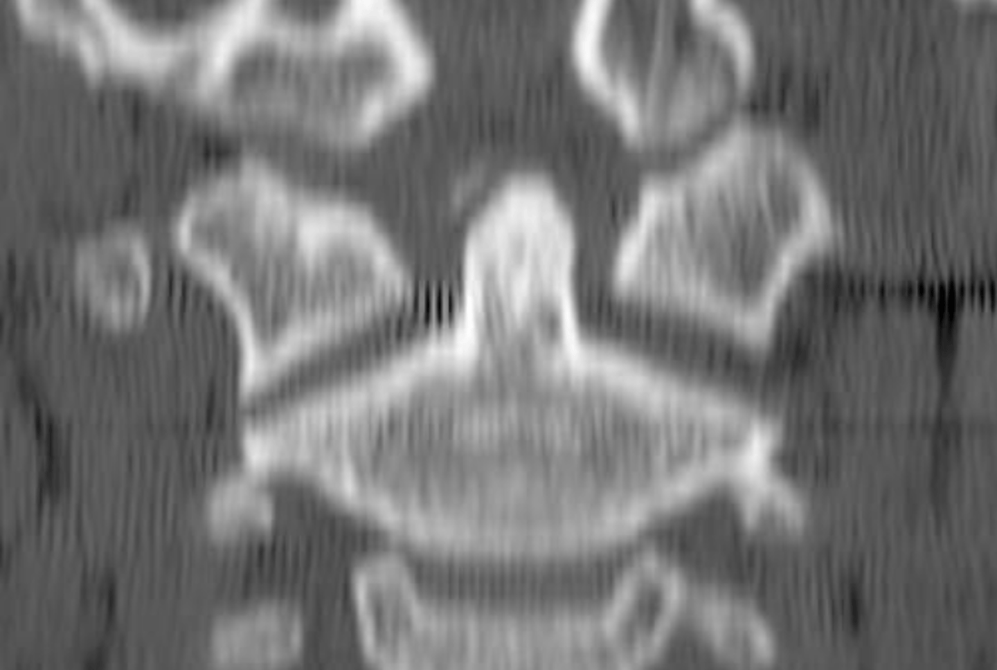
Type 1 Odontoid Fracture at C2
What is the diagnosis?
5
In a type 2 odontoid fracture if it is displaced greater than ___ mm it is likely to never fuse (100% nonunion)
True
T/F: Most os odontoideum are old ununited dens fx's?
Type 2 fracture of the odontoid at C2 (probably from hyperflexion)
What is the diagnosis?
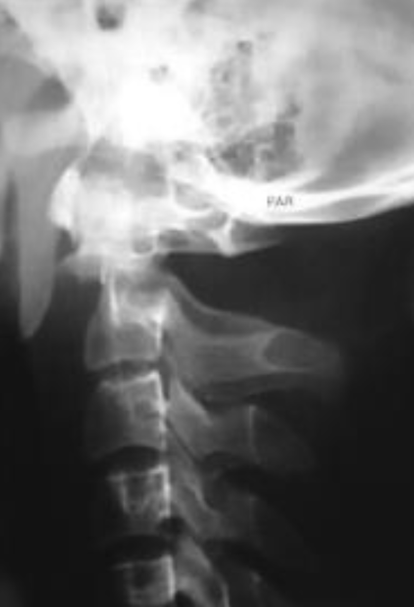
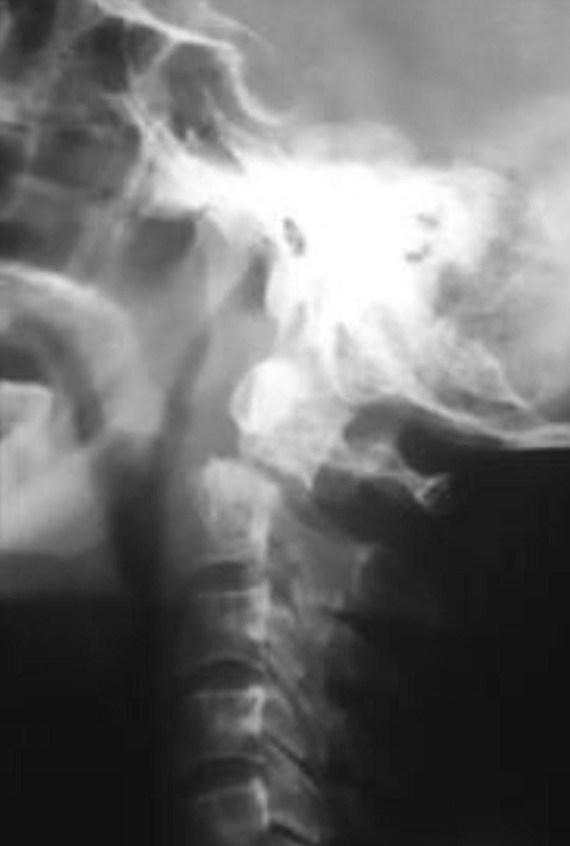
Type 2 fracture of the odontoid at C2 (probably from hyperextension)
What is the diagnosis?
Type 3 Odontoid Fracture
What odontoid fracture occurs below the junction of the dens and C2 body and may disrupt Harris' Ring?
Type 3 Odontoid Fracture
What is the diagnosis?
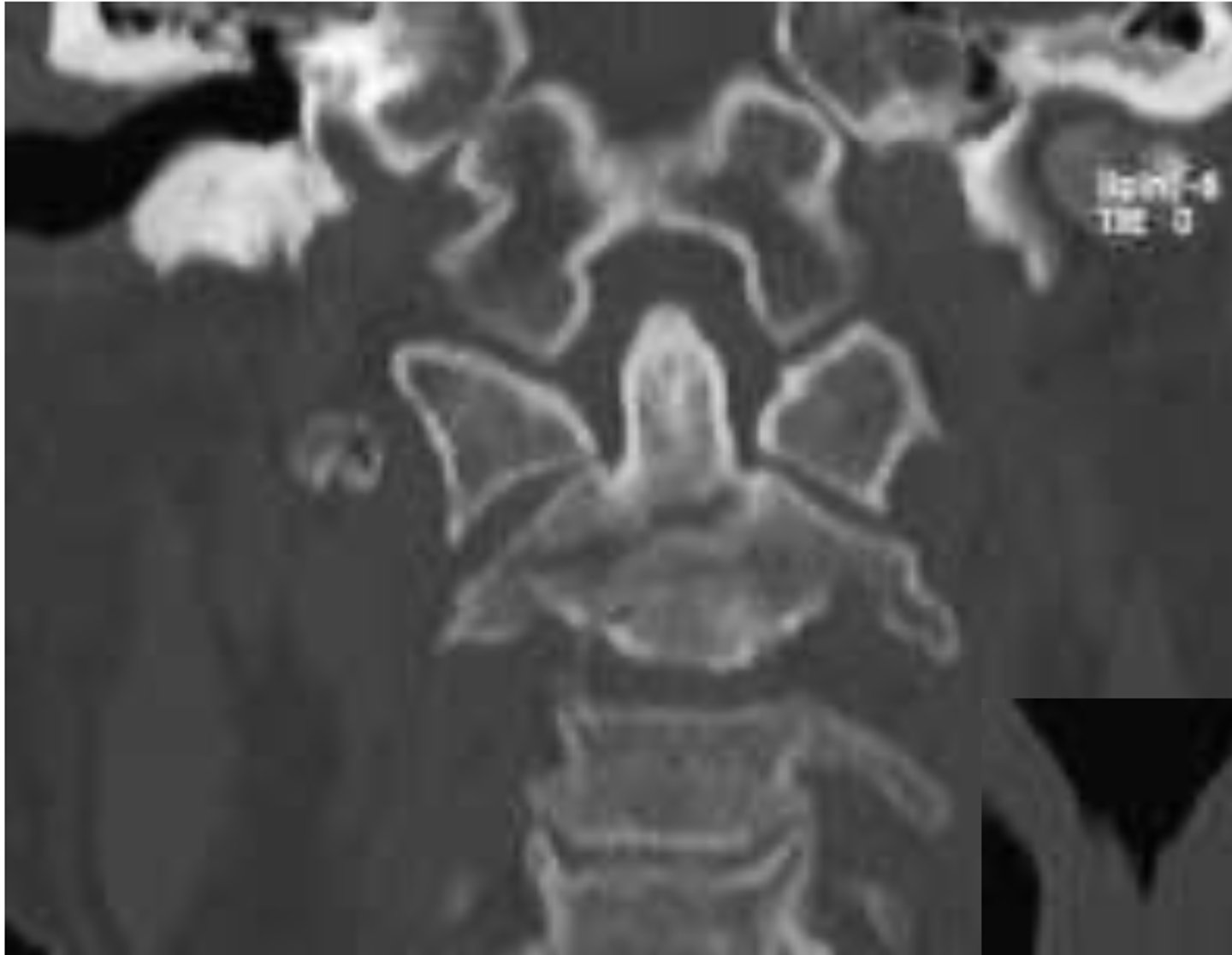
Type 3 Odontoid Fracture
What is the diagnosis?
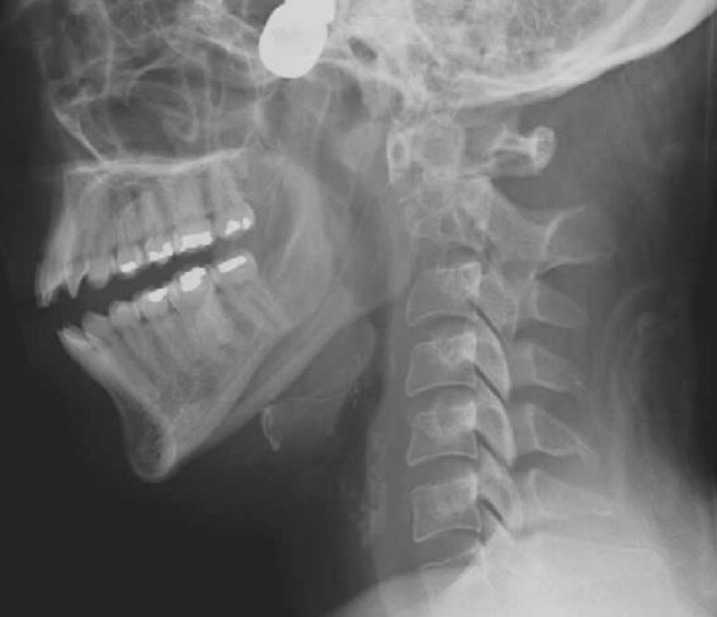
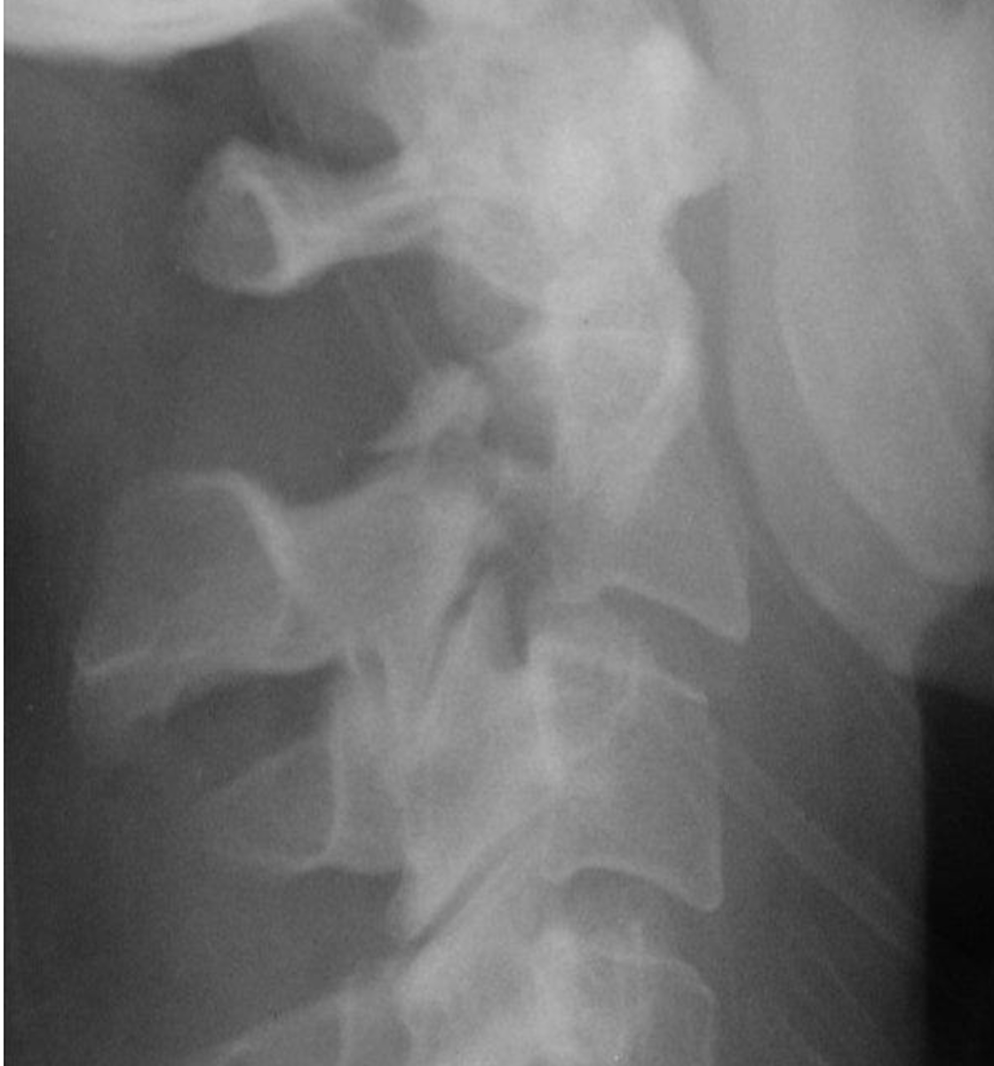
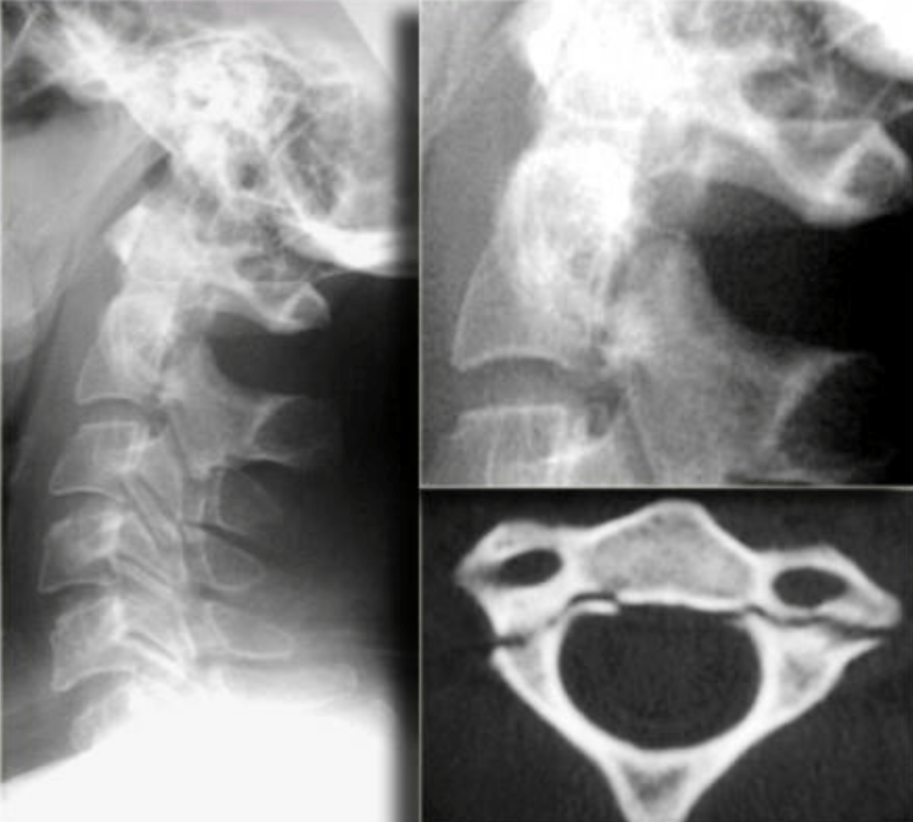
Hangman's Fracture
What is a Traumatic Spondylolisthesis of C2?
Hangman's Fracture
What is a bilateral pedicle fracture of C2 resulting in anterior displacement of C2 due to a hyperextension injury?
Hangman's Fracture
What is the diagnosis?
Hangman's Fracture
What is the diagnosis?
Compression Fracture
What is decreased anterior 1/3 to 2/3 of body height with preservation of posterior body height?
Compression Fracture
Whatis the most common injury to the axial skeleton?
Superior Endplate
What endplate is predominantly deformed in a compression fracture?
True
T/F: A compression fracture is usually stable.
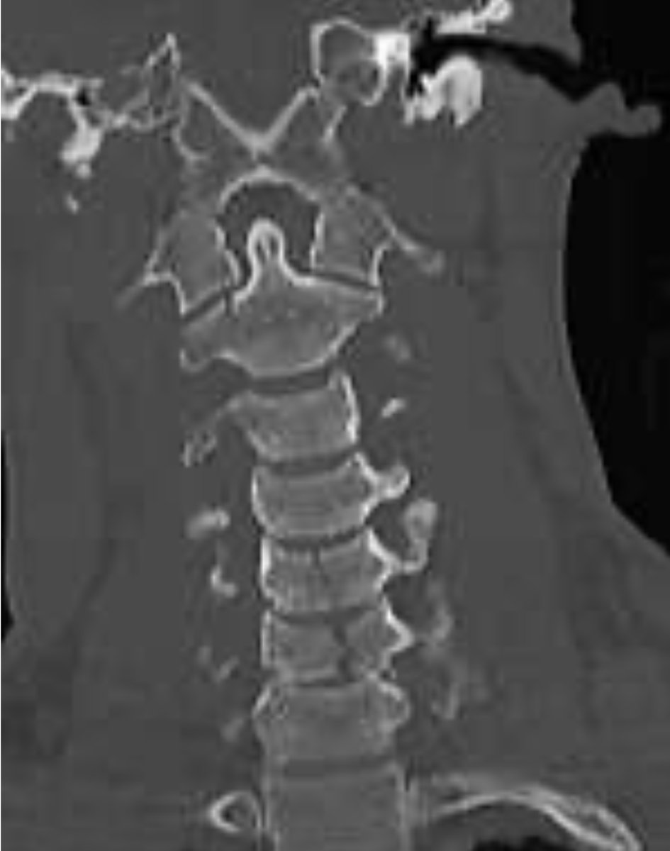
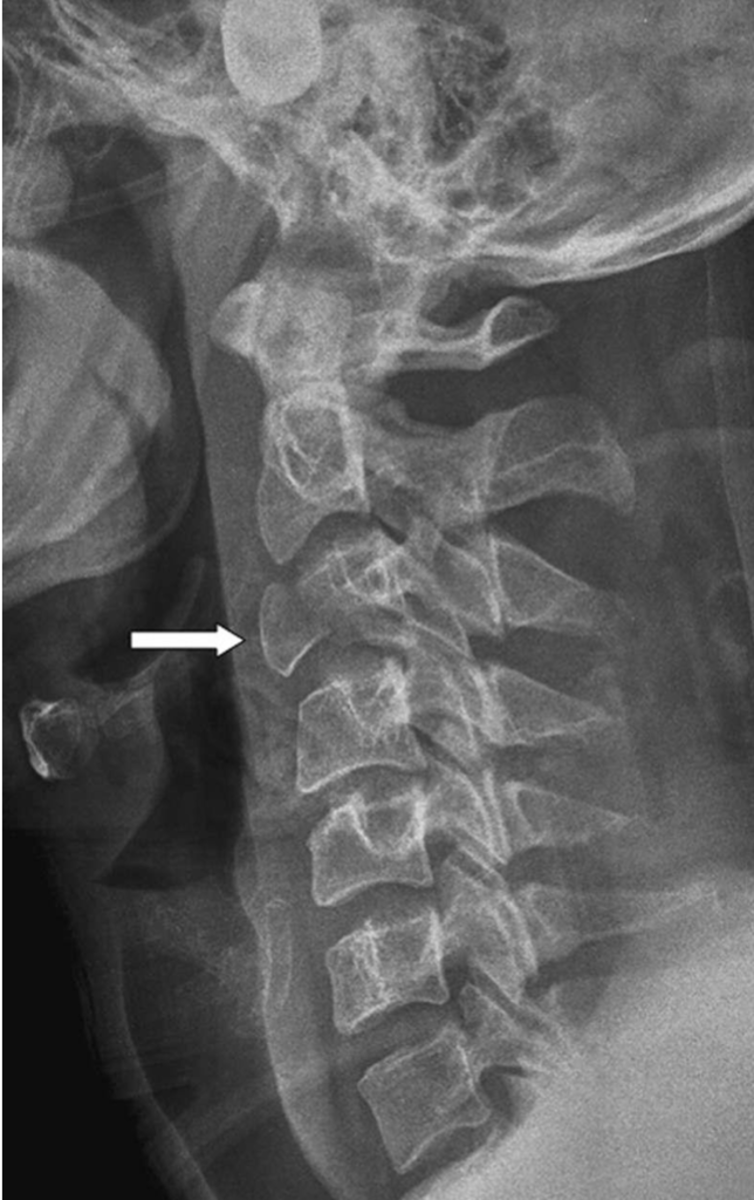
Burst Fracture
What is it when a vertebral body "explodes" into several fragments usually due to axial compression?
False
T/F: A burst fracture is stable.
Burst Fracture
What might you expect if you see a vertical split of the vertebral body on the A-P film, and the lateral shows a comminuted flattened body centrally?
Type 3 Dens fracture & Burst Fractures at C5 & 6
What is the diagnosis?
Burst Fracture at C3
What is the diagnosis?
Flexion & Rotation
What is the mechanism of trauma for a Unilateral Facet Dislocation?
Unilateral Facet Dislocation
If a patient comes in and has loss of rotation of one side what's a likely diagnosis?
Unilateral Facet Dislocation
"Bow Tie Sign" is a finding for what diagnosis?
decrease
A ________ of lamina length is seen in Unilateral Facet Dislocation?
Cervical Oblique
What cervical view is helpful for determining a Unilateral Facet Dislocation?
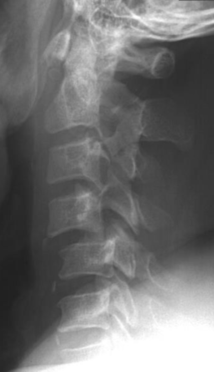
Finding: Bow Tie sign at C4
Diagnosis: C4 Unilateral Facet Dislocation on C5
What is the finding & diagnosis
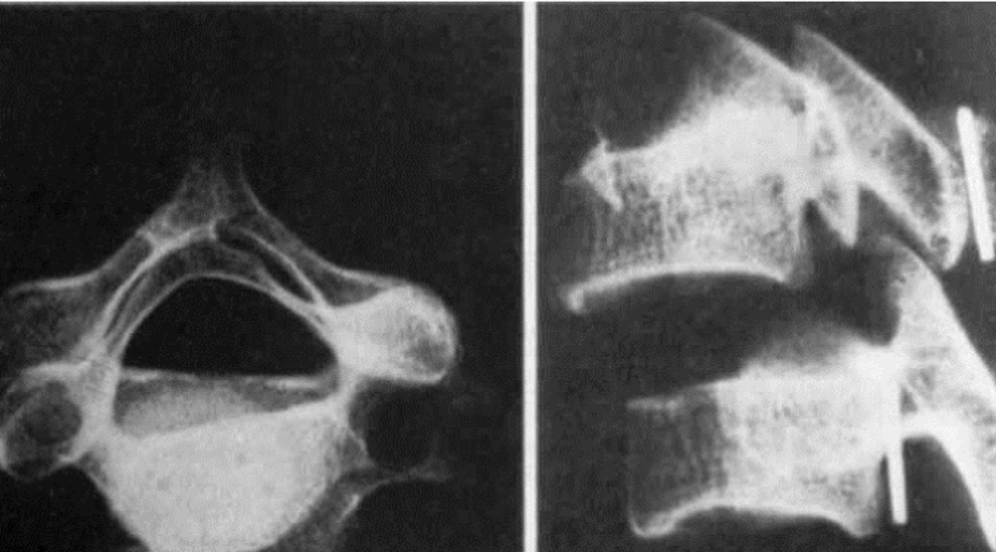
Finding: Bow Tie Sign
Diagnosis: Unilateral Facet Dislocation
What is the finding & diagnosis?
True
T/F: In Unilateral Facet Dislocation the spinous rotates to the same side of the dislocation
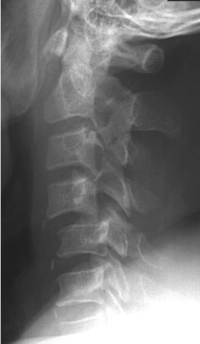
Finding: Bow Tie Sign
Diagnosis: Unilateral Facet Dislocation of C4
What is the finding & diagnosis?
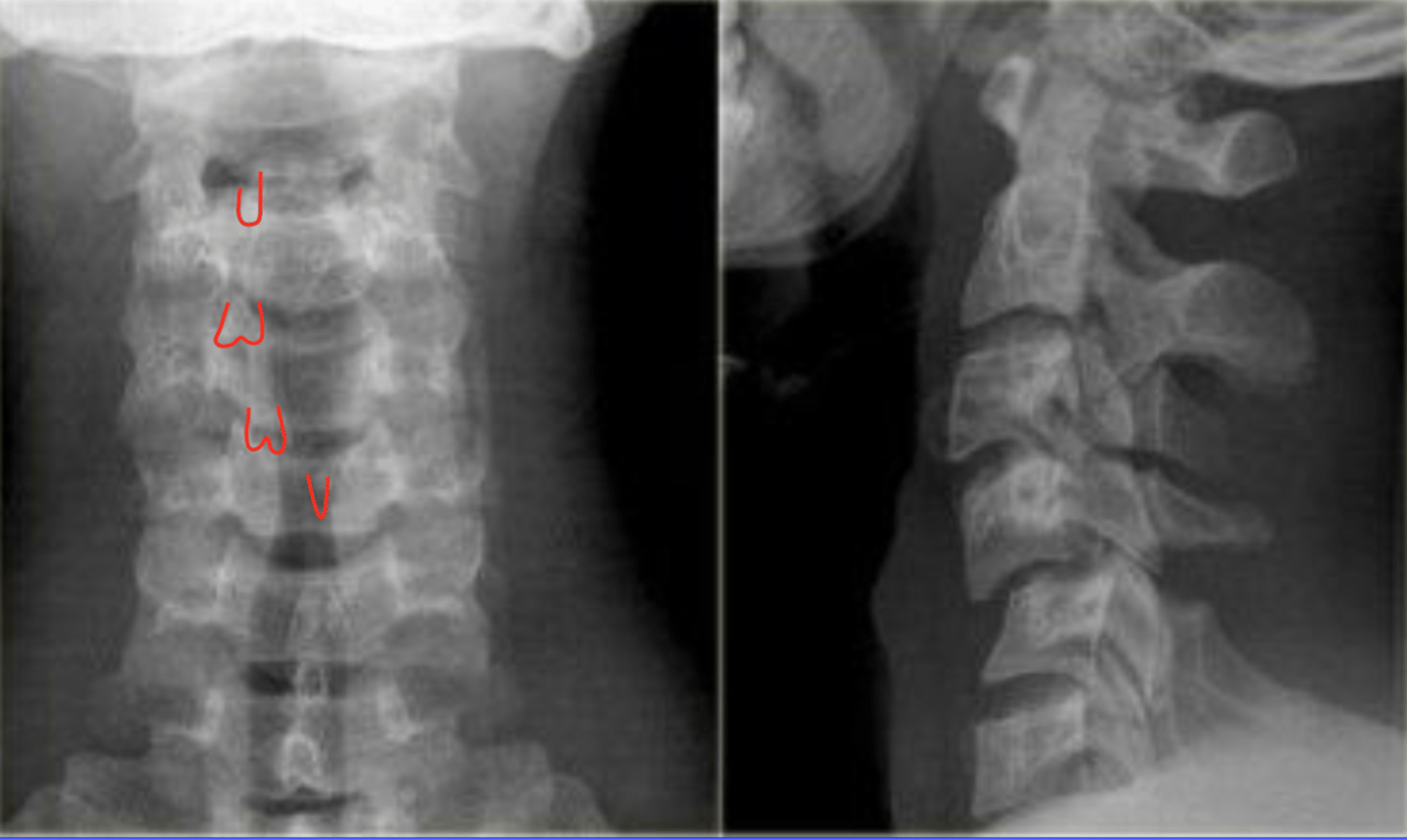
Unilateral Facet Dislocation
If a patient shows you a CT with a "Inverted Hamburger Sign" what is the likely diagnosis?
Hyperflexion
A bilateral facet dislocation is due to what mechanism of injury?
Bilateral Facet Dislocation
What is the diagnosis?
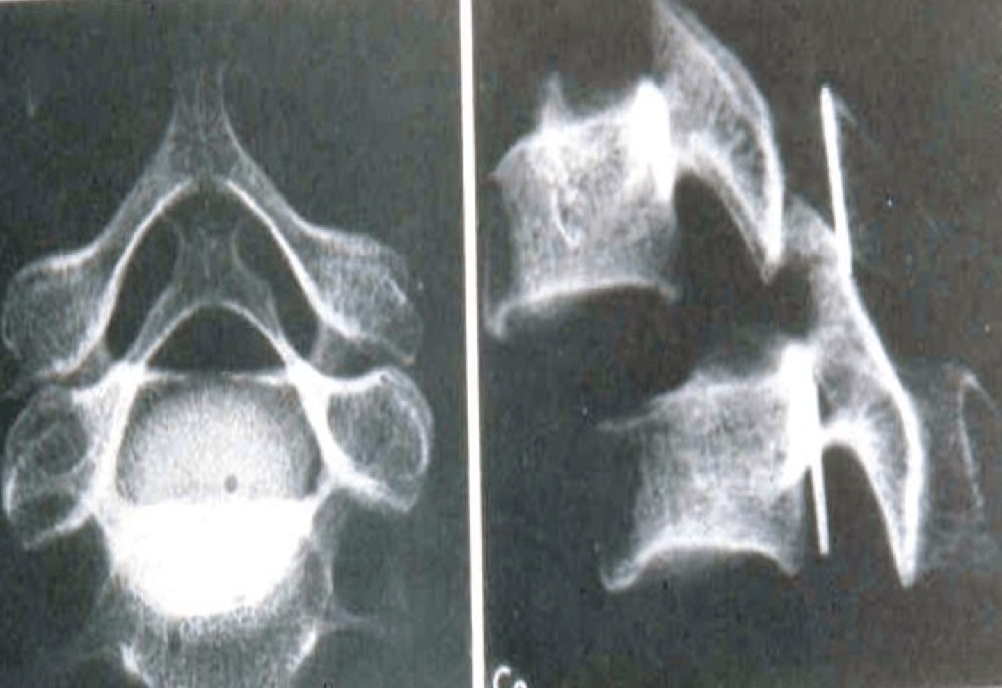
Bilateral Facet Dislocation of C5
What is the diagnosis?
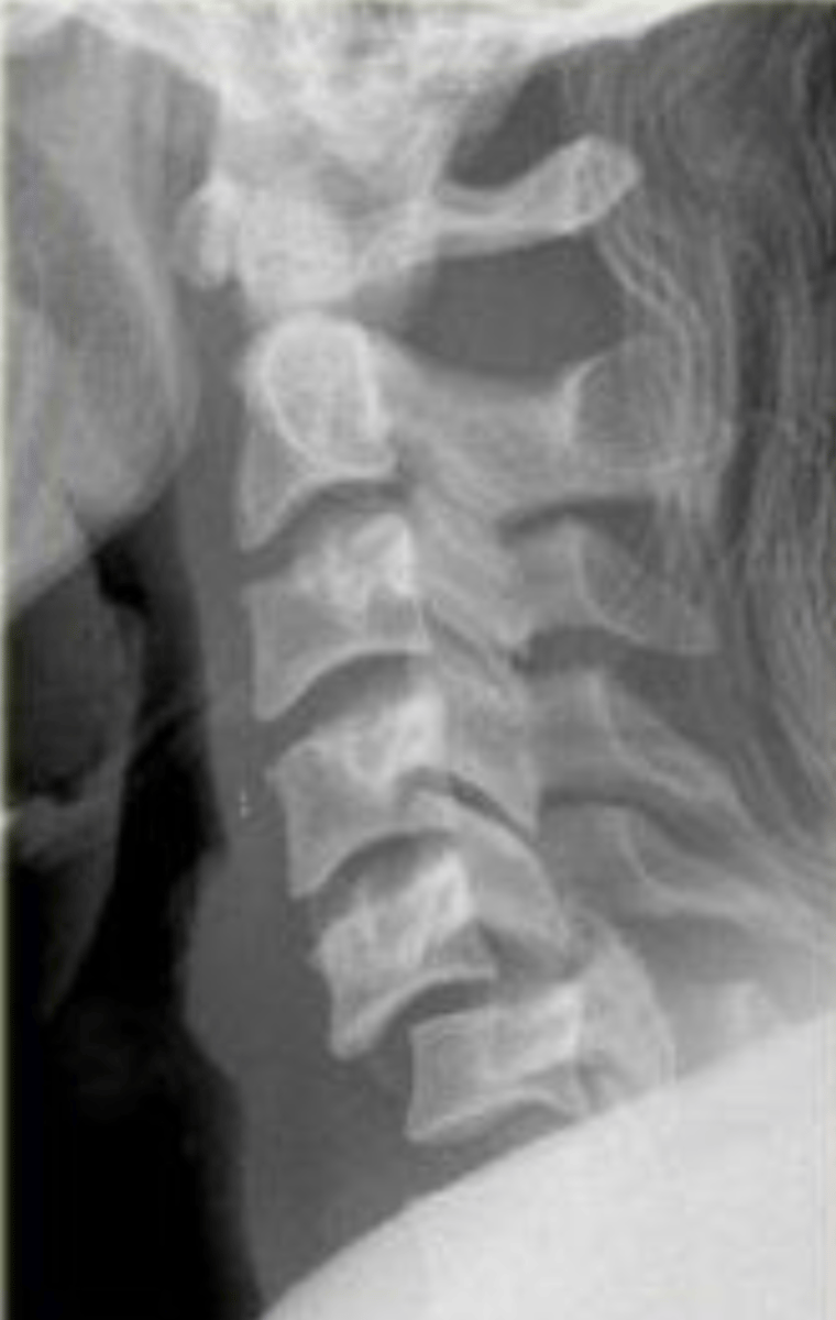
Flexion Teardrop Fracture
What is the most severe injury of the cervical spine?
Flexion Teardrop Fracture
Retropulsion of the vertebral body and a triangular fragment at the anterior aspect is common in what injury?
Flexion Teardrop Fracture at C4
What is the diagnosis?
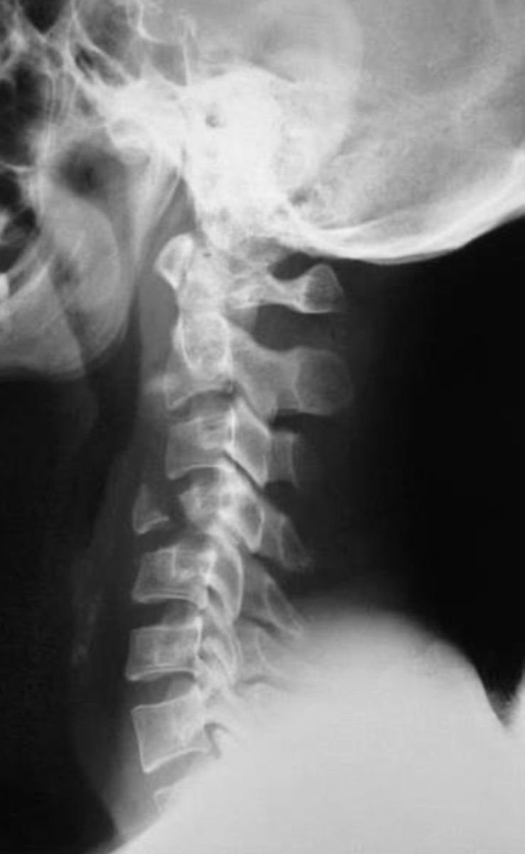
Both will have that anterior fragment, however in a Flexion Teardrop the vertebral body will move posterior.
How do you tell a Flexion Teardrop Fx from an Extension Teardrop Fx?
Ligamentum Flavum
An Extension Teardrop Fracture is usually unstable and due to buckling of what ligament?
Extension Teardrop Fracture of C2
What is the diagnosis?
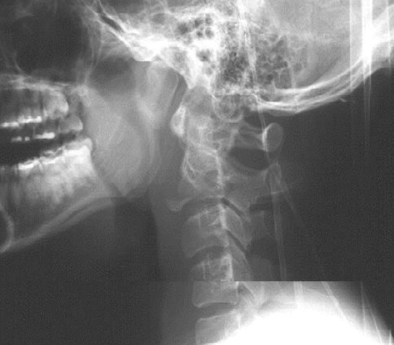
Extension Teardrop Fracture of C3
What is the diagnosis?
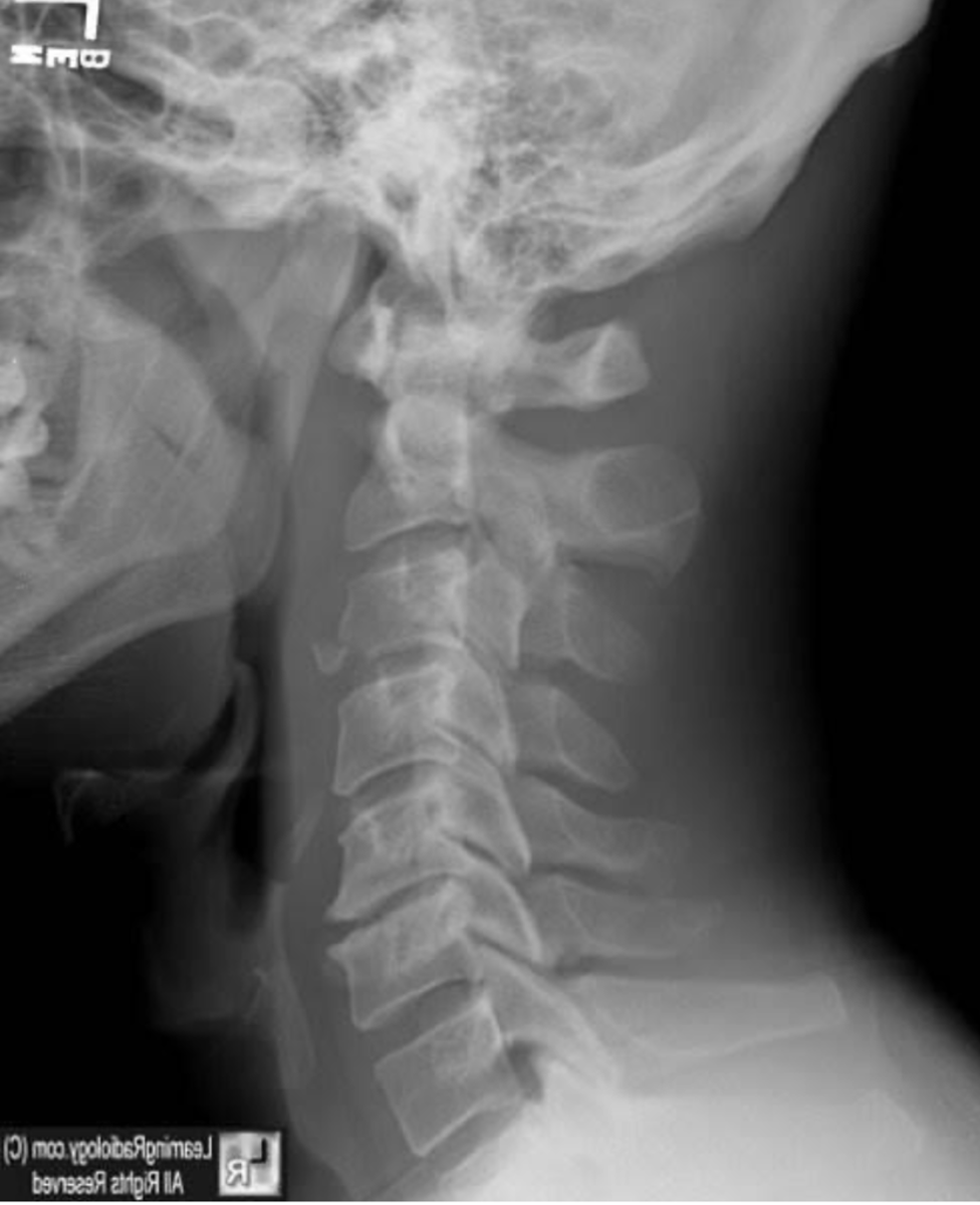
Clay Shoveler's Fracture
What is an avulsion fracture from flexion usually at the base of the spinous of C6, 7 or T1? (aka Root Puller's Fx)
Clay Shoveler's Fracture
A double spinous sign on an AP film is a sign of what?
Finding: Double Spinous Sign
Diagnosis: Clay Shoveler's Fracture at C6
What is the finding & diagnosis?
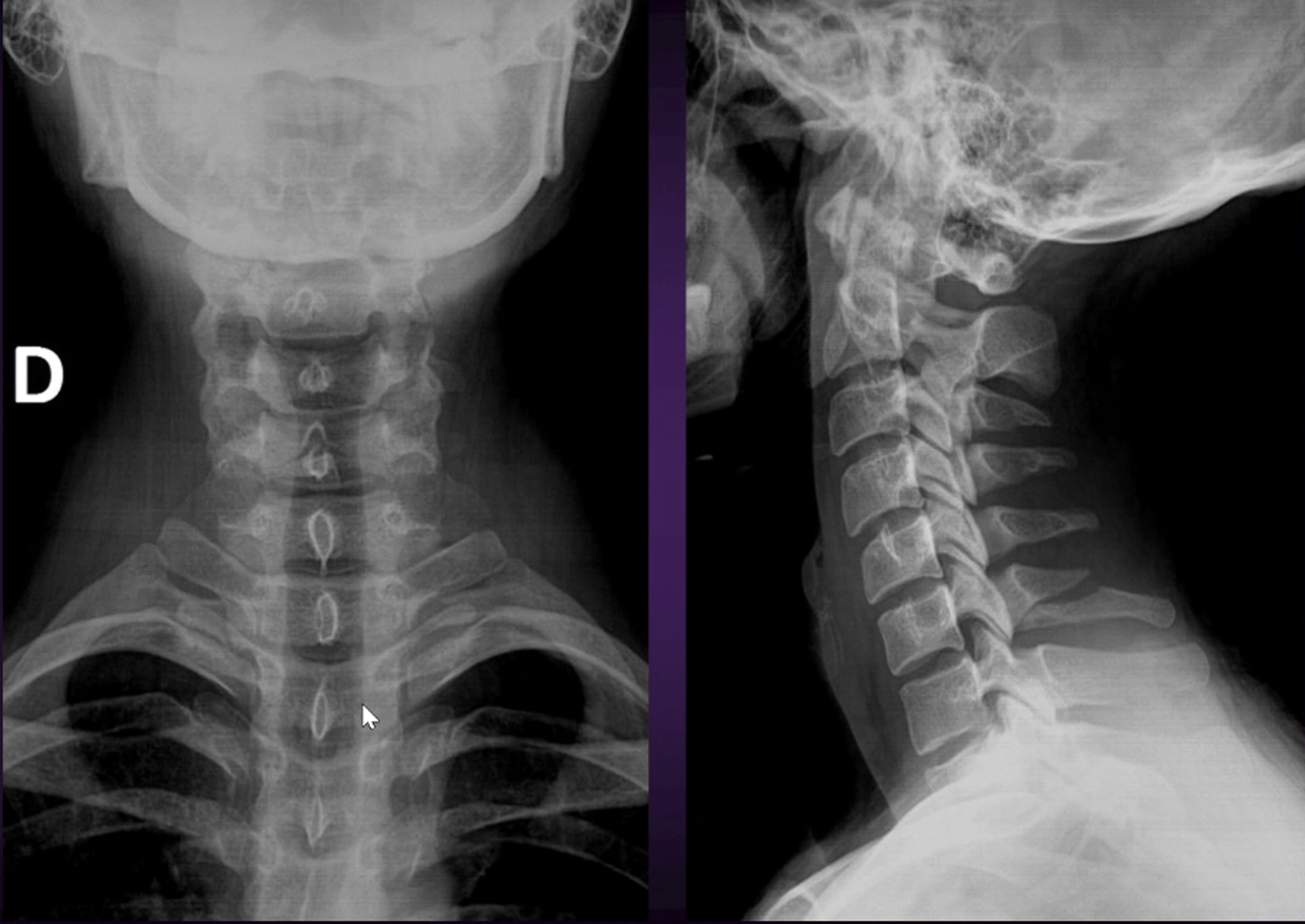
Finding: Double Spinous Sign
Diagnosis: Clay Shoveler's Fracture at C7
What is the Finding & Diagnosis?
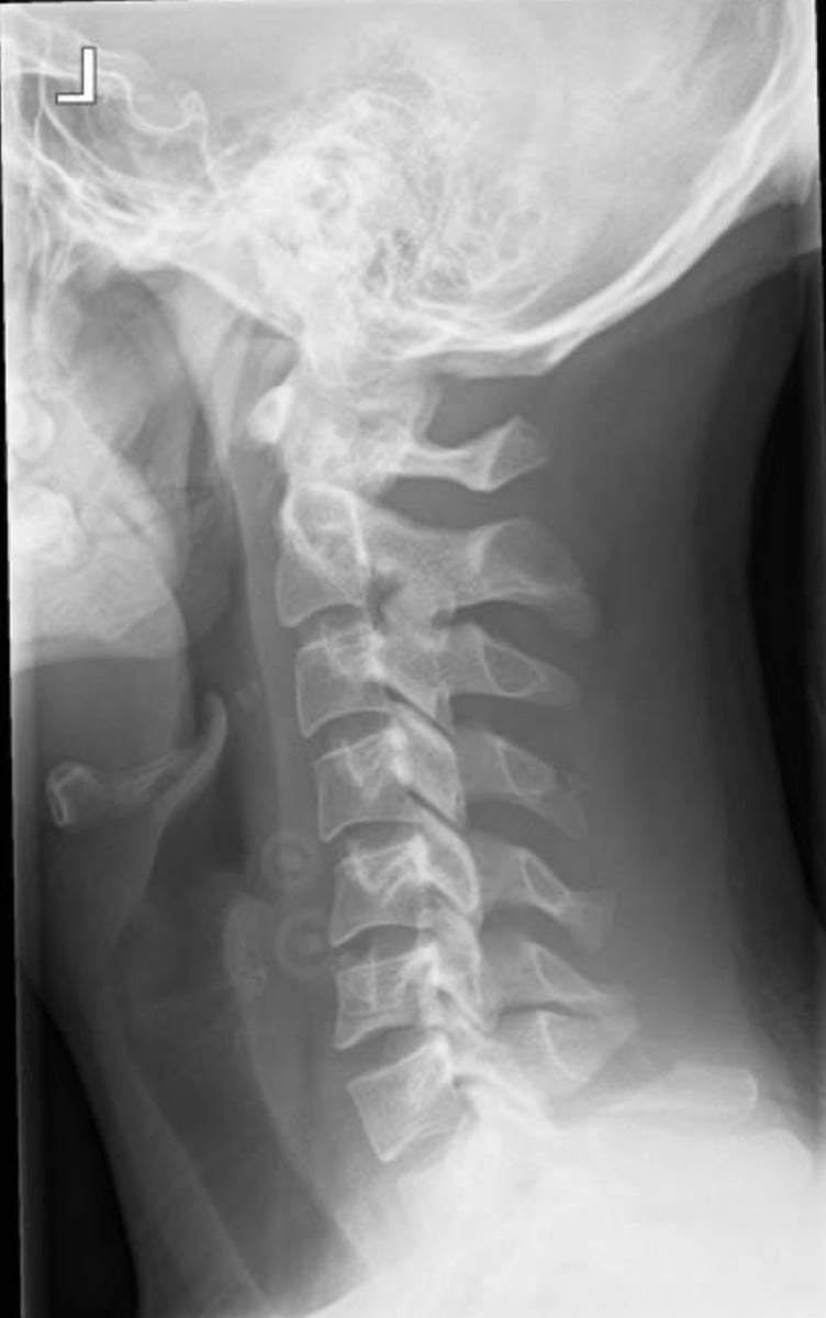
Spinous Fracture at C3
If the mechanism was extension or direct trauma what is the likely diagnosis?
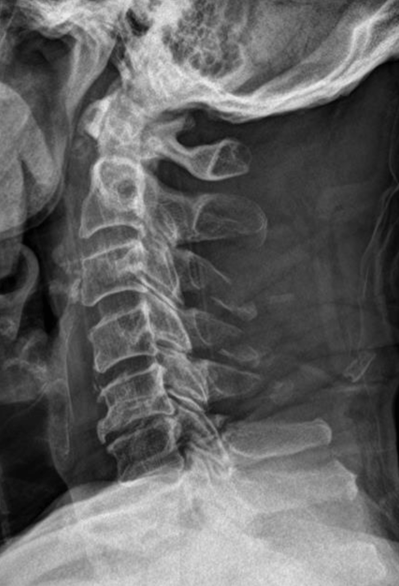
CT
If you suspect a lamina fracture what imaging should you do?
Lamina Fracture
What is the diagnosis?
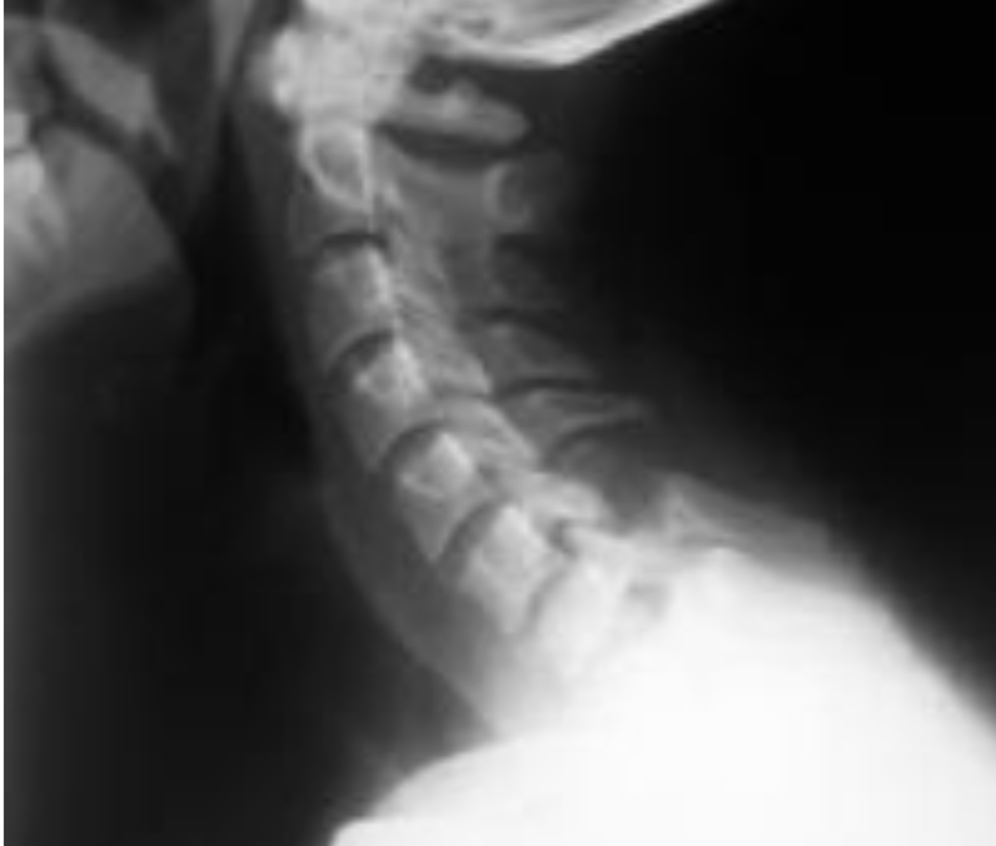
Extension-Compression
A Pillar Fracture is due to what mechanism?
T11-L2
Most thoracic and lumbar fractures occur between what segments?
Compression
What is the most common mechanism of fractures in the Thoracics & Lumbars?
Compression Fracture
If you see anterior wedging in the thoracics and lumbar what is the likely diagnosis?
Superior
A depression of the ______ endplate is usually noticed in a Compression Fracture
Zone of Impaction/ Line of Condensation
The "_________," will present radiographically as a dense white band just below the compressed endplate.
step defect
The "________" is seen on the lateral projection and represents failure of the anterior superior cortex of the vertebral body.
Finding: Zone of condensation and Step Defect on L2
Diagnosis: Acute compression fracture of L2
What are the findings & diagnosis?
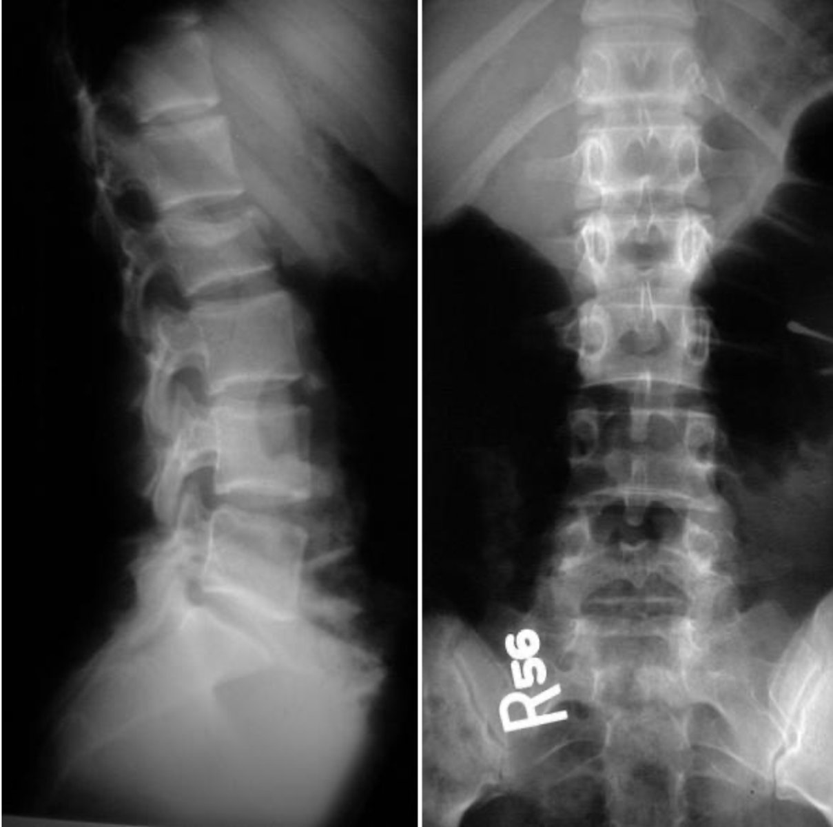
Finding: Step defect and zone of impaction of T11-L2
Diagnosis: Acute compression fractures of T11-L2
What is the findings & Diagnosis?
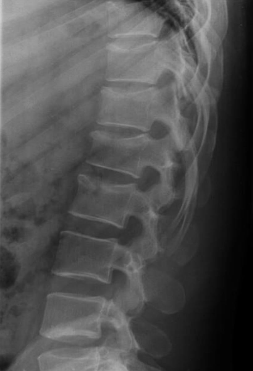
True
T/F: Any posterior body convexity into the canal is a bad thing!
Kyphosis (dowager's hump)
Osteoprototic compression fractures are more common after 50 in females and may increase what normal curvature?