7. Cardiovascular
1/62
There's no tags or description
Looks like no tags are added yet.
Name | Mastery | Learn | Test | Matching | Spaced | Call with Kai |
|---|
No analytics yet
Send a link to your students to track their progress
63 Terms
What are 3 components of cardiovascular system?
Blood, blood vessel and heart
What are blood vessels and what is their primary function?
Blood vessels are tubes that transport blood throughout the body. Their primary function is to circulate blood, carrying essential substances
What types of substances do blood vessels transport?
Blood vessels transport several substances, including oxygen (O2) and carbon dioxide (CO2) for respiratory processes, hormones, immune cells, clotting proteins for regulation and protection, absorbed nutrients from digestion, metabolic wastes to the liver and kidneys, and help in thermoregulation and blood clotting.
What type of organ is the heart, and where is it located?
The heart is a single muscular organ located in the mediastinum, which is the central part of the chest cavity between the lungs.
How many pumps does the heart have, and what is their purpose?
The heart functions as two pumps. The right side pumps deoxygenated blood to the lungs for oxygenation, while the left side pumps oxygenated blood to the rest of the body to supply tissues with oxygen and nutrients.
How many chambers does the heart have?
Four chambers
How much blood is in the average human body, and what percentage of body weight does it represent?
The average human body contains about 5 liters of blood, which represents approximately 8% of a person's body weight.
What is arterial blood, and how does it differ from venous blood?
Arterial blood is blood leaving the heart and is bright red in color due to its high oxygen content (oxyhaemoglobin). In contrast, venous blood is returning to the heart, has a darker color, and contains lower oxygen levels (deoxyhaemoglobin).
What is the cellular portion of blood, and what does it consist of?
The cellular portion of blood makes up about 45% of the blood volume. It includes:
Erythrocytes (red blood cells): These cells transport oxygen from the lungs to the tissues.
Leukocytes (white blood cells): These cells are involved in immune responses and protecting the body against infections.
Platelets: These are involved in blood clotting to prevent excessive bleeding.
What is plasma, and what are its main components?
Plasma makes up about 55% of blood volume. It is mostly water and contains dissolved solutes such as ions, plasma proteins (e.g., albumin, globulins, fibrinogen), and other components like metabolites, hormones, enzymes, and antibodies.
What is the size and weight of the heart?
The heart is approximately the size of a fist and weighs between 250-350 grams (less than 1 pound).
Where is the heart located in the body?
The heart is located in the mediastinum, which is the region between the two lungs. It rests on the diaphragm and is positioned slightly tilted to the left.

What structures protect the heart?
The heart is protected by rigid structures including the ribs, sternum, and vertebral column. These offer protection from physical impact, and the ribs, in particular, allow for the necessary movement during CPR.
Which part of the heart is tilted to the left?
The heart is slightly tilted to the left, with the apex (the tip) located at the left ventricle.
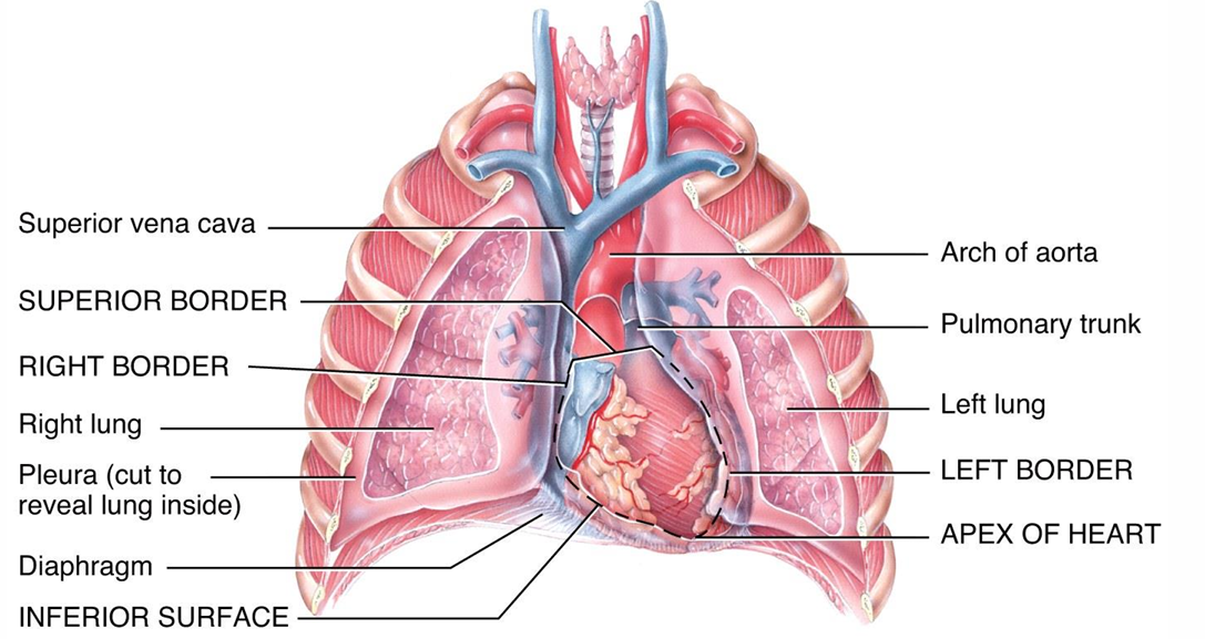
What forms the posterior surface of the heart?
The posterior (towards the back) surface of the heart is primarily formed by the atria (the upper chambers of the heart).
Where is the anterior surface of the heart located?
The anterior surface of the heart is deep to the sternum and ribs, positioned towards the front of the chest.
What is the base of the heart, and where does it face?
The base of the heart is the upper portion, and it faces upward, backward, and to the right. The inferior surface (bottom part of the heart) faces the diaphragm, and it has a fibrous attachment to it.
Where is the right border of the heart located?
The right border of the heart is almost vertical and lies along the right lung.

Where is the left border of the heart located?
The left border of the heart extends from the base of the heart to the apex (tip of the left ventricle).

What type of muscle is the myocardium, and how does it function?
The myocardium is involuntary striated muscle
What is the myocardium, and where is it located in the heart?
The myocardium is the middle layer of the heart wall, positioned between the outer epicardium (outer layer) and the inner endocardium (inner lining). It is the thickest layer and plays the primary role in the heart's contraction.

What types of cells does the myocardium consist of?
99% contractile cells
1% autorhythmic cells
What is the pericardium, and what are its layers?
The pericardium is a double-walled sac that encloses the heart and the roots of the great vessels. It has two layers:
Serous layer: A thin, inner layer that produces serous fluid.
Fibrous layer: A tough, outer layer that provides structural support and protection.

What is the epicardium, and what tissues make it up?
The epicardium is the visceral layer of the pericardium that directly covers the heart. It consists of adipose (fat) tissue and fibro-elastic tissue, which help cushion and protect the heart.

How does the pericardium help protect the heart?
The pericardium protects the heart by fixing it in place within the mediastinum, providing protection against infection, and offering lubrication to reduce friction as the heart beats.
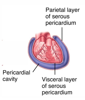
What is the function of the pericardial cavity and pericardial fluid?
The pericardial cavity contains pericardial fluid, which provides lubrication to reduce friction between the heart and surrounding tissues during the heart's contraction and relaxation.

What is the myocardium composed of, and what is its role?
The myocardium is made up of muscle tissue that contracts to pump blood through the heart. It forms the thick middle layer of the heart wall and plays a crucial role in the heart's pumping action.

What is the endocardium, and where is it located?
The endocardium is the inner lining of the heart, composed of endothelial cells. It provides a smooth surface for blood to flow through and also covers the heart valves, ensuring smooth and efficient circulation.
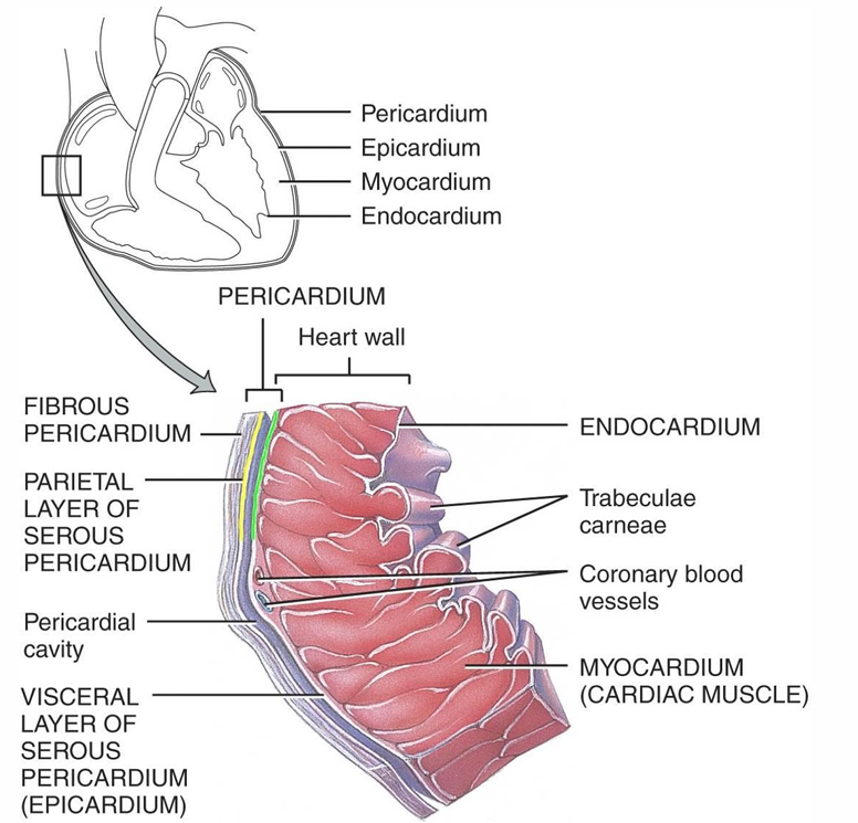
What is the fibrous pericardium, and where is it located?
The fibrous pericardium is the outermost layer of the pericardium. It is made of dense connective tissue and is located around the heart, anchoring it to the diaphragm and great vessels.
What is the primary function of the fibrous pericardium?
The fibrous pericardium provides protection to the heart and prevents overfilling of the heart by limiting its expansion.
What is the serous pericardium, and how is it structured?
The serous pericardium consists of two continuous layers separated by the pericardial cavity, which contains serous pericardial fluid
The parietal layer is attached to the fibrous pericardium.
The visceral layer is attached directly to the surface of the heart (also called the epicardium).
What is pericarditis, and what types exist?
Pericarditis is the inflammation of the pericardium. It can be classified as:
Acute pericarditis: Sudden onset inflammation of the pericardium.
Chronic pericarditis: Long-lasting inflammation of the pericardium.
Cardiac tamponade: A life-threatening condition caused by fluid accumulation in the pericardial cavity, which compresses the heart and impairs its ability to pump.
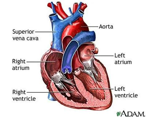
What type of blood does the atrium receive in pulmonary circulation, and where does this blood come from?
In pulmonary circulation, the right atrium receives deoxygenated blood from the systemic and coronary circulation
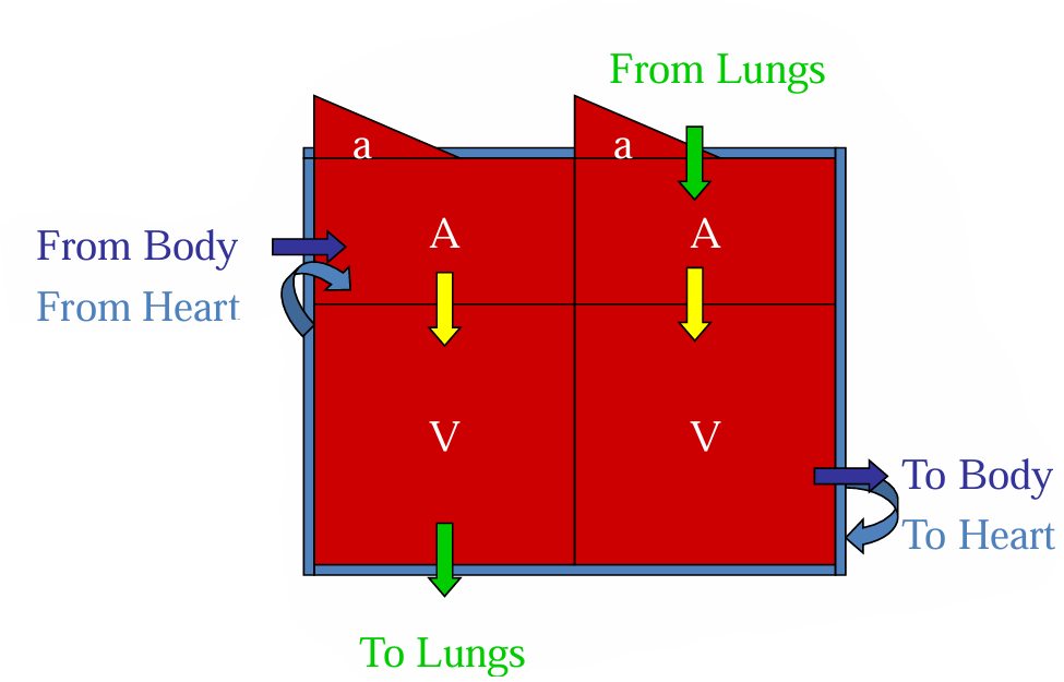
Where does blood go after it passes through the atrium in pulmonary circulation?
After deoxygenated blood passes through the right atrium, it moves into the right ventricle. From there, it is pumped to the lungs for oxygenation.

What role does the left side of the heart play in systemic and coronary circulation?
The left side of the heart functions as the pump for systemic and coronary circulation.
Where does the left atrium receive blood from, and what type of blood is it?
The left atrium receives oxygenated blood from the pulmonary circulation (the lungs).
After passing through the left atrium, where does blood flow next, and what happens to it?
After passing through the left atrium, blood flows into the left ventricle. The left ventricle then pumps oxygenated blood to the body and heart muscle via the systemic and coronary circulation.

What is coronary circulation?
Coronary circulation is the flow of blood through the blood vessels of the heart muscle (myocardium). This circulation supplies the myocardium with oxygenated blood and removes deoxygenated blood from it.
What are the coronary arteries, and what is their function?
Coronary arteries are the blood vessels that deliver oxygen-rich blood to the myocardium (heart muscle), ensuring it has the oxygen needed for contraction.
What are cardiac veins, and what role do they play in coronary circulation?
Cardiac veins are the vessels that remove deoxygenated blood from the heart muscle (myocardium), carrying it back to the right atrium.
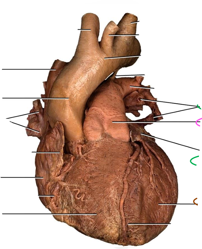
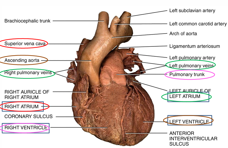
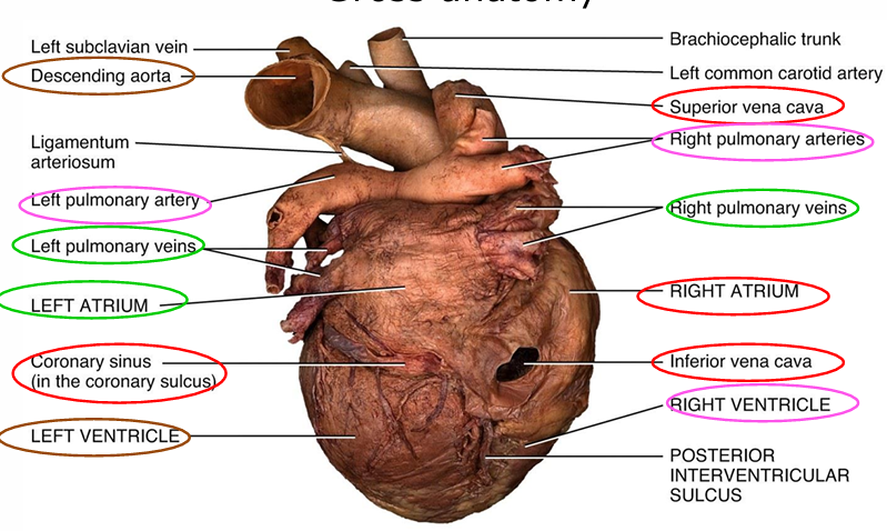
Name different part from (anterior external view)
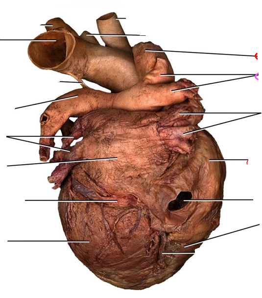
Name the different parts from (posterior external view)
Superior vena cava
Coronary sinus
Pulmantory arteries
What are the main structural features of the right atrium?
The right atrium has a smooth posterior wall, while the rest of the wall is ridged by pectinate muscles. It also has a small outpouching called the auricle and receives blood from the superior vena cava, inferior vena cava, and coronary sinus.
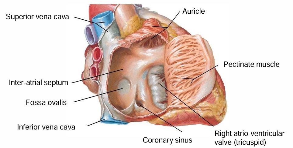
What is the fossa ovalis, and where is it located?
The fossa ovalis is a depression in the inter-atrial septum (wall between the right and left atria) and is a remnant of the embryonic inter-atrial circulation, where it allowed blood flow between the atria before birth.

What valve regulates the opening between the right atrium and right ventricle?
The right atrio-ventricular (tricuspid) valve regulates the opening between the right atrium and right ventricle. It ensures unidirectional blood flow from the atrium to the ventricle.

What is the role of the tricuspid valve, and how does it function?
The tricuspid valve separates the right atrium and right ventricle and consists of three cusps of connective tissue. It opens when papillary muscles contract, pulling on the chordae tendinae to allow blood flow from the atrium to the ventricle.
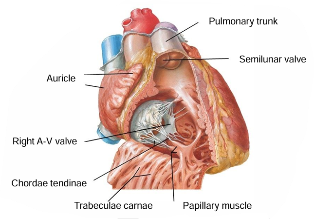
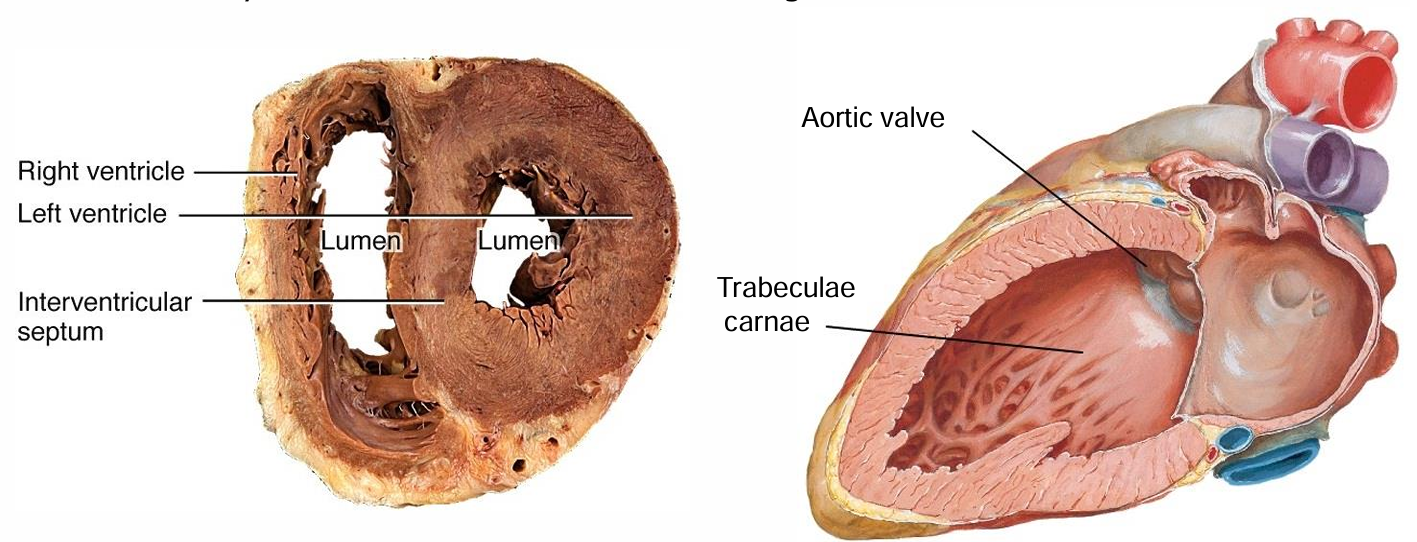
What are trabeculae carnae, and where are they located?
Trabeculae carnae are ridged muscle bands located in the inflow tract of the right ventricle. They provide structural support and help prevent the walls from sticking together during contraction.

What type of blood does the right ventricle receive, and where does it pump this blood?
The right ventricle receives deoxygenated blood from the right atrium and pumps it through the pulmonary valve into the pulmonary trunk, which carries the blood to the lungs for oxygenation.

What is the function of the pulmonary valve, and where does it lead?
The pulmonary valve is a semilunar valve that controls blood flow from the right ventricle into the pulmonary trunk. It prevents backflow of blood into the ventricle after it is pumped out.

How is the pulmonary trunk related to the pulmonary arteries, and what is unique about these arteries?
The pulmonary trunk splits into the right and left pulmonary arteries, which carry deoxygenated blood to the lungs. These are the only arteries in the body that carry deoxygenated blood.
What are the main structural features of the left atrium?
The left atrium has a smooth posterior and anterior wall. It also has a small outpouching called the auricle, which contains pectinate muscles. The left atrium receives oxygenated blood from the four pulmonary veins—two from each lung.

What valve regulates the flow of blood between the left atrium and the left ventricle?
The left atrio-ventricular (mitral or bicuspid) valve regulates blood flow between the left atrium and left ventricle. It ensures one-way flow from the atrium to the ventricle.
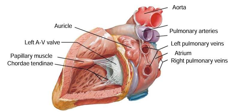
How does the wall thickness of the left ventricle compare to other parts of the heart, and why is it thicker?
The wall of the left ventricle is the thickest part of the heart. It needs to be thicker to generate the higher pressure required to pump blood through the systemic circulation, reaching all parts of the body.
What structures are found in the inflow and outflow tracts of the left ventricle?
The inflow tract of the left ventricle contains trabeculae carnae (rugged muscle ridges), while the outflow tract is smooth to facilitate blood flow into the aorta.
What role do the coronary arteries play, and where do they originate?
The coronary arteries branch off from the ascending aorta and supply oxygenated blood to the heart muscle (myocardium), ensuring it has the oxygen and nutrients needed to function effectively.
What is the structure of the mitral valve, and how does it function?
The mitral valve (also called the bicuspid valve) has two cusps made of connective tissue. It opens when the papillary muscles contract, pulling on the chordae tendinae, which prevents the valve from inverting during ventricular contraction.

What is the function of the aortic valve, and where is it located?
The aortic valve is a semilunar valve that controls the flow of blood from the left ventricle into the aorta. It prevents backflow of blood into the left ventricle after it is pumped out.

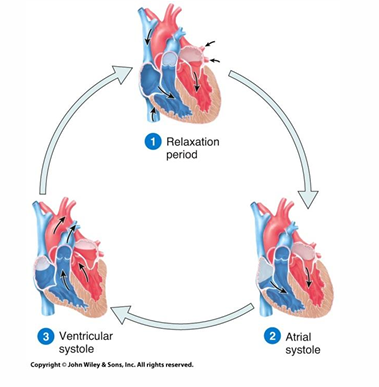
What happens during the relaxation period of the cardiac cycle?
During the relaxation period, the ventricles are relaxed and blood flows passively from the atria into the ventricles.

What is atrial systole, and what is its purpose in the cardiac cycle?
Atrial systole is the phase where the atria contract to completely fill the ventricles with blood before ventricular contraction.

Describe ventricular systole and its role in the cardiac cycle.
During ventricular systole, the ventricles contract to push blood out of the heart into the arteries, sending oxygenated blood to the body and deoxygenated blood to the lungs.
What is the SA (sinoatrial) node, and what function does it serve in the heart's conduction system?
The SA (sinoatrial) node is the heart's natural pacemaker, generating an action potential that initiates and synchronizes the contraction of both atria.
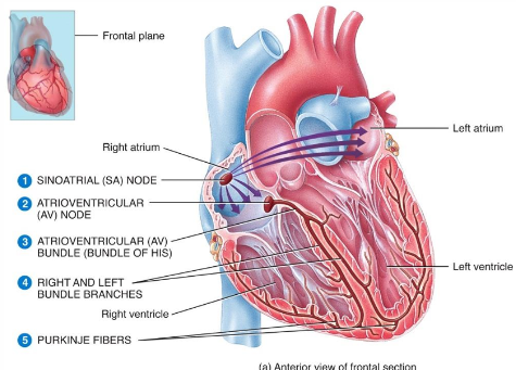
What is the purpose of the delay in the signal from the SA node to the AV (atrioventricular) node?
The delay allows the atria to fully contract and transfer blood into the ventricles before the ventricles contract, ensuring efficient blood flow through the heart.
How does the AV (atrioventricular) node signal affect the ventricles?
After the delay, the AV node sends the electrical signal throughout the ventricles, causing them to contract and expel blood out of the heart.
