Looks like no one added any tags here yet for you.
The cell envelope
Consists of a series of layered structures that surround the cytoplasm and govern cellular interactions with the external environment. Consists of:
Cytoplasmic membrane
Cell wall
Outer membrane
S-layers
The cytoplasmic membrane
Surrounds the cytoplasm
Is selectively permeable
In order for a cell to grow, nutrients must be transported inwards and waste products outwards.
Bacterial cytoplasmic membrane
Is a phospholipid bilayer containing embedded proteins
Is 8-10 nanometers wide
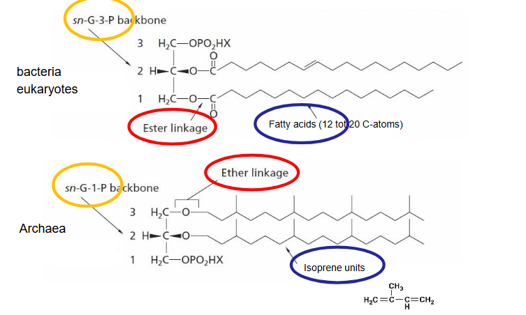
Phospholipids in bacteria cytoplasmic membrane.
Contain a hydrophobic and hydrophilic part.
Hydrophobic part consists of fatty acid chains
The hydrophilic part consists of a glycerophosphate (glycerol molecule bound to a phosphate) and one of several other functional groups also bonded to the phosphate.
Hydrophobic part and hydrophilic part are connected through an ester linkage.
Membrane proteins in bacteria cytoplasmic membrane
Transmembrane proteins: Extend completely across the membrane
Peripheral proteins: are more loosely attached.
Some peripheral membrane proteins are lipoproteins, proteins that contain a hydrophobic lipid tail that anchors the protein into the membrane.
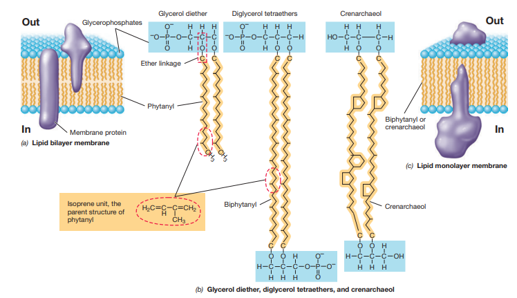
Archaeal cytoplasmic membranes
Similar to those of bacteria and Eukarya (phospholipid membrane)
Chemistry is slightly different.
Hydrophobic part is instead isoprenoid, which are bound to glycerol by ether bonds.
Isoprenoid is formed from repeating units of the five-carbon hydrocarbon isoprene
Archaeal mono vs. bilayer
When its a monolayer the hydrophobic part is two strands of phytanyl
Some Archaea can also have lipid monolayers composed of diphosphoglycerol tetraether lipids or crenarchaeol.
Cytoplasmic membrane function
Permeability barrier: preventing the passive leakage of solutes into or out of the cell
The cytoplasmic membrane anchors several proteins that catalyze a suite of key cell functions
The cytoplasmic membrane of Bacteria and Archaea plays a major role in energy conservation and consumption.
Selective permeability in the cytoplasmic membrane
Is a barrier to the diffusion of most substances, especially polar or charged molecules.
Charged molecules can’t go through because hydrophilic part is charged
Most substances that enter or leave the cell must be carried in or out by transport proteins.
Can go against concentration gradient
Three types of active transporters
Simple transport system
Group translocation
ABC transport systems
Are all energy driven
Simple transport system
Are driven by the energy inherent in the proton motive force
Can either have symport reactions (a solute and a proton are cotransported in the same direction)
Or an antiport reaction (where a solute and a proton are transported in opposite directions)
e.g. uptake of lactose by lac permease which is a symporter in E. coli.
How is group translocation different from simple transport?
The transported substance is chemically modified during the transport process
An energy-rich organic compound drives the transport event (rather than proton motive force)
e.g. uptake of glucose, mannose and fructose in E. coli.
These compounds are phosphorylated by the phosphotransferase system.
ABC transporter system
Are modular systems that have three components:
A binding protein
a transmembrane protein channel and
an ATP hydrolyzing protein.
ABC stands for: ATP, binding, cassette. which is a structural feature of proteins that bind ATP
Peptidoglycan
Is found in Bacteria cell walls.
Unique to bacteria
Is a polysaccharide
Structure peptidoglycan
The sugar backbone of peptidoglycan is composed of alternating repeats of two modified glucose residues called N-acetylglucosamine and N-acetylmuramic acid.
These are joined by a Beta-1,4 linkage.
Attached to the latter residue is a short peptide side chain. The amino acid composition can vary considerably.
Strands of peptidoglycan run parallel to each other around the circumference of the cell.
The side chains of adjacent peptidoglycan strands are cross-linked together by covalent peptide bonds.
In this way, the peptidoglycan forms one single enormous molecule.
Cell wall in the gram-negative cell envelope
Is 2-7 nm thick
A single layer of peptidoglycan although it can be up to three layers thick in some places
The peptidoglycan mesh so formed is flexible and porous but strong enough to resist turgor pressure and prevent rupture of the cytoplasmic membrane and cell lysis.
Does not have a pentaglycine bridge
Cell wall in the gram-positive cell envelope
Contains a thick peptidoglycan cell wall, (20-35 nm)
Is much thicker than the wall of gram negative organisms
As much as 90% of the gram-positive cell envelope can consist of peptidoglycan - which is 15 or more layers (the gram-negative cell wall typically contains only a single layer of peptidoglycan)
Is stabilized by three dimensional peptide cross-links, which form between adjacent peptidoglycan strands both horizontally and vertically.
With pentaglycine between chains
What gives strength and stability to the membrane in eukaryotes and prokaryotes?
Eukaryotes: Sterols (Cholesterol)
Prokaryotes: Hopanoids
Membrane bacteria vs. archaea
What is “Proton Motive Force”?
A source of energy resulting from the transport of protons across the cytoplasmic membrane, generating a membrane electrochemical potential.
Teichoic acids
Found in gram-positive bacteria embedded in the ell wall.
Are composed of glycerol phosphate or ribitol phosphate with attached molecules of glucose or D-alanine.
Individual alcohol molecules are then connected through their phosphate groups to form long strands, and these are then covalently linked to peptidoglycan
Are responsible for the overall negative charge of the cell surface. They give flexibility to the cell wall.
Lysozyme
Peptidoglycan can be destroyed by lysozyme
An enzyme that cleaves the glycosidic bond between N-acetylglucosamine and N-acetylmuramic acid.
This weakens the peptidoglycan and can cause cell lysis.
Lysozyme is present in human secretions including tears, saliva and other bodily fluid, and functions as a major line of defense against bacterial infection.
Many antibiotics target Ppeptidoglycan
Periplasm
Found in gram negative cell wall
Is the space between membranes
Contains many extracellular proteins all synthesized in the cytoplasm:
Hydrolytic enzymes
Binding protein
Chemoreceptors.
Lipopolysaccharide (LPS)
Found on top of cell wall
Give negative charge
Give rigidity to membrane structure
Protect the cell against attacks from outside
Lipid A is oftne poisonous (endotoxine)
Cell wall in Archaea
Have no peptidoglycan and no outer membrane
Some methanogenic Archaea have pseudomureine.
Many Archaea have S-layers - a paracrystalline structure of proteins or glycoproteins.
Cell wall of some methanogenic Archaea
Has pseudomureïne
Pseudomureïne is a polysaccharide similar to peptidoglycan composed of acetylglucosamine and N-acetyltalosaminuronic acid
Has beta 1-3 glycosidic bonds instead of beta 1-4
Slime layer
A slime layer may be present on the outside of the cell wall
Consists of polysaccharides
There are 2 types:
Capsule: if tightly attached, tight matrix
Slime layer: loosely attached, easily deformed
Function of slime layer
Protection dependent on environmental conditions
Assists in attachment to surfaces
Role in development of biofilms
Prevent dehydration/desiccation
Pili
Think (2-10 nm diameter) filamentous structures made of protein that extend from the surface of a cell and can have many functions.
Play a role in DNA exchange between cells (conjugation)
Play a role in transport of electrons
Type IV pili needed for “twitching motility”
Fimbriae
Short pili that mediate attachment
Enable bacterial cells to stick to surfaces to form pellicles (thin sheets of cells on a liquid surface) or biofilms on solid surfaces.
Hami
Certain archaea have a special type of pilus, called hamus
Resemble type IV pili, but with a spiky end (hook)
Used for attachment to surfaces and each other (making biofilms)
What are cell inclusions and what are their functions?
Cell inclusions are considered various nutrients or pigments that can be found within the cell, but do not have activity like other organelles
Their functions include:
Energy reserves
Carbon resevoirs
And/or have special functions
Enclosed by thin membrane
Reduce osmotic stress
Carbon storage polymers
A type of inclusion body in a prokaryotic organism
An example: poly-B-hydroxybutyric acid (PHB)
Another example: glycogen: polymer of glucose
Polyphosphate granules
Many prokaryotic and eukaryotic microbes accumulate inorganic phosphate in the form of polyphosphate granules.
These granules are formed when phosphate is in excess and can be drawn upon as a source of phosphate for nucleic acid and phospholipid biosynthesis when phosphate is limiting
In some organisms, polyphosphate can be broken down to synthesize ATP from ADP
Sulfur globules
Many gram-negative bacteria and several archaea oxidize reduced sulfur compounds, such as hydrogen sulfide (H2S)
These organisms are sulfur bacteria → discovered by Winogradsky
Oxidation of the sulfide generates electrons for use in energy metabolism (chemolithotrophy) or CO2 fixation (autotrophy)
Found in the periplasm
Magnetosomes
Magnetic iron oxides
Allow cell to orient using the magnetic field of the earth
Magnetotactic bacteria are all motile (they have flagella)
Respond to O2 concentration
Mostly micro-aerophilic (Survive best in environments with little oxygen)
These structures are biomineralized particles of the magnetic iron oxides magnetite [Fe(II)Fe(III)2O4] or greigite [Fe(II)Fe(III)2S4]
gas vesicles
Some bacteria and archaea can float because they contain gas vesicles.
Allows cells to position themselves in regions of the water column that best suit their metabolisms.
Example: Cyanobacteria that form massive accumulations called blooms in lakes.
These are usually near the lake surface where sunlight is most intense and photosynthesis can occur at maximal rates.
They take up gas from surrounding water phase
The gas is in structures made of protein, which is impermeable to water and solutes
Gas vesicles are composed of two proteins GvpA and GvpC
Endospores
Certain species of Bacteria produce specialized spores called endospores
Endospores are highly differentiated dormant cells that function as survival structures and can tolerate harsh environmental conditions
Are a dormant stage of a bacterial life cycle: vegetative cell → endospore → vegetative cell.
Sporulation
The process of cellular differentiation in endospores.
Is triggered when some nutrient becomes limiting
It happens in three steps:
Activation: heated for several minutes at elevated but sublethal temperature
Germination: rapid loss of refractility and loss of resistance to heat and chemicals
Swelling from water uptake and synthesis of RNA, proteins and DNA
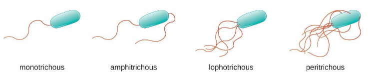
Flagella
Long, thin appendages (15-20 nm wide); helical shape; composed of flagellin
Different arrangements: polar, lophotrichous, amphitrichous, peritrichous
Tiny rotating machines (reversible)
Increase or decrease rotational speed relative to strength of proton motive force
Flagellar synthesis
More than 50 genes
Filament grows from tip
Build from cytoplasmic membrane
Flagelline produced in cytoplasm.
Archaella
Archaea version of flagellum
Half the diameter of bacterial flagella
Moves by rotation
Composed of several different filament proteins with little homologous bacterial flagellin
Speeds vary from 0.1 - 10x
Similar structure to type IV pili
Drive by ATP (not the PMF)
Surface motility
bacteria only
requires surface contact
slower and smoother than swimming
different mechanisms
excretion of polysaccharide slime
type IV pili/twitching motility
gliding-specific proteins
Chemotaxis
Taxis: directed movement in response to chemical or physical gradients
chemotaxis: response to chemicals
phototaxis: response to light
aerotaxis: response to oxygen
osmotaxis: response to ionic strength
hydrotaxis: response to water
How is a gradient sensed in chemotaxis?
By specific receptor proteins (MCP)
The signal is transmitted to the flagellum
Measuring chemotaxis
Measured by inserting a capillary tube containing an attractant or a repellent in a medium of motile bacteria
Can also be seen under a microscope
Phototaxis
Green algae were not sensitive to red light
They were sensitive to blue-green light as they orientated towards it.