Unit 9- Renal and Variants (Elie)
1/45
There's no tags or description
Looks like no tags are added yet.
Name | Mastery | Learn | Test | Matching | Spaced |
|---|
No study sessions yet.
46 Terms
What are the four parts and branches of the Renal Artery?
- Main Renal Artery
-Segmental Arteries
-Interlobar Arteries
-Arcuate Arteries
Where does the RRA extend?
From lateral AO to hilum of kidney
-10:00 position
-Posterior to IVC
On a longitudinal image, what does the RRA look like?
Seen as circular structure posterior to IVC
The RRA is _______ than the LRA
Longer
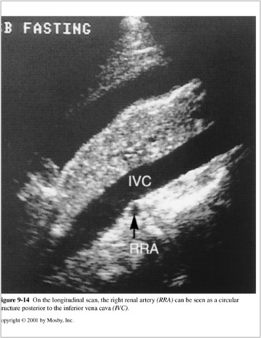

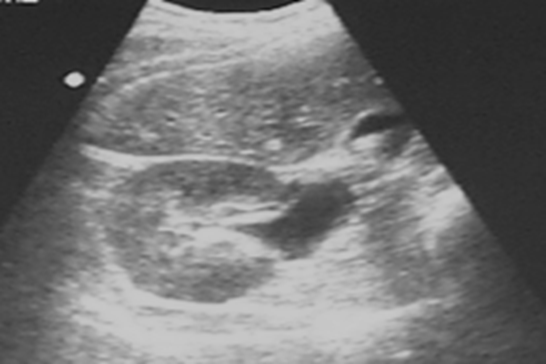
What are these images showing?
Right Renal Artery(RRA)
Where does the RRV extend?
From renal sinus to IVC
The RRV is ______ than the LRV
Shorter
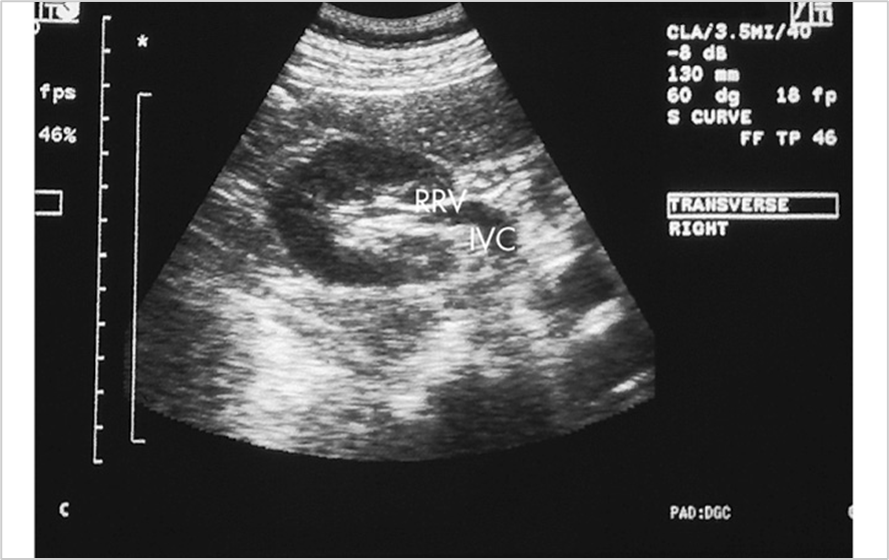
What is this image showing?
Right Renal Vein (RRV)
Where does the LRA extend?
From posterolateral AO to hilum of kidney
-4:00 position
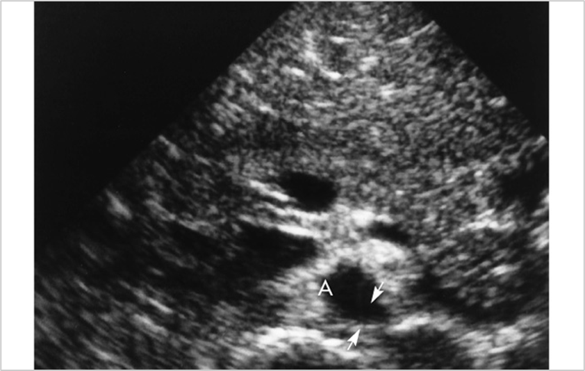
What is this image showing?
Left Renal Artery (LRA)
Where does the LRV extend?
From renal sinus, anterior to AO, posterior to SMA to join IVC
The LRV is _______ than RRV
Longer
What is a variant of the Left Renal Vein?
Retroaortic LRV
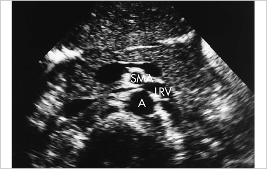
What is this image showing?
Left Renal Vein (LRV)
Where does the Crura of Diaphragm run?
-Transversely in paraaortic region
Where is the crura of diaphragm located?
Posterior to renal arteries
How can the Crura of Diaphragm be identified?
-Lack of pulsations
-No doppler flow
What is the echogenicity of the Crura of diaphragm?
-Hypoehoic
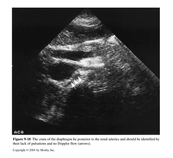
What is this image showing?
Crura of Diaphragm
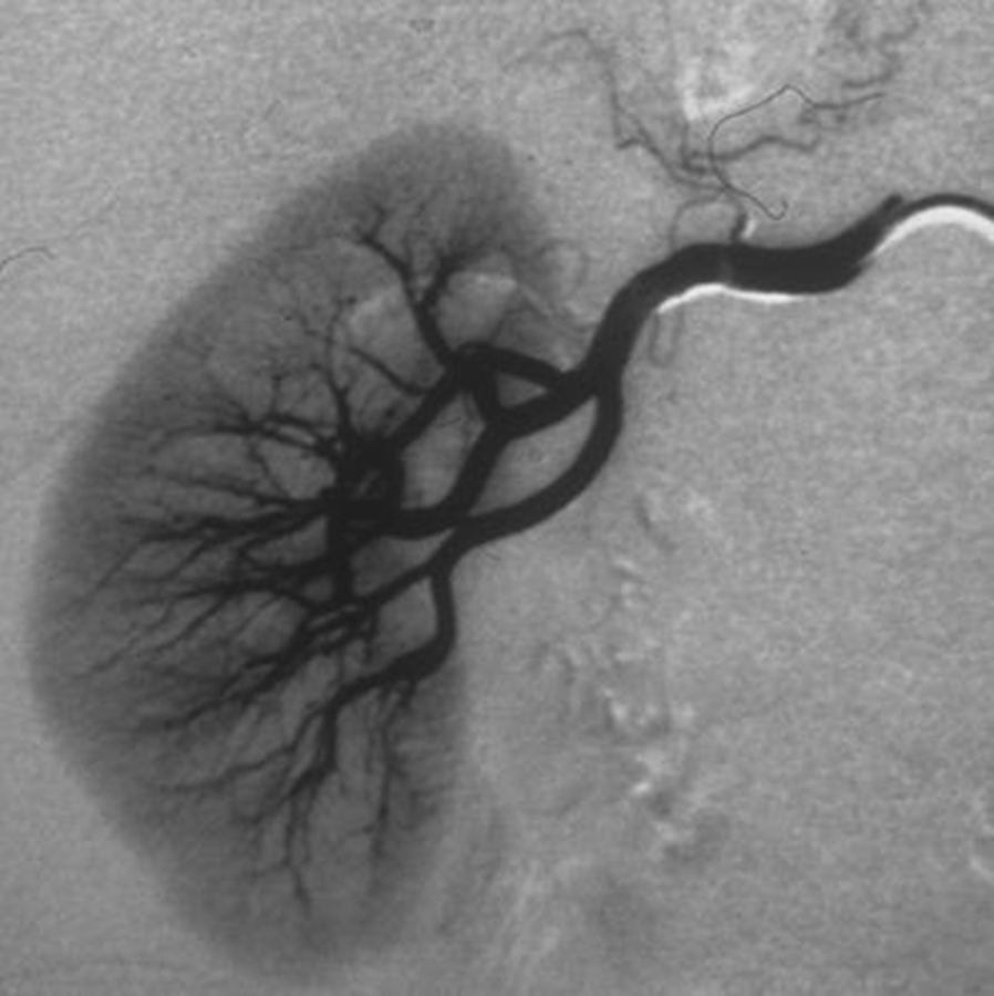
What is this image demonstrating?
Renal Angiogram
What is included in the Renal Medulla?
-Hypoechoic Pyramids
Where are the pyramids located in the renal medulla?
-At junction between renal cortex and central sinus
What do the renal pyramids look like sonographically?
-Uniform in size, shape & distribution
-Interlobar vessels run alongside the pyramids
-Arcuate vessels lie at base of pyramids
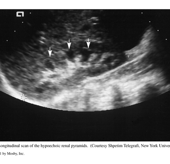
What is this image showing?
Renal Pyramids
What are normal variants?
Slight alterations in anatomy
What is the variant Column of Bertin?
- Cortex located at varying depths within medulla
-Contiguous with normal cortex tissue
-Same echogenicity as renal parenchyma
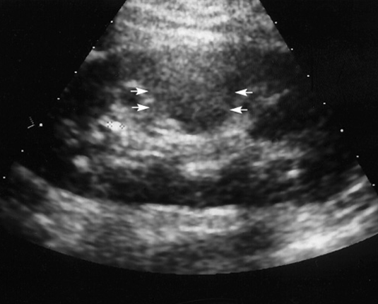
What is this image showing?
Column of Bertin
What is the Dromedary Hump variant?
-Cortical Bulge along lateral border
Where is a Dromedary Hump more common?
Left > Right
What is the echogenicity of a dromedary hump?
-Identical echogenicity to renal cortex
What can a dromedary hump resemble?
-Neoplasm = pseudotumor
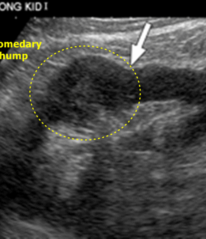
What is this image showing?
Dromedary Hump
What is the variant Junctional Parenchymal Defect?
-Triangular, echogenic area
-Upper pole, anteriorly
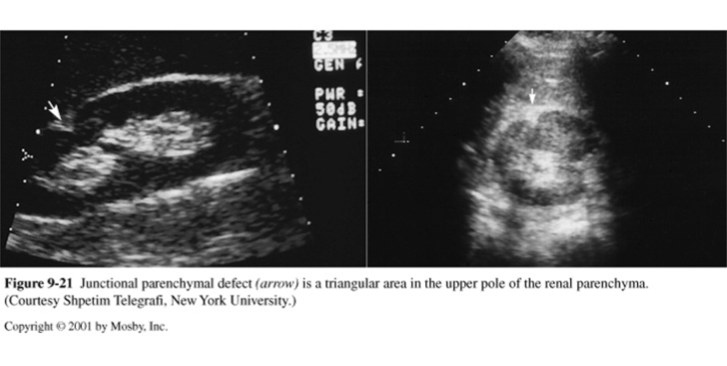
What is this image showing?
Junctional Parenchymal Defect
What is the renal variant Lobar Dysmorphism?
-Lobar fusion variant with malrotation of renal lobe
Where is the Lobar dysmorphism located?
-Middle & upper calyces may be splayed & displaced; lower calyx deviated posteriorly
What may lobar dysmorphism resemble?
-Mass or prominent Column of Bertin
What is the variant Fetal Lobulations?
-Fusion of two embryonic parenchymatous masses
-Indentation along border or surface giving kidney a lobulated appearance
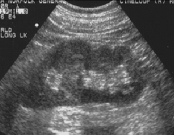
What is this image showing?
Fetal Lobulations
What is the variant Sinus Lipomatosis?
-Abundant fibrofatty tissue in renal sinus
-Enlargement of sinus region
What is the echogenicity of Sinus Lipomatosis?
Increased echogenicity
What is this image showing?
Sinus Lipomatosis
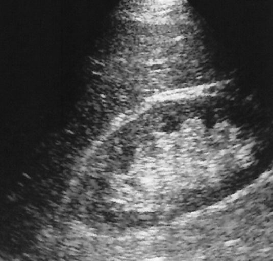
What is the variant Extrarenal Pelvis?
-Larger Pelvis
-Transverse view shows continuity with renal sinus
Where does the Variant extrarenal pelvis extend?
-Extends medially from confines of renal tissue
Why should you use color on a Extrarenal Pelvis?
-To prove its not RA or RV
What is this image showing?
Extrarenal Pelvis