Pathology Final Practice
 Call Kai
 Learn
Learn Practice Test
 Spaced Repetition
 Match
Match Flashcards
 Knowt Play
1/127

Earn XP
Description and Tags
Flashcards 1-71 were before the midterm. Flashcards 72- are after the midterm.
Name | Mastery | Learn | Test | Matching | Spaced | Call with Kai | Chat |
|---|
No analytics yet
Send a link to your students to track their progress
128 Terms
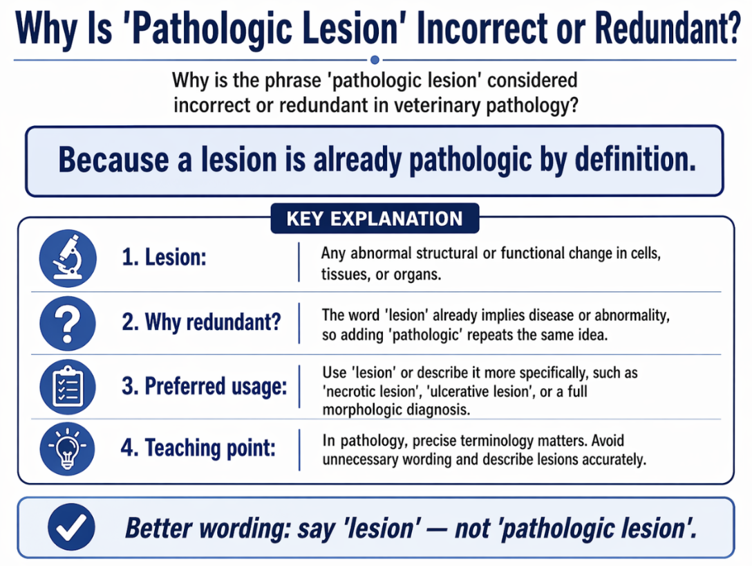
Why is the phrase "pathologic lesion" considered incorrect or redundant in veterinary pathology?
A) Lesions only refer to functional changes, while pathology refers to structural changes.
B) All lesions are by definition pathologic, making the term "pathologic lesion" a redundant repetition.
C) The term "pathology" should only be used for living animals, not postmortem findings.
D) A lesion implies a benign process, while pathology implies a malignant one.
Correct Answer: B.
Pathology is the study of disease, and a lesion is defined as a structural abnormality in a tissue or organ. Because every lesion is, by definition, a pathologic deviation from normal homeostasis, saying "pathologic lesion" is redundant.
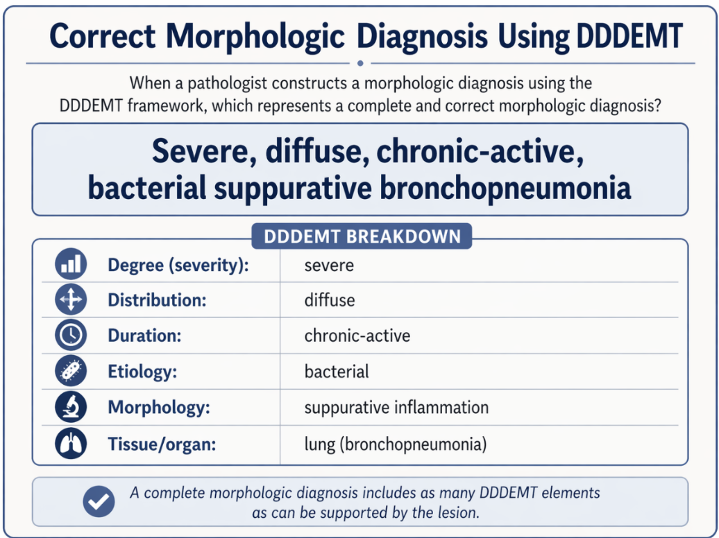
When a pathologist constructs a morphologic diagnosis using the DDDEMT framework, which of the following represents a complete and correct morphologic diagnosis?
A) Liver disease
B) Necrosis
C) Severe, acute, multifocal necrotizing hepatitis
D) Tyzzer's disease (Clostridium piliforme)
Correct Answer: C.
This is a complete morphologic diagnosis because it categorizes the lesion and provides the required DDDEMT descriptors: Degree (Severe), Duration (acute), Distribution (multifocal), Modifier (necrotizing), and Tissue (hepatitis).
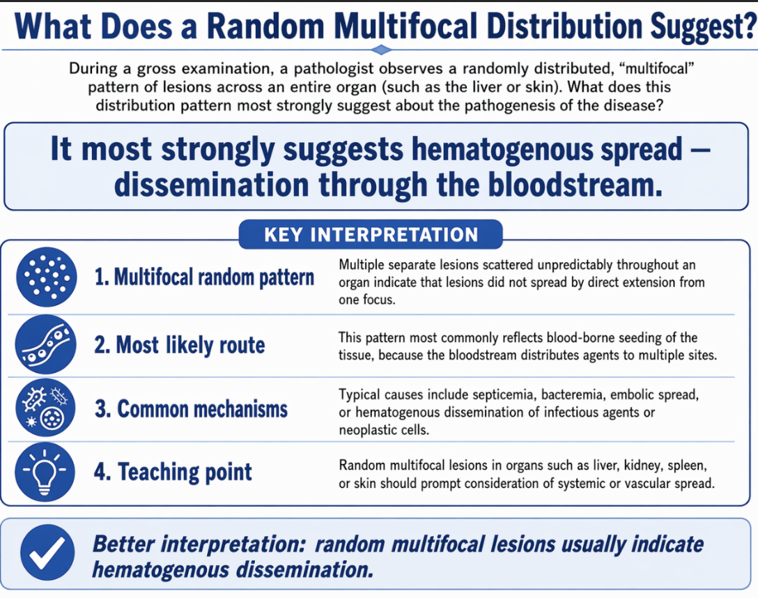
During a gross examination, a pathologist observes a randomly distributed, "multifocal" pattern of lesions across an entire organ (such as the liver or skin). What does this specific distribution pattern most strongly suggest about the pathogenesis of the disease?
A) The stimulus or pathogen arrived in the tissue via a systemic, blood-borne (hematogenous) route.
B) The lesion is the result of an inhaled pathogen settling in the cranioventral lobes.
C) The injury is due to a single direct penetrating trauma to the organ.
D) The entire organ is suffering from a uniform metabolic or toxic insult.
Correct Answer: A.
A multifocal, random distribution pattern generally implies that the disease-causing stimulus (such as bacterial sepsis, viral pox, or cancer metastasis) spread systemically and arrived in the tissue via the bloodstream.
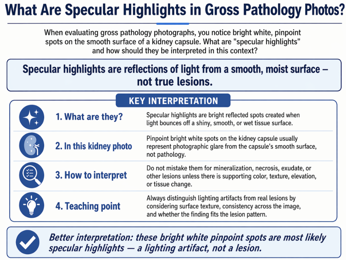
When evaluating gross pathology photographs, you notice bright white, pinpoint spots on the smooth surface of a kidney capsule. What are "specular highlights" and how should they be interpreted in this context?
A) They are areas of acute fibrin depositionindicatingsevere surface inflammation.
B) They are focal areas of mineralization and calcium accumulation.
C) They are purely optical artifacts caused by a light sourcereflecting offwet, shiny surfaces, and are not actual lesions.
D) They are pathognomonic patterns of tissue necrosis caused by ischemic infarcts.
Correct Answer: C.
Specular highlights are bright white areas (RGB 255) caused by the reflection of a light source (like a camera flash or room lights) off smooth, wet, or shiny surfaces (like serosae or a renal capsule). They are photographic artifacts and must not be confused with true pathological changes.
A veterinary student is examining a bovine kidney during a necropsy laboratory. The kidney surface shows several pale areas separated by normal-appearing dark red tissue. Some of the pale areas are touching and beginning to merge. Which pair of terms most accurately describes the distribution of the lesions?
a. Focal and diffuse
b. Diffuse and generalized
c. Multifocal with some coalescing
d. Generalized and widespread
c. Correct.
Multiple discrete lesions separated by normal tissue = multifocal. When some of those lesions begin to merge together, the correct term is coalescing. This scenario describes both conditions simultaneously, requiring students to apply both terms from the distribution vocabulary triad. Note that the lesions have not yet become diffuse — normal tissue is still visible between non-merging areas. This distinction is critical preparation for Case Set 1.
A pathologist examining a tissue sample describes: 'The hepatocytes show cytoplasmic vacuolation and nuclear pyknosis, with loss of normal cord architecture at the cellular level.' Which level of pathologic assessment is being applied?
a. Gross (organ-level) assessment
b. Histologic (tissue/cellular) assessment
c. Clinical-pathologic correlation
d. Etiologic interpretation
b. Correct.
A first-year student submits the following description of a kidney lesion: 'The kidney contains an infarct in the cortex due to vascular occlusion, producing coagulative necrosis.' A faculty member returns the description with a note saying it violates a fundamental rule of pathologic observation. Which statement best explains why?
a. The description assigns a mechanism and a diagnosis before establishing what is observed at the morphologic level
b. The description uses incorrect terminology because 'infarct' is not a recognized pathologic term
c. The description is too brief and should include more detail about organ size and color
d. The description is acceptable because naming a diagnosis is the goal of pathologic assessment
a. Correct.
The fundamental discipline of pathologic observation is: describe first, interpret later. The student's submission names the lesion ('infarct'), assigns a cause ('vascular occlusion'), and identifies the type of necrosis ('coagulative') — all of which are interpretations — without first providing a neutral morphologic description of what is actually visible. A correct starting description would be: 'The kidney contains a sharply demarcated, wedge-shaped, pale tan region in the cortex extending from the capsule.' Mechanism and diagnosis follow after observation is established.
A pathology report reads: 'The liver contains several abnormal pale areas.' A supervising pathologist marks this as inadequate. Which revised description best corrects the deficiency?
a. The liver appears diseased with multiple pale hepatic lesions suggesting inflammatory disease
b. The liver contains pale areas that are probably necrotic based on their appearance
c. The liver has many lesions of varying sizes distributed throughout
d. The liver contains multifocal, 0.5–1 cm, round to nodular, pale tan, firm areas scattered throughout the parenchyma
d. Correct.
This description systematically applies all five core morphologic descriptor categories: distribution (multifocal, scattered throughout the parenchyma), size (0.5–1 cm), shape (round to nodular), color (pale tan), and consistency (firm). No interpretation or mechanism is introduced. This is the professional standard that students must apply in the Case Set 2 application exercise.
A pathologist observes that a wedge-shaped pale lesion in the kidney has a depressed capsular surface, while nearby tissue appears normal. Which explanation best accounts for the depressed surface?
a. The capsule has thickened over the lesion, pulling the surface inward
b. Tissue within the lesion has been lost or collapsed, reducing the volume that normally supports the capsular surface
c. The normal adjacent tissue has swollen outward, making the lesion appear depressed by contrast
d. The lesion contains excess fluid that compresses the tissue, flattening the surface
b. Correct.
The kidney capsule is a fibrous envelope that conforms to the shape of the tissue beneath it. When parenchymal cells within the lesion die and are resorbed or collapse, the volume of tissue supporting that region of capsule is reduced. The capsule then sinks inward over the area of tissue loss, producing the characteristic depressed surface. This is the anatomy-physiology-pathology link: normal anatomy (capsule conforms to parenchyma) + pathologic change (cell death and volume loss) = observable morphologic finding (depressed capsular surface).
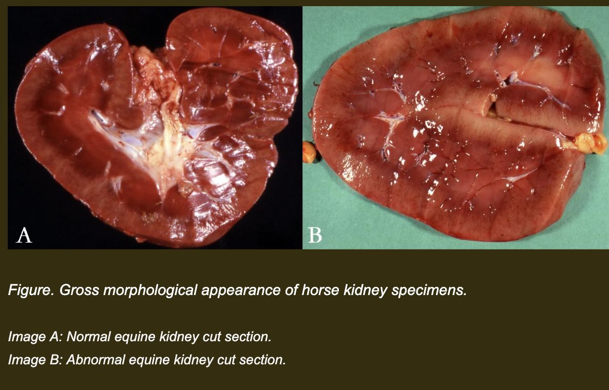
You are examining equine kidneys during a necropsy laboratory. Three specimens are available: one normal kidney (Image A) and one abnormal kidney (Image B) from two different horses. Both are cut sections showing the parenchyma.
Image A: Normal equine kidney cut section.
Image B: Abnormal equine kidney cut section.
Before answering the questions, your team reviews Image A (normal kidney):
• The cortex is uniformly dark red-brown throughout
• The corticomedullary junction is clearly visible as a distinct boundary
• The surface is smooth and convex with no discrete lesions
• No pale areas, nodules, or depressions are present
Image B (abnormal kidney) — describe what you observe before answering.
Which description best matches the gross appearance of Image B (the abnormal kidney) using standardized, neutral morphologic terminology — without interpreting the cause?
a. The kidney is diffusely swollen and pale due to toxic or ischemic injury, which has caused loss of intracellular ion and fluid homeostasis resulting in severe cellular swelling.
b. The parenchyma is diffusely pale red-tan with a slightly bulging contour and rounded capsular margins. The change involves the entire organ without discrete separate lesions, with a moist, glistening texture.
c. The kidney contains multifocal to coalescing, well-demarcated, pale tan, raised nodules scattered throughout the cortex, giving the organ an irregular contour.
d. The kidney is diffusely expanded by severe edema and a purulent inflammatory exudate that obscures the normal architecture, indicating an acute bacterial infection.
b. Correct.
This description uses standardized morphologic language: distribution (diffuse — the entire organ, no discrete separate lesions), color (pale red-tan), contour (slightly bulging, rounded capsular margins), and texture (moist, glistening). No mechanism or cause is introduced. Every term refers to what can be directly observed with the naked eye. This is the standard students must apply throughout the course.
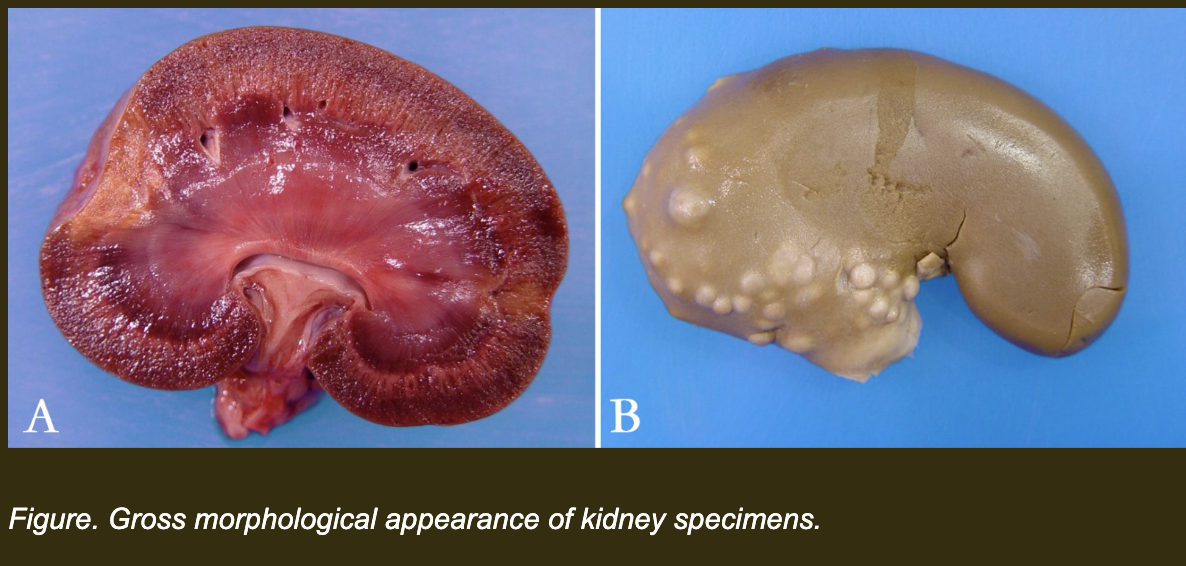
Now examine two additional kidney specimens — Kidney A and Kidney B. Both are abnormal. Your team must compare them using neutral morphologic language only.
Which description most accurately compares Kidney A and Kidney B using neutral morphologic language — without diagnostic interpretation?
a. Kidney A contains a focal, wedge-shaped, pale, dry lesion with a depressed capsular surface. Kidney B contains multifocal, round, pale nodules of varying sizes that are raised above the capsular surface, giving the organ an irregular contour.
b. Kidney A contains an infarct caused by vascular occlusion. Kidney B contains bacterial abscesses from embolic seeding.
c. Kidney A contains a large abnormal damaged area. Kidney B contains irregular lumpy tissue.
d. Both kidneys contain diffuse pale lesions extending through the entire organ.
a. Correct.
Both descriptions apply the core morphologic descriptor categories systematically — distribution (focal vs multifocal), shape (wedge-shaped vs round), color (pale in both), consistency (dry for Kidney A), and contour change (depressed vs raised capsule). No mechanism, diagnosis, or interpretation is introduced. The contour observation (depressed vs raised) is particularly important because it reflects a real morphologic difference with mechanistic implications — but those implications are saved for the discussion, not embedded in the description.
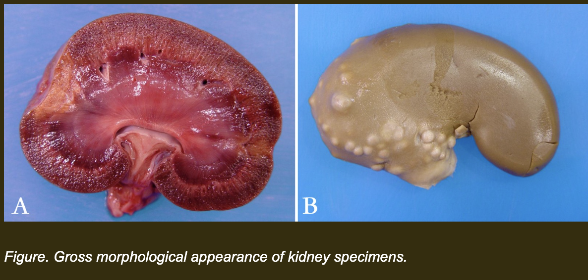
A junior pathology resident submits the following descriptions of Kidney A and Kidney B. Which statement violates the rule of objective morphologic description?
a. Kidney A contains a single pale lesion near the pole. Kidney B contains multifocal pale round lesions scattered throughout.
b. The capsular surface of Kidney A is indented. The capsular surface of Kidney B is raised and irregular.
c. Kidney A contains ischemic coagulative necrosis due to vascular occlusion. Kidney B contains multifocal bacterial granulomas from embolic spread.
d. The lesion in Kidney A maintains tissue density. The lesions in Kidney B contain dry, friable material replacing normal tissue.
c. Correct.
This statement violates the rule. Every component of this description is interpretation: 'ischemic coagulative necrosis' is a histopathologic and mechanistic conclusion, 'due to vascular occlusion' is an etiologic assignment, 'bacterial granulomas' is a diagnostic entity, and 'from embolic spread' is a mechanistic narrative. None of these can be established from gross observation alone without microscopic examination and additional diagnostic workup. The resident has bypassed description entirely and produced a diagnostic narrative instead.
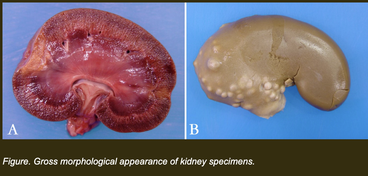
Using the same two kidneys, which complete morphologic description best represents professional pathologic reporting standards — applying all five descriptor categories and correctly linking the contour observation to the underlying tissue state?
a. Kidney A: A space-occupying inflammatory process has caused capsular distension, while Kidney B has experienced diffuse parenchymal atrophy producing irregular contour.
b. Kidney A: A single, focal, wedge-shaped, pale, dry lesion occupying the cortex with a sharply demarcated border and depressed capsular surface. Kidney B: Multifocal, variably sized, round, pale, raised nodules scattered throughout the cortex, producing an irregular capsular contour.
c. Kidney A: Classic infarct pattern. Kidney B: Granulomatous abscesses with embolic distribution.
d. Kidney A: A damaged area in the cortex. Kidney B: Abnormal lumps throughout the organ.
b. Correct.
Kidney A's description covers all five categories: distribution (focal), shape (wedge-shaped), color (pale), consistency (dry), and contour (depressed capsular surface), plus the sharpness of demarcation. Kidney B's description covers distribution (multifocal), size (variably sized), shape (round), color (pale), and contour (raised, irregular). Both descriptions are free of interpretation. The contour observations — depressed for A and raised for B — are morphologic observations, not diagnoses. This is the complete professional standard.
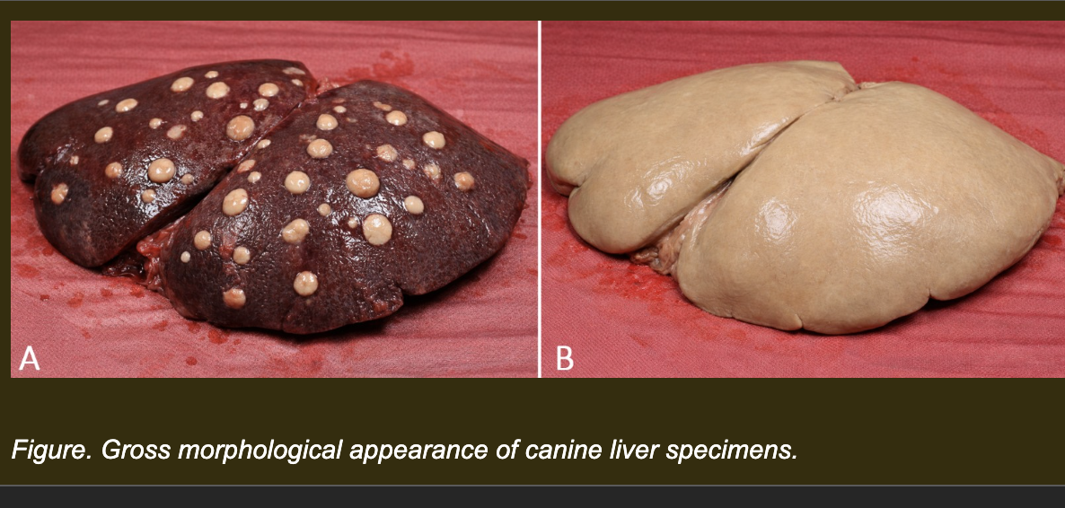
You are now examining two canine livers at necropsy. Both are abnormal.
Liver A: Multiple discrete pale tan round areas 0.5–1 cm in diameter scattered throughout the liver. Normal dark red-brown tissue is visible between the pale areas.
Liver B: The entire liver is uniformly pale tan throughout. No distinct separate pale areas are visible. The texture is uniform across the entire organ.
Which option provides the most accurate and complete morphologic descriptions of both Liver A and Liver B — using all five descriptor categories without introducing diagnosis or mechanism?
a. Liver A: Diffuse pale tan liver disease throughout. Liver B: Multifocal pale tan lesions in the parenchyma.
b. Liver A: Multifocal, numerous, 0.5–1 cm, round, pale tan areas scattered throughout the parenchyma, with normal tissue visible between lesions. Liver B: Diffuse, uniform, pale tan discoloration of the entire organ without discrete separate lesions.
c. Liver A: Numerous small nodules. Liver B: Severe liver inflammation.
d. Liver A: Multifocal hepatic infection. Liver B: Diffuse hepatic necrosis.
b. Correct.
Liver A's description covers all five categories: distribution (multifocal, scattered), size (0.5–1 cm), shape (round), color (pale tan), and the critical qualifier that normal tissue is visible between lesions (which is what makes multifocal the correct term rather than diffuse or coalescing). Liver B's description correctly identifies diffuse distribution and uniform involvement without discrete lesions. Both descriptions are free of diagnosis or mechanism. This is the complete professional standard for comparative morphologic reporting.
Which of the following cellular adaptations involves the reversible replacement of one mature, differentiated cell type by another of the same germline in order to withstand chronic irritation (e.g., ciliated respiratory epithelium changing to stratified squamous epithelium), acting as a protective but potentially maladaptive change?
A) Hyperplasia
B) Hypertrophy
C) Metaplasia
D) Atrophy
Correct Answer: C.
Metaplasia is a phenotypic adaptation where a fragile, specialized epithelium is replaced by a tougher, more resilient cell type to withstand chronic stress. However, it is potentially maladaptive because it causes functional degeneration; for instance, the respiratory tract loses its mucociliary clearance mechanism, making the tissue more prone to secondary infections.
During hypoxia and ischemia, cells suffer a precipitous drop in oxidative phosphorylation and subsequent ATP depletion. This energy loss leads to the earliest morphological manifestation of reversible cell injury, known as acute cell swelling (hydropic degeneration). What is the primary biochemical mechanism driving this swelling?
A) Influx of calcium into the mitochondria, opening the permeability transition pore
B) Failure of energy-dependent cell membrane Na⁺/K⁺-ATPase pumps, leading to intracellular sodium and water accumulation
C) Rupture of lysosomal membranes and the release of acid hydrolases into the cytosol
D) Massive lipid peroxidation of the plasma membrane by reactive oxygen species (ROS)
Correct Answer: B.
Under normal homeostatic conditions, Na⁺/K⁺ pumps actively extrude sodium and import potassium using ATP. When hypoxia causes ATP depletion, these pumps fail. Sodium moves down its concentration gradient into the cytosol, and water follows passively via osmosis, causing the cell and its organelles to swell.
A calf is diagnosed with "white muscle disease" due to a severe dietary deficiency in vitamin E and selenium. Upon necropsy, chalky white, gritty deposits are observed specifically within the areas of necrotic skeletal muscle, while the animal's systemic serum calcium levels are tested and found to be completely normal. What type of pathologic accumulation does this represent?
A) Metastatic calcification
B) Amyloidosis
C) Saponification (Fat necrosis)
D) Dystrophic calcification
Correct Answer: D.
Dystrophic calcification occurs exclusively in areas of local tissue necrosis (such as the dead muscle in white muscle disease or caseous tuberculosis nodules) despite the animal having perfectly normal systemic serum calcium levels. It happens because dying cells lose the ability to regulate calcium, causing it to precipitate in their mitochondria.
As animals age, their long-lived, postmitotic cells (such as cardiac myocytes and neurons) gradually accumulate a yellow-brown, autofluorescent endogenous pigment. This harmless "wear-and-tear" pigment is the indigestible lipid-protein residue resulting from decades of lipid peroxidation and autophagy. Which pigment is this?
A) Hemosiderin
B) Lipofuscin
C) Melanin
D) Anthracosis
Correct Answer: B.
Lipofuscin is the classic "aging" or "wear-and-tear" pigment. It accumulates over time in the secondary lysosomes of postmitotic cells as a benign consequence of continuous cellular metabolism, autophagy, and lipid peroxidation.
A 4-year-old Quarter Horse in active training develops enlarged hindquarter musculature over several months of conditioning work. Biopsy shows increased fiber diameter with no change in fiber number. ATP levels are normal and cell membranes are intact. Which adaptive response best explains these findings?
a. Hyperplasia -- increased fiber number in response to training demand
b. Hypertrophy -- increased fiber size in response to increased mechanical demand
c. Atrophy -- reduced metabolic demand prompts fiber size reduction
d. Metaplasia -- mechanical stress causes substitution of fiber type
b. Correct.
Hypertrophy is defined as an increase in cell size without an increase in cell number, occurring in response to increased functional demand. The biopsy findings precisely match this definition: increased fiber diameter, unchanged fiber number, normal ATP, intact membranes. The training stimulus provides the mechanical trigger. This is the textbook presentation of exercise-induced physiologic hypertrophy in skeletal muscle.
A dairy cow experiences a period of systemic hypoxia during anesthesia recovery. Hepatocytes show decreased ATP production, cellular swelling, and failure of sodium-potassium pump activity. Plasma membranes remain intact. Which statement best characterizes the current cellular state and predicts what would happen if the hypoxia were relieved at this point?
a. The cells have crossed into irreversible injury -- relief of hypoxia cannot prevent cell death
b. The cells are undergoing adaptive atrophy -- hypoxia is reducing metabolic demand appropriately
c. The cells have activated caspase pathways and are undergoing programmed cell death
d. The cells are in reversible injury -- intact membranes indicate the injury threshold has not been crossed and cells can recover if oxygen is restored
d. Correct.
Intact plasma membranes are the key indicator that the injury remains reversible. ATP depletion and pump failure produce cellular swelling -- a hallmark of early hypoxic reversible injury -- but these changes can be corrected if oxygen delivery is restored before membrane integrity is lost. The plasma membrane is the last line of defense; once it ruptures, cell death is irreversible. This threshold concept is the critical readiness anchor for Cases 3 and 4 (Clover the dairy cow).
A 9-year-old cat has been housed in a heavily smoke-exposed environment for three years. Tracheal biopsy shows that the normal ciliated columnar epithelium has been replaced by stratified squamous epithelium. The basement membrane is intact and tissue organization is preserved. Which response best explains this change and identifies what function is lost?
a. Hypertrophy -- epithelial cells have enlarged in response to chronic irritation
b. Hyperplasia -- repeated irritation has increased the number of epithelial cells
c. Metaplasia -- chronic irritation has caused replacement of the normal epithelial phenotype with one better suited to withstand mechanical stress, at the cost of mucociliary clearance function
d. Irreversible injury -- the loss of cilia indicates the epithelium has undergone necrotic cell death
c. Correct.
Metaplasia is the replacement of one differentiated mature cell type with another differentiated mature cell type that is better suited to tolerate a persistent environmental stress. Stratified squamous epithelium is more resistant to mechanical and chemical irritation than ciliated columnar epithelium. However, this adaptation is not cost-free -- cilia are lost, which eliminates mucociliary clearance. This trade-off (durability gained, function lost) is the key concept of metaplasia as adaptive but potentially maladaptive. The intact basement membrane confirms the change is adaptive, not neoplastic.
Two animals present with incidental findings on post-mortem examination. Animal A is a dairy cow in early lactation whose liver shows diffuse cytoplasmic vacuolation on histology. Nuclei are preserved and membranes are intact. Animal B is an 11-year-old dog whose previously injured skeletal muscle contains focal mineral deposits. Serum calcium and phosphorus are within normal reference ranges in Animal B. Which pair of interpretations best explains both findings?
a. Animal A: lipid accumulation from negative energy balance mobilizing hepatic fat. Animal B: dystrophic calcification in previously damaged tissue despite normal systemic mineral metabolism
b. Animal A: glycogen accumulation from excess dietary carbohydrate intake. Animal B: metastatic calcification from systemic hypercalcemia
c. Animal A: protein accumulation from increased hepatic synthesis. Animal B: lipofuscin deposition from oxidative cellular aging
d. Animal A: reversible hypoxic swelling from anemia-related ischemia. Animal B: dystrophic calcification from systemic hypercalcemia
a. Correct.
Early lactation dairy cows mobilize peripheral fat stores to meet the energy demands of milk production. Free fatty acids flood the liver faster than they can be processed, producing lipid accumulation in hepatocytes -- visible as cytoplasmic vacuolation with preserved nuclei and membranes. Dystrophic calcification occurs in previously injured tissue regardless of systemic mineral levels; the key distinguishing feature is that serum calcium and phosphorus are normal, ruling out metastatic calcification. Location of injury determines this type of calcification.
A 16-year-old dog presents with progressive weakness and reduced exercise tolerance. Post-mortem examination reveals golden-brown granular pigment in cardiac myocytes, focal areas of myocyte loss replaced by fibrous tissue, and reduced overall myocyte diameter compared with age-matched controls from younger adults. No prior cardiac disease has been documented. Which interpretation best explains the combined findings as a unified process?
a. Repeated episodes of reversible hypoxic injury have produced cellular swelling and subsequent membrane rupture across all findings
b. Metaplastic replacement of cardiac myocytes by fibroblasts has occurred in response to chronic mechanical stress
c. Cellular aging has produced lipofuscin accumulation from oxidative damage, focal cell loss replaced by repair fibrosis, and reduced myocyte size from decreased trophic stimulation -- representing the cumulative cellular consequences of normal biological aging
d. Dystrophic calcification has replaced lost myocytes and the brown pigment represents calcium mineral deposits in damaged tissue
c. Correct.
This option integrates three distinct aging-related findings into a coherent unified interpretation. Lipofuscin is the golden-brown wear-and-tear pigment that accumulates in post-mitotic cells (cardiac myocytes, neurons) as a byproduct of incomplete lysosomal degradation of oxidatively damaged cellular components -- it is the cellular marker of aging. Focal myocyte loss with fibrous replacement represents the limited regenerative capacity of terminally differentiated cells. Reduced myocyte diameter reflects decreased trophic stimulation and protein synthesis in aging cells. Together these findings define the cellular aging phenotype.
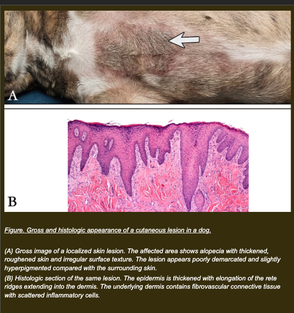
A dog has worn a poorly fitted harness during daily exercise for several weeks. The skin over the sternum becomes thickened. Histologic examination shows:
- Increased number of epithelial cell layers (thickness 2-3x normal)
- Epithelial cell size: unchanged
- Cell type: remains stratified squamous epithelium throughout
- Basement membrane: intact
- No membrane rupture or inflammatory infiltration within the epithelium
Which interpretation best explains the observed skin changes?
a. Mechanical stress enlarges epithelial cells, producing thickening through hypertrophy
b. Mechanical irritation stimulates increased epithelial proliferation, producing thickening through hyperplasia
c. Mechanical stress decreases oxygen delivery, producing epithelial swelling from reversible injury
d. Mechanical irritation replaces squamous epithelium with another cell type through metaplasia
b. Correct.
Hyperplasia is an increase in cell number in response to a stimulus. Stratified squamous epithelium is a proliferative tissue capable of regulated proliferation. Chronic mechanical irritation provides a sustained proliferative signal. Cell size unchanged, cell type unchanged, basement membrane intact -- all consistent with regulated hyperplasia. Growth is stimulus-dependent and would be expected to partially resolve if the harness is removed.
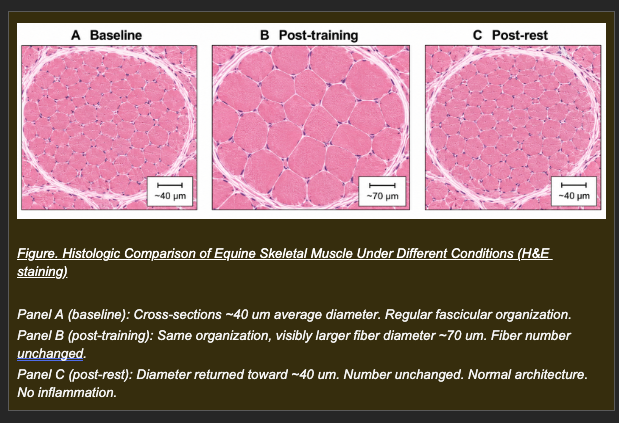
A performance horse completes 6 months of structured strength training. Biopsy at end of training:
- Fiber diameter: increased (~40 to ~70 um)
- Fiber number: unchanged
- ATP: normal. Membranes: intact.
The horse then sustains a fracture and is stall-rested for 3 months. Follow-up biopsy at end of rest:
- Fiber diameter: reduced (~40 um, returning toward baseline)
- Fiber number: unchanged. Membranes: intact. No fibrosis or inflammation.
Which pair of interpretations correctly identifies the adaptive response at each time point and explains why the same tissue produces different responses in opposite stimulus directions?
a. Post-training: hyperplasia from new fiber formation. Post-rest: atrophy from fiber loss
b. Post-training: reversible injury from exercise stress. Post-rest: recovery from that injury
c. Post-training: hypertrophy -- post-mitotic fibers respond to increased demand by enlarging through additional contractile protein synthesis. Post-rest: atrophy -- reduced demand decreases trophic signals maintaining protein synthesis, reducing fiber size while preserving fiber number and viability
d. Post-training: metaplasia from sustained mechanical stress converting fiber type. Post-rest: reversal of metaplasia
c. Correct.
This completes the adaptive triad in a single case. Post-training: fibers cannot divide so enlargement is the only available adaptive response. Diameter increases; number and membranes unchanged; ATP normal -- regulated adaptation confirmed. Post-rest: reduced mechanical stimulation decreases trophic signals, reducing protein synthesis. Fibers shrink through controlled myofilament autophagy. Number and viability preserved -- atrophy is a viable cell response, not injury.
Clover | Dairy cow | Two liver biopsies during anesthesia recovery hypoxia
Clover experiences systemic hypoxia during anesthesia recovery. Liver biopsies are taken at two time points as hypoxia progresses.
Stage 1: Early hypoxia
- Decreased ATP production
- Failure of sodium-potassium ATPase activity
- Cellular swelling
- Plasma membranes: INTACT
Which interpretation best characterizes Clover's hepatocyte state at Stage 1?
a. Hypoxia stimulates adaptive atrophy to reduce hepatocyte metabolic demand
b. Hypoxia has caused caspase activation and apoptotic cell death is underway
c. Hypoxia has caused irreversible membrane rupture -- Clover's hepatocytes cannot recover
d. Hypoxia has caused ATP depletion and pump failure producing cellular swelling -- this is reversible injury and Clover's hepatocytes can recover if oxygen is restored before membranes fail
d. Correct.
Hypoxia has caused ATP depletion and pump failure producing cellular swelling -- this is reversible injury and Clover's hepatocytes can recover if oxygen is restored before membranes fail
Correct. The sequence: hypoxia depletes ATP, impairing the sodium-potassium ATPase, allowing sodium accumulation, drawing water into the cell by osmosis, producing swelling. This is classic reversible hypoxic injury. The critical indicator of reversibility: plasma membranes remain intact. Restoring oxygen at this stage allows pump recovery and reversal of the cellular changes.
Clover | Dairy cow | Two liver biopsies during anesthesia recovery hypoxia
Clover experiences systemic hypoxia during anesthesia recovery. Liver biopsies are taken at two time points as hypoxia progresses.
Stage 2: Prolonged hypoxia (same patient, 30 minutes later)
- Plasma membranes: RUPTURED
- Intracellular enzymes (AST, ALT) detectable in surrounding tissue
- Inflammatory cells accumulating in the region
Which interpretation best explains the transition from Stage 1 to Stage 2?
a. Continued hypoxia stimulates adaptive atrophy preserving hepatocyte survival
b. Prolonged ATP depletion disrupted membrane integrity, producing irreversible injury with enzyme leakage and secondary inflammatory recruitment
c. Hypoxia activated caspase pathways producing apoptotic body formation and inflammation
d. Membrane rupture caused cells to undergo adaptive hypertrophy as a compensatory stress response
b. Correct.
Prolonged ATP depletion disrupted membrane integrity, producing irreversible injury with enzyme leakage and secondary inflammatory recruitment
Membrane rupture is the threshold event. When ATP depletion is sustained beyond the cell's capacity to maintain membrane integrity, the membrane fails. Intracellular contents -- enzymes, organelle fragments, DAMPs -- leak into surrounding tissue. These act as inflammatory signals recruiting leukocytes. Once membranes rupture, injury is irreversible: no oxygen restoration can reconstitute a dead cell.
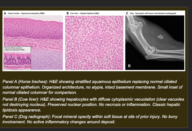
Animal A (horse): Tracheal biopsy following years in a poorly ventilated stable. Normal ciliated columnar respiratory epithelium replaced by stratified squamous epithelium. Basement membrane intact. Tissue organization preserved. Cilia absent.
Animal B (dairy cow, peak lactation): Liver biopsy. Diffuse cytoplasmic vacuolation in hepatocytes. Nuclei preserved and centrally positioned. Cell membranes intact. No inflammatory infiltrate. Cow is in negative energy balance.
Animal C (older dog): Incidental radiograph finding -- focal mineral deposits in hindlimb region that sustained severe muscle injury 2 years ago. Serum calcium and phosphorus: within normal reference ranges.
Which set of interpretations correctly identifies the process in each animal and explains the mechanism?
a. Animal A: metaplasia -- chronic irritation has replaced ciliated columnar epithelium with stratified squamous epithelium that resists chemical stress but lacks mucociliary clearance. Animal B: lipid accumulation -- negative energy balance mobilizes peripheral fat stores faster than the liver can export them, producing vacuolation in viable hepatocytes. Animal C: dystrophic calcification -- mineral deposition in previously damaged tissue driven by local tissue factors, not systemic mineral excess (confirmed by normal serum calcium and phosphorus)
b. Animal A: reversible injury from irritant exposure. Animal B: hypertrophic response to increased metabolic demand. Animal C: metastatic calcification from systemic hypercalcemia
c. Animal A: hyperplasia from chronic irritation. Animal B: cellular swelling from hypoxic reversible injury. Animal C: dystrophic calcification
d. Animal A: dysplasia from chronic irritant exposure. Animal B: lipid accumulation. Animal C: reversible hypoxic injury producing mineral deposits
a. Correct.
A: metaplasia is phenotype substitution -- one differentiated cell type replacing another better suited to the environmental stress. Intact basement membrane and preserved organization confirm adaptive rather than neoplastic change. The cost: loss of mucociliary clearance. B: free fatty acids from peripheral fat mobilization flood the liver faster than they can be packaged and exported, accumulating as lipid droplets in viable cells (preserved nuclei, intact membranes, no inflammation). C: dystrophic calcification is driven by local tissue damage, not systemic mineral excess -- confirmed by normal serum calcium and phosphorus. Calcium deposits form in previously damaged tissue regardless of systemic mineral levels.
A pathologist examines a tissue biopsy and observes a zone of cell death characterized by profound cellular swelling, loss of plasma membrane integrity, and a robust infiltration of neutrophils. Which of the following statements best differentiates this process (oncotic necrosis) from apoptosis?
A) Apoptosis results in severe cellular swelling (oncosis),whereas necrosis results in cellular shrinkage.
B) In apoptosis, the plasma membrane remains intact as the cell fragments into apoptotic bodies, eliciting no inflammatory response.
C) Necrosis is typically an energy-dependent, tightly programmed "suicide" process, while apoptosis is always an accidental death.
D) Necrosis frequently occurs as a normal physiologic process during embryogenesis, whereas apoptosis is exclusively pathologic.
Correct Answer: B.
In apoptosis, the cell shrinks and fragments into membrane-bound "apoptotic bodies." Because the plasma membrane remains structurally continuous throughout this process, intracellular contents do not spill into the extracellular matrix, meaning apoptosis is a "clean" death that does not elicit a local inflammatory response.
Following a severe ischemic event in the central nervous system (brain), the affected tissue undergoes rapid enzymatic digestion that completely predominates over protein denaturation. This process ultimately transforms the solid neural tissue into a viscous fluid mass containing lipid-laden macrophages (gitter cells). What is this specific morphologic pattern of necrosis called?
A) Coagulative necrosis
B) Caseous necrosis
C) Liquefactive necrosis
D) Fat necrosis
Correct Answer: C.
Liquefactive necrosis is characterized by the complete enzymatic lysis of cells. It is the default, end-stage pattern of ischemic necrosis within the central nervous system (clinically termed malacia) due to the brain's lack of fibrous connective tissue and high lipid/lytic enzyme content.
Which of the following statements most accurately describes the mechanisms and biological roles of apoptosis in a multicellular organism?
A) Apoptosis is an energy-dependent, highly regulated pathway driven by caspase enzymes to eliminate redundant physiologic cells or cells with irreparable DNA damage.
B) Apoptosis is exclusively a pathologic process triggered only by massive ischemic injury or severe physical trauma.
C) The intrinsic pathway of apoptosis is initiated by the rupture of lysosomes and the unregulated release of acid hydrolases into the cytosol.
D) Both physiologic and pathologic apoptosis consistently trigger a severe local inflammatory response to help clear the dead tissue.
Correct Answer: A.
Apoptosis is an energy-dependent, programmed pathway executed by a family of proteases called caspases. It serves both physiologic roles (such as fetal tissue remodeling and maintaining adult cell populations) and pathologic roles (such as destroying cells with irreparable DNA damage or viral infections).
Cellular vulnerability to ischemic injury varies significantly depending on the cell's metabolic requirements and its ability to utilize anaerobic glycolysis for energy. Which of the following cell types is most exquisitely sensitive to hypoxia, crossing the "point of no return" and undergoing irreversible cell death within just 3 to 5 minutes?
A) Cardiac myocytes
B) Fibroblasts
C) Skeletal myocytes
D) Neurons
Correct Answer: D. Neurons have a massive energy requirement and rely almost entirely on continuous aerobic respiration. Because they cannot switch to anaerobic glycolysis effectively, they are the most susceptible to hypoxia and undergo irreversible cell death within 3 to 5 minutes of oxygen deprivation.
Following a period of severe exertion, skeletal muscle cells in a performance horse show the following: ruptured plasma membranes, leakage of intracellular contents into the surrounding tissue, and a dense accumulation of inflammatory cells in the affected region. ATP levels are severely depleted. Which mechanism best explains these findings?
a. Regulated caspase activation producing controlled cell shrinkage and apoptotic body formation
b. Adaptive hypertrophy of muscle fibers from exercise-induced mechanical demand
c. Unregulated cell death from membrane integrity failure, with release of intracellular contents triggering secondary inflammation
d. Reversible cellular swelling from transient pump failure, expected to resolve with rest
c. Correct.
This describes necrosis: unregulated cell death driven by severe injury that exceeds the cell's capacity to maintain membrane integrity. When membranes rupture, intracellular contents -- including enzymes, organelle fragments, and damage-associated molecular patterns (DAMPs) -- are released into the extracellular space. These released contents act as inflammatory signals, recruiting leukocytes to the area. This is the fundamental difference between necrosis and apoptosis: necrosis triggers inflammation; apoptosis does not.
During recovery from a conditioning program, individual skeletal muscle cells in a horse demonstrate the following: cell shrinkage, nuclear chromatin condensation, membrane blebbing with formation of membrane-bound apoptotic bodies, and phagocytic uptake of apoptotic bodies by neighboring cells. No surrounding inflammatory infiltrate is present. ATP levels are normal in the affected cells at the time of these changes. Which mechanism best explains this process?
a. Regulated caspase-mediated programmed cell death executed without membrane rupture or inflammatory recruitment
b. Necrosis triggered by exercise-induced ATP depletion and membrane failure
c. Reversible hypoxic injury producing cellular swelling that will resolve with adequate oxygenation
d. Metaplastic replacement of muscle cells by fibroblasts in response to repetitive mechanical stress
a. Correct.
Apoptosis is a precisely regulated, energy-dependent cell death pathway characterized by a specific morphologic sequence: cell shrinkage (opposite of necrotic swelling), chromatin condensation and nuclear fragmentation, membrane blebbing, and packaging of cellular contents into membrane-bound apoptotic bodies that are phagocytosed without membrane rupture. Because the plasma membrane remains intact throughout, intracellular contents are never released into the extracellular space -- so no inflammatory signal is generated. Normal ATP levels confirm energy availability for this process, distinguishing it from the ATP-depleted necrosis scenario.
A pathology resident is examining two adjacent regions of tissue from a single biopsy. Region 1 shows cells with ruptured membranes, cytoplasmic enzyme leakage, and dense neutrophil infiltration. Region 2 shows cells with intact membranes, nuclear condensation, and packaged apoptotic bodies being engulfed by neighboring macrophages. The resident must determine which single histologic feature most reliably distinguishes the cell death mechanism in Region 1 from that in Region 2.
a. Nuclear condensation is present in Region 2 but absent in Region 1
b. Cell shrinkage is the key feature distinguishing Region 2 from Region 1
c. Cytoplasmic eosinophilia is greater in Region 1 than Region 2
d. The inflammatory infiltrate in Region 1 and its absence in Region 2 is the most reliable single discriminating feature between the two death pathways
d. Correct.
The presence or absence of inflammatory recruitment is the most biologically fundamental and reliable discriminator between necrosis and apoptosis. Necrosis invariably triggers inflammation because membrane rupture releases DAMPs and intracellular contents that act as inflammatory signals. Apoptosis invariably avoids inflammation because membrane integrity is maintained throughout, preventing release of intracellular contents. This relationship -- necrosis causes inflammation; apoptosis does not -- reflects the underlying mechanism and is consistent regardless of the tissue type, species, or stage of the process.
In normal intestinal mucosa of a healthy dog, individual epithelial cells at the villus tip regularly show chromatin condensation, cell shrinkage, and apoptotic body formation. These cells are phagocytosed by neighboring cells without generating an inflammatory response. The surrounding epithelium is intact and architecturally normal. Which interpretation best explains this observation?
a. Pathologic apoptosis -- the finding is abnormal and indicates early mucosal disease
b. Physiologic apoptosis -- regulated cell removal maintains epithelial homeostasis and barrier continuity in normal tissue turnover
c. Necrosis -- the nuclear condensation and cell shrinkage indicate ATP depletion and irreversible injury
d. Reversible injury -- the cells will recover normal morphology when mucosal oxygenation is restored
b. Correct.
The intestinal epithelium undergoes continuous renewal -- new cells are produced in the crypts, migrate up the villus, and are shed at the tip through apoptosis. This turnover is essential for maintaining epithelial barrier function and replacing senescent or DNA-damaged cells. The morphologic features described (chromatin condensation, cell shrinkage, apoptotic bodies, phagocytosis without inflammation) are exactly those of apoptosis. The key contextual feature confirming this is physiologic rather than pathologic is that normal architecture is maintained and no inflammation is present -- the surrounding tissue is healthy.
Two animals of different species experience equivalent degrees of systemic hypoxia for the same duration. In Animal A, neurons in the cerebral cortex begin to die within 3-4 minutes of hypoxia onset. In Animal B, skeletal muscle cells in the limbs sustain several hours of equivalent hypoxia before irreversible injury begins. Both animals experience the same degree of oxygen deprivation. Which explanation best accounts for the difference in vulnerability?
a. Animal A's neurons are larger than Animal B's muscle cells, and larger cells deplete oxygen faster
b. Animal B's muscle cells are incapable of undergoing necrosis under any circumstance
c. Animal A's neurons depend almost entirely on continuous aerobic metabolism with minimal energy reserves, while Animal B's muscle cells have greater metabolic flexibility and anaerobic capacity, giving them more time before ATP depletion reaches the injury threshold
d. Animal A's immune system mounts a faster inflammatory response, causing more rapid tissue damage
c. Correct.
Tissue vulnerability to hypoxia is determined primarily by the degree of dependence on continuous aerobic ATP production and the availability of alternative energy sources. Neurons have extremely high ATP demands, no glycogen stores to speak of, and cannot sustain function through anaerobic glycolysis for more than seconds to minutes. Skeletal muscle, by contrast, can utilize glycogen stores and tolerate anaerobic glycolysis for extended periods, providing a buffer before ATP depletion reaches the injury threshold. The same degree of oxygen deprivation therefore has very different time courses to irreversible injury in these two tissues.
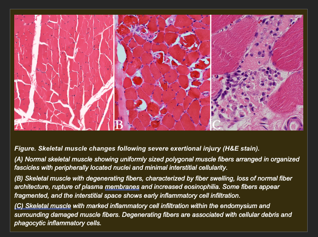
A performance horse collapses following extreme exertion. Muscle biopsies taken from the gluteal region show:
- Fiber swelling followed by rupture of plasma membranes
- Leakage of intracellular enzymes (creatine kinase, AST) into surrounding tissue
- Dense infiltration of inflammatory cells (neutrophils) into the affected region
- Ghost outlines of fiber architecture visible in some areas despite cell death
- ATP levels severely depleted in affected fibers
Which interpretation best explains the mechanism responsible for the observed cellular injury and its consequences?
a. ATP-dependent signaling activates a regulated cell death pathway that maintains membrane integrity and produces no inflammatory response
b. Reduced oxygen delivery produces reversible cellular swelling without loss of viability, expected to recover with reperfusion
c. Severe cellular injury exceeds the repair capacity of affected fibers, producing loss of plasma membrane integrity with release of intracellular contents and recruitment of inflammatory cells -- the hallmarks of necrosis
d. Controlled intracellular signaling pathways produce programmed cell shrinkage and apoptotic body formation without inflammatory recruitment
c. Correct.
This describes necrosis: unregulated, irreversible cell death driven by injury that exceeds the cell's capacity to maintain homeostasis. The sequence is: severe ATP depletion from exertion disrupts membrane maintenance, membranes fail, intracellular enzymes leak into the extracellular space, leaked cellular components act as damage-associated molecular patterns (DAMPs) that recruit neutrophils. The ghost outlines of fiber architecture visible in some areas are an early morphologic feature of coagulative necrosis -- the architecture is retained briefly before inflammatory resorption destroys it.
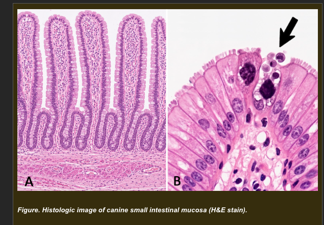
A healthy adult dog dies of unrelated trauma. Routine post-mortem histologic sampling of the small intestinal mucosa shows the following at the villus tips:
- Individual epithelial cells demonstrating cell shrinkage
- Nuclear chromatin condensation into smooth crescent-shaped masses
- Membrane blebbing and formation of membrane-bound apoptotic bodies
- Apoptotic bodies being phagocytosed by neighboring epithelial cells and macrophages
- No inflammatory infiltrate in the surrounding mucosa
- The rest of the villus epithelium is intact and architecturally normal
Which interpretation best explains the significance of the observed cellular changes at the villus tip?
a. Pathologic apoptosis indicating early viral enteritis affecting the villus tip epithelium selectively
b. Physiologic apoptosis maintaining epithelial homeostasis through regulated removal of senescent cells at the villus tip, consistent with normal intestinal epithelial turnover
c. Necrosis at the villus tip caused by relative ischemia from the post-traumatic hemodynamic changes before death
d. Reversible cellular injury at the villus tip that will recover if the underlying cause is identified and treated
b. Correct.
Intestinal epithelium has one of the highest cell turnover rates in the body. Cells are produced in the crypts, migrate up the villus over approximately 3-5 days, and are shed at the tip by apoptosis. This turnover is essential for maintaining barrier function, replacing DNA-damaged cells, and preventing accumulation of senescent epithelium. The morphology (cell shrinkage, chromatin condensation, apoptotic bodies, phagocytosis without inflammation) is precisely what apoptosis looks like regardless of context. The context -- healthy animal, normal architecture, appropriate location -- establishes this as physiologic.
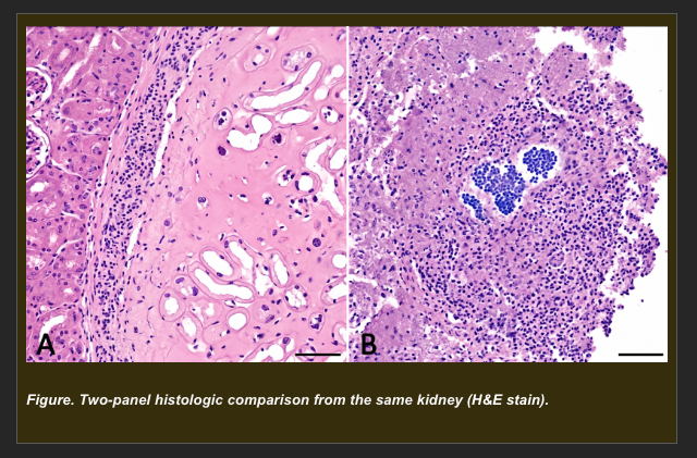
A cat presents with acute renal failure following suspected vascular thromboembolism. Post-mortem examination shows a pale, wedge-shaped cortical lesion with a depressed capsular surface (from Session 1A). Histologic examination of the pale region reveals:
- Dead cells with preserved outlines of tissue architecture -- tubule outlines and glomerular shapes are still recognizable
- Ghost cell forms visible where tubular epithelium has died
- Nuclear changes: pyknosis, karyorrhexis, karyolysis in affected cells
- Dense neutrophilic infiltration at the margin of the lesion
- Intact surrounding kidney architecture outside the lesion boundary
A second region from the same kidney, adjacent to a focus of suppurative bacterial infection, shows:
- Complete loss of tissue architecture -- no cell outlines, no tubule shapes recognizable
- Liquefied, granular, structureless debris filling the area
- Dense neutrophilic infiltration throughout the debris
Which explanation best accounts for the DIFFERENCE in morphologic appearance between the two necrotic regions?
a. Region 1 (pale wedge) shows coagulative necrosis where ischemic protein denaturation preserves the ghost architecture of dead cells. Region 2 (bacterial focus) shows liquefactive necrosis where proteolytic enzymes from neutrophils and bacteria overwhelm any structural preservation, converting the dead tissue to liquid debris
b. Region 1 shows reversible injury with preserved architecture, while Region 2 shows irreversible injury with complete architectural loss
c. Region 1 shows apoptosis (organized cell death with preserved architecture), while Region 2 shows necrosis (disorganized death with architectural loss)
d. Region 1 and Region 2 represent the same process at different time points -- Region 2 is simply more advanced necrosis that has had longer to develop
a. Correct.
Coagulative necrosis is the predominant pattern in solid organs following ischemic injury. Ischemia causes protein denaturation, which paradoxically preserves the basic architectural scaffold of the tissue for days to weeks -- glomeruli, tubule outlines, and cellular borders remain recognizable as ghost forms even though the cells are dead. This preservation occurs because the same protein denaturation that kills the cells also inactivates the lysosomal enzymes that would otherwise digest the tissue structure. Liquefactive necrosis occurs when enzymatic digestion dominates -- either because the tissue itself is enzyme-rich (as in the brain) or because large numbers of neutrophils release proteolytic enzymes that liquefy the dead tissue, converting it to pus. Both regions show necrosis; the difference is whether structural preservation or enzymatic liquefaction dominates the morphologic outcome.
Two animals experience equivalent systemic hypoxia following cardiovascular collapse. Post-mortem examination is performed after the same elapsed time post-collapse.
Animal A (dog): Cerebral cortical neurons show membrane rupture, loss of nuclear detail, and dense neutrophilic infiltration.
Animal B (horse): Skeletal muscle fibers in the limbs show cellular swelling with intact membranes and no inflammatory infiltration.
Additional information:
- Neurons rely almost entirely on continuous aerobic glucose oxidation with minimal energy reserves
- Skeletal muscle can utilize glycogen stores and tolerate periods of anaerobic metabolism
Which explanation best accounts for the difference in injury severity between the two tissues at the same post-collapse time point?
a. Animal A's neurons are incapable of necrosis -- the findings represent a different cell death process specific to neural tissue
b. Animal B's muscle cells have undergone apoptosis rather than necrosis, which explains why they appear less damaged
c. Animal A received a more severe hypoxic insult than Animal B, which is why neuronal injury is more advanced
d. Neurons have minimal energy reserves and depend on continuous aerobic metabolism, so they cross the irreversible injury threshold much faster than skeletal muscle cells, which have metabolic flexibility and anaerobic capacity to extend their window before membrane failure
d. Correct.
The vulnerability of a tissue to hypoxic injury is determined by its degree of dependence on continuous aerobic ATP production and its capacity to sustain function through alternative energy sources. Neurons have extremely high ATP demands, no significant glycogen reserves, and cannot sustain function through anaerobic glycolysis for more than seconds to minutes. Their irreversible injury threshold is therefore crossed very rapidly during hypoxia. Skeletal muscle can mobilize glycogen and tolerate anaerobic conditions, extending the window before ATP depletion reaches membrane-threatening levels. The same duration of hypoxia therefore produces irreversible necrosis in neurons while muscle remains in reversible injury.
A dog develops progressive mesenteric ischemia due to a developing vascular obstruction. Intestinal tissue is sampled at four time points as the ischemia evolves. The following findings are observed:
Stage 1: Intestinal smooth muscle cells show decreased contractile activity and reduced cell size. ATP normal. Membranes intact. No inflammation.
Stage 2: Cells now show swelling, ATP depletion, and pump failure. Membranes still intact.
Stage 3: Membranes have ruptured. Intracellular enzymes detectable in surrounding tissue. Dense neutrophilic infiltration.
Stage 4: Adjacent cells (not the primary ischemic population) show cell shrinkage, chromatin condensation, and apoptotic body formation. No inflammation around these specific cells. ATP normal in these cells.
Which sequence best describes the process occurring at each stage?
a. Stage 1: Hyperplasia -- Stage 2: Reversible injury -- Stage 3: Necrosis -- Stage 4: Metaplasia
b. Stage 1: Atrophy (ischemia-induced reduction in cell size with preserved viability) -- Stage 2: Reversible injury (ATP depletion with intact membranes) -- Stage 3: Necrosis (membrane rupture with inflammatory recruitment) -- Stage 4: Apoptosis in adjacent stressed but viable cells (regulated death without inflammation)
c. Stage 1: Reversible injury -- Stage 2: Necrosis -- Stage 3: Apoptosis -- Stage 4: Atrophy
d. Stage 1: Metaplasia -- Stage 2: Necrosis -- Stage 3: Apoptosis -- Stage 4: Reversible injury
b. Correct.
This sequence maps the full Module 2 conceptual arc onto a single progressive scenario. Stage 1: reduced blood flow reduces metabolic demand and substrate delivery, producing atrophy -- a viable adaptive response. Stage 2: ischemia deepens, ATP depletion produces pump failure and cellular swelling -- reversible injury with intact membranes. Stage 3: sustained ATP depletion exceeds membrane maintenance capacity, membranes rupture, enzymes leak, inflammation is recruited -- necrosis. Stage 4: adjacent cells stressed by the inflammatory microenvironment and partial ischemia undergo regulated apoptosis -- they have sufficient ATP to execute the apoptotic program but are signaled to die. The absence of inflammation around Stage 4 cells confirms apoptosis rather than necrosis.
Following acute tissue injury, a highly orchestrated series of events takes place to deliver defense cells to the site of damage. Which of the following best describes the correct sequence and relationship of the early vascular and cellular events in acute inflammation?
A) Leukocyte margination and migration occur immediately upon injury, physically causing the subsequent vasodilation and increased vascular permeability.
B) Vasodilation leads to increased blood flow, followed by increased vascular permeability; the resulting slowing of blood flow (stasis) allows leukocytes to marginate, roll, and migrate through the endothelium.
C) Phagocytosis by tissue macrophages triggers a sudden drop in vascular permeability, trapping circulating neutrophils inside the blood vessels to prevent systemic infection.
D) Increased vascular permeability occurs strictly in the arterial system, preventing leukocyte margination and forcing cells to migrate exclusively through the lymphatic system.
Correct Answer: B.
The hallmark of early acute inflammation is vasodilation (which increases local blood flow and causes redness/heat) accompanied by increased vascular permeability (which allows protein-rich fluid to escape, causing edema). The loss of fluid concentrates the red blood cells, slowing blood flow (stasis). This stasis alters hemodynamics, pushing leukocytes to the periphery of the vessel (margination), allowing them to adhere to the endothelium and migrate out into the tissue to begin phagocytosis.
Chemical mediators are essential to the inflammatory response. Which of the following statements best describes the high-level conceptual role of these mediators (such as vasoactive amines, cytokines, and arachidonic acid metabolites) during inflammation?
A) They act as intercellular messengers that orchestrate the entire inflammatory response by regulating vascular caliber, inducing endothelial permeability, and recruiting leukocytes via chemotaxis.
B) They function exclusively as direct structural components that immediately cross-link to form a physical barrier (scar tissue) over the site of injury.
C) They are lysosomal enzymes that strictly cause the autolysis of damaged cells without influencing vascular dynamics or leukocyte movement.
D) They suppress the immune response to ensure that acute inflammation immediately transitions into a chronic state without a resolution phase.
Correct Answer: A. Chemical mediators are the molecular "directors" of inflammation. Released from local cells (like mast cells or macrophages) or derived from plasma proteins, they bind to specific receptors to trigger and amplify vascular changes (vasodilation and permeability) and cellular events (chemotaxis, leukocyte activation, and phagocytosis).
Depending on the nature of the inciting agent and the extent of tissue damage, acute inflammation can lead to several distinct outcomes. Which of the following correctly contrasts two common outcomes of acute inflammation?
A) Resolution involves the complete clearance of inflammatory cells and restoration of normal tissue architecture, whereas abscess formation involves extensive localized liquefactive necrosis walled off by fibrous tissue.
B) Abscess formation represents the successful regeneration of normal parenchymal cells, while resolution represents a failure of healing that leads to septicemia.
C) Resolution exclusively follows dry gangrenous necrosis, while abscess formation is the default outcome of strictly viral infections.
D) Abscess formation is characterized by a severe lack of neutrophils, whereas resolution is characterized by the permanent persistence of pus within the tissue.
Correct Answer: A.
Resolution is the ideal outcome, occurring when the injury is limited, the tissue is capable of regeneration, and cellular debris is completely cleared, restoring perfect homeostasis. Conversely, if the injury involves pyogenic (pus-forming) bacteria or massive tissue destruction, the influx of neutrophils leads to liquefactive necrosis, creating an abscess that is eventually walled off by a fibrous capsule.
A tissue biopsy from a patient with a prolonged, persistent bacterial infection reveals chronic inflammation. Which of the following microscopic and conceptual features is the defining hallmark of chronic inflammation?
A) An intense, rapid influx of pure neutrophils accompanied by severe cytotoxic edema and immediate tissue resolution.
B) The simultaneous presence of active inflammation, tissue destruction, and attempts at healing (fibrosis and angiogenesis), dominated by a mononuclear cell infiltrate.
C) A strictly vascular response characterized by intense reactive hyperemia and fluid exudation, with a complete absence of cellular infiltrates.
D) The exclusive accumulation of intracellular amyloid and lipofuscin pigments without any accompanying tissue destruction or leukocyte activity.
Correct Answer: B.
Chronic inflammation is defined by its prolonged duration in which active inflammation, ongoing tissue destruction, and attempts at repair (such as fibrosis and the formation of granulation tissue) occur simultaneously. Unlike the neutrophil-rich acute phase, the cellular infiltrate in chronic inflammation is predominantly mononuclear, consisting of macrophages, lymphocytes, and plasma cells.
Immediately following a contaminated skin laceration, the wound margins of a dog become bright red and warm to the touch. A veterinary student correctly identifies these signs as rubor and calor. Which vascular event directly produces these findings?
a. Venous outflow obstruction causing accumulation of deoxygenated blood in the tissue
b. Arteriolar dilation increasing local blood flow, producing active hyperemia with oxygenated blood
c. Lymphatic obstruction preventing drainage of inflammatory fluid from the tissue
d. Arteriolar constriction reducing perfusion and triggering local tissue ischemia
b. Correct.
The first vascular event in acute inflammation is arteriolar dilation driven by inflammatory mediators (histamine, prostaglandins, nitric oxide) released at the injury site. Dilation increases blood flow to the affected area, filling the microcirculation with oxygenated blood. This active hyperemia produces the characteristic bright red color (rubor) because the increased blood volume is oxygenated, and the heat (calor) because increased blood flow delivers metabolic heat to the surface. This is the physiologic basis of the first two cardinal signs of inflammation.
Two hours after a contaminated skin wound, a dog develops visible swelling around the wound site. The swelling contains protein-rich fluid. Arterial blood flow to the area is normal. Which mechanism most directly produces inflammatory edema?
a. Arteriolar dilation alone forces additional fluid into the interstitium by pressure
b. Leukocyte accumulation increases tissue mass sufficiently to produce visible swelling
c. Inflammatory mediators cause endothelial cell contraction, opening intercellular gaps that allow plasma proteins and fluid to exit the vessel
d. Lymphatic obstruction caused by the laceration prevents normal fluid clearance
c. Correct.
Inflammatory mediators -- particularly histamine, bradykinin, and leukotrienes -- bind receptors on endothelial cells and cause cytoskeletal contraction. This pulls endothelial cells apart, opening intercellular gaps in the vessel wall (especially in postcapillary venules). Through these gaps, plasma proteins such as albumin, fibrinogen, and immunoglobulins leave the vessel along with fluid. The escaped proteins establish an oncotic gradient in the interstitium that draws additional fluid out of the vessel. The resulting protein-rich fluid is an exudate -- the hallmark of inflammatory edema, distinct from the protein-poor transudate of hemodynamic edema.
A contaminated wound in a dog develops appropriate redness and swelling within the first hour. However, a microbiology student notes that without effective leukocyte delivery to the wound, bacterial clearance will fail. Which sequence correctly describes how circulating leukocytes normally reach bacteria in the infected tissue?
a. Leukocytes marginate along the vessel wall, adhere to activated endothelium, migrate through intercellular gaps into the tissue, and phagocytose bacteria at the site of infection
b. Leukocytes are produced locally within the tissue in response to bacterial products and do not need to cross the vessel wall
c. Leukocytes enter the tissue exclusively through lymphatic vessels that open directly into the infected tissue
d. Leukocytes passively diffuse through intact vessel walls in response to the concentration gradient of bacterial products
a. Correct.
Leukocyte recruitment to infected tissue follows a defined sequence: (1) margination -- leukocytes slow and move to the periphery of the blood vessel as blood flow slows in the inflamed microvasculature; (2) adhesion -- selectin and integrin molecules on activated endothelium capture and hold leukocytes to the vessel wall; (3) diapedesis (extravasation) -- leukocytes squeeze through intercellular gaps in the endothelium and into the extravascular tissue; (4) chemotaxis -- leukocytes follow chemical gradients (bacterial products, complement components, cytokines) to the site of infection; (5) phagocytosis -- leukocytes engulf and destroy bacteria. This entire sequence is required for effective innate immune defense.
A contaminated wound releases bacterial products that are recognized by tissue-resident cells. Within seconds to minutes, the surrounding blood vessels dilate, endothelial permeability increases, and circulating leukocytes begin to adhere to vessel walls. A student asks: 'What links the recognition of bacteria at the tissue level to these vascular and cellular responses?' Which statement best explains the conceptual role of chemical mediators in this sequence?
a. Chemical mediators are produced by bacteria themselves and directly cause vessel dilation by toxin activity
b. Chemical mediators are structural proteins of the vessel wall that passively deform when exposed to bacteria
c. Chemical mediators are produced by bacteria-activated tissue cells and circulating precursors; they act on blood vessels and leukocytes to coordinate the vascular and cellular responses of inflammation
d. Chemical mediators are signaling molecules released from injured and activated host cells that act on vascular endothelium to increase permeability, on smooth muscle to produce vasodilation, and on leukocytes to trigger adhesion and migration -- translating local injury recognition into coordinated vascular and cellular inflammatory responses
d. Correct.
This is the conceptual role of chemical mediators at the level required for first-semester students. Key mediators include: histamine (from mast cells and platelets -- causes rapid vasodilation and permeability increase), prostaglandins (from arachidonic acid metabolism -- sustain vasodilation, produce pain and fever), leukotrienes (increase permeability, promote leukocyte chemotaxis), and cytokines (coordinate systemic responses). Students are not expected to know all mediators, but they must understand that mediators are the molecular bridge between injury recognition and the vascular/cellular events they observe. Without mediators, the cascade cannot proceed.
A bovine mammary gland develops a bacterial infection. Animal A shows redness, swelling, and pain with neutrophil-dominated tissue infiltration that resolves within 10 days after antibiotic treatment. Animal B develops persistent tissue infiltration with macrophages and lymphocytes, progressive connective tissue deposition, and structural remodeling of the gland that continues for weeks after the inciting bacteria are cleared. Which statement best distinguishes the inflammatory pattern in Animal A from that in Animal B?
a. Animal A has chronic inflammation because resolution takes 10 days, while Animal B has acute inflammation because it involves macrophages
b. Animal A shows acute inflammation with neutrophil dominance and resolution when the stimulus is removed; Animal B shows chronic inflammation with mononuclear cell dominance and progressive tissue remodeling that persists beyond stimulus clearance
c. Animal A has a more severe inflammatory response because neutrophils are more destructive than macrophages
d. Both animals show the same type of inflammation at different stages -- Animal B simply has more advanced acute inflammation
b. Correct.
The distinction between acute and chronic inflammation rests on three features: duration and stimulus dependence (acute resolves when stimulus is removed; chronic persists), dominant cell population (acute = neutrophils; chronic = macrophages and lymphocytes), and tissue consequences (acute = edema, exudate, possible resolution; chronic = mononuclear infiltration, fibrosis, structural remodeling). Animal A fits the acute pattern precisely. Animal B fits the chronic pattern -- notably, the tissue remodeling persists even after bacteria are cleared, which is a key feature of chronic inflammation: the tissue response has taken on a life of its own beyond the original stimulus.
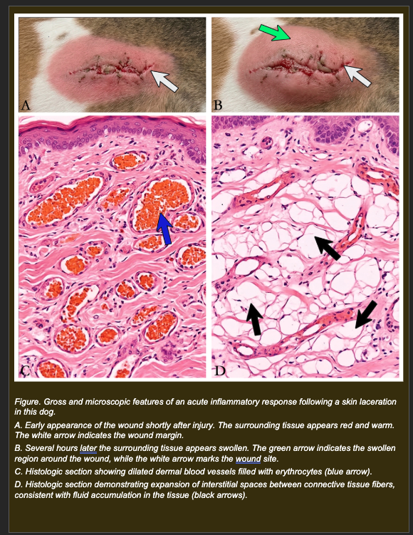
A dog sustains a contaminated skin laceration. Within minutes the wound margins become bright red and warm. Two hours later the surrounding tissue is visibly swollen.
Histologic examination shows:
- Dilated dermal blood vessels filled with erythrocytes (Panel C)
- Expansion of interstitial spaces between connective tissue fibers consistent with fluid accumulation (Panel D)
- Early neutrophil presence at the vessel margins
Which explanation most accurately accounts for both the early redness/warmth AND the later swelling -- using the vascular event sequence of acute inflammation?
a. Venous obstruction traps blood producing redness, while increased arterial inflow expands tissue producing swelling
b. Arteriolar dilation increases blood flow producing active hyperemia (redness and warmth); inflammatory mediators then increase vascular permeability allowing protein-rich fluid to exit vessels into tissue producing edema (swelling)
c. Lymphatic obstruction prevents fluid drainage producing redness, while leukocyte accumulation within tissue produces swelling by increasing tissue mass
d. Tissue hypoxia from reduced arterial perfusion causes cell death producing redness; the resulting necrotic debris accumulates causing swelling
b. Correct.
The vascular event sequence of acute inflammation proceeds in two linked steps. First: inflammatory mediators (histamine, prostaglandins, nitric oxide) released at the injury site cause arteriolar dilation, increasing local blood flow. This active hyperemia delivers oxygenated blood, producing the bright red color (rubor) and increased surface temperature (calor). Second: the same and additional mediators (histamine, bradykinin, leukotrienes) act on postcapillary venule endothelium, causing cell contraction and opening intercellular gaps. Plasma proteins escape through these gaps, establishing an oncotic gradient in the interstitium that draws fluid out of the vessel -- producing the protein-rich exudate responsible for swelling (tumor) and contributing to pain (dolor) from increased tissue pressure.
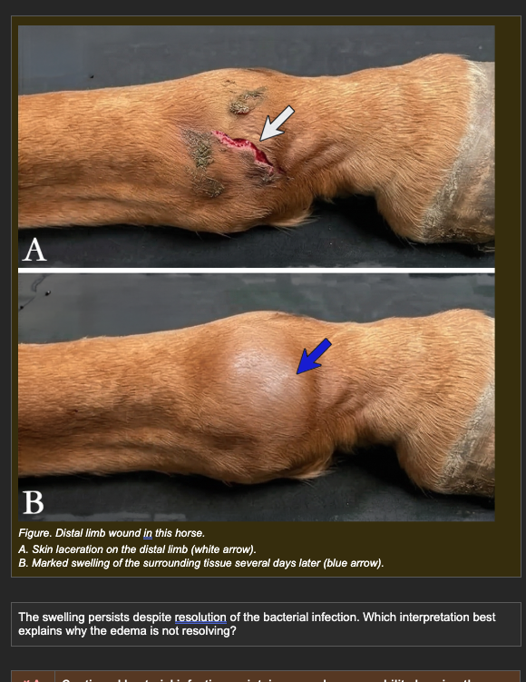
A horse develops a contaminated wound on the distal forelimb. After appropriate treatment, the infection resolves and bacterial cultures are negative. However, swelling of the distal limb persists several days after resolution of infection.
Additional clinical findings:
- Arterial blood flow to the limb: normal on Doppler
- Venous pressure: within normal range
- Plasma albumin concentration: normal
- Regional lymph nodes: enlarged on palpation
The swelling persists despite resolution of the bacterial infection. Which interpretation best explains why the edema is not resolving?
a. Continued bacterial infection maintains vascular permeability keeping the inflammatory exudate active
b. Decreased plasma albumin concentration reduces plasma oncotic pressure, drawing fluid into the interstitium
c. Inflammatory fluid has accumulated normally but reduced lymphatic clearance capacity in the distal limb allows interstitial fluid to persist beyond the resolution of the acute permeability event
d. Increased venous pressure prevents reabsorption of interstitial fluid into the venous end of the capillary
c. Correct.
Inflammatory edema formation during the acute phase was appropriate and expected. Once vascular permeability returns to baseline after infection resolution, normal fluid dynamics should gradually reabsorb the interstitial fluid -- but this requires functional lymphatic drainage. The horse distal limb has limited lymphatic clearance capacity relative to the volume of fluid that can accumulate, and inflammation itself can damage or overload regional lymphatics. The enlarged regional lymph nodes suggest ongoing lymphatic engagement. With normal arterial flow, normal venous pressure, and normal plasma albumin, lymphatic clearance failure is the remaining mechanistic explanation for persistent edema.
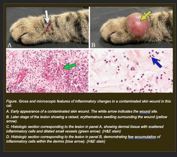
A cat develops a contaminated skin wound. Redness and swelling develop appropriately within the first few hours, indicating that the vascular events of acute inflammation are proceeding normally. Despite this, the bacterial infection worsens over the following 48 hours.
Histologic examination shows:
- Dilated dermal vessels with marginating leukocytes visible at vessel walls
- Very few leukocytes within the dermis or wound tissue -- leukocytes are present in blood but not migrating into tissue
- Bacteria visible within the wound tissue
- No significant phagocytic activity in the wound
Which interpretation best explains why the bacterial infection is worsening despite normal vascular events?
a. The vascular events have created appropriate conditions for leukocyte delivery, but leukocytes are failing to migrate from blood vessels into the infected tissue -- preventing phagocytosis and bacterial clearance
b. Increased vascular permeability is diluting the antibacterial proteins in the wound, allowing bacteria to survive in the protein-rich exudate
c. The bacteria have developed resistance to the mediators released during the vascular phase, preventing inflammatory recognition
d. Lymphatic obstruction is preventing clearance of bacteria from the wound site, allowing them to accumulate
a. Correct.
This case demonstrates that the vascular limb of the inflammatory response (dilation, hyperemia, permeability) is necessary but not sufficient for bacterial clearance. The leukocytes are present in the circulation and are marginating appropriately at the vessel wall, but the extravasation step is failing. Leukocytes cannot reach the bacteria in the wound tissue. Without extravasation, phagocytosis cannot occur. Without phagocytosis, bacteria persist and proliferate. This is a failure of the cellular limb of inflammation despite an intact vascular limb.
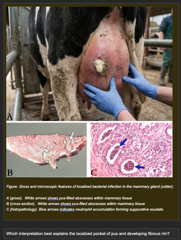
A dairy cow develops acute bacterial mastitis. Treatment is delayed. Post-mortem examination of the mammary gland shows:
- Localized pocket of creamy yellow material within the parenchyma
- Histology: dense neutrophil accumulation with central liquefactive necrosis
- A developing fibrous capsule at the periphery of the necrotic core
- Viable mammary tissue immediately outside the fibrous rim
Which interpretation best explains the localized pocket of pus and developing fibrous rim?
a. Reduced vascular permeability prevents inflammatory cells from spreading, producing containment by limiting fluid escape
b. Reduced arterial blood flow limits the area of leukocyte delivery, confining infection and inflammation to a localized region
c. Increased lymphatic drainage removes inflammatory cells from the surrounding tissue, confining infection to the primary site
d. Persistent bacteria maintain ongoing neutrophil recruitment and tissue destruction, producing a localized core of liquefactive necrosis, while surrounding fibroblasts lay down a fibrous capsule that walls off the infection -- forming an abscess
d. Correct.
Abscess formation is the outcome when bacteria persist at a localized site but are contained by the inflammatory response rather than spreading systemically. Continued bacterial products maintain chemotactic signaling, recruiting wave after wave of neutrophils. These neutrophils die in large numbers, releasing proteolytic enzymes that produce liquefactive necrosis of the tissue core -- creating the pus. Surrounding fibroblasts respond to the chronic tissue damage by depositing collagen, forming the fibrous capsule. The capsule contains the infection but also walls off antibiotic penetration, which is why abscesses often require surgical drainage. This is the containment outcome of persistent localized acute inflammation.
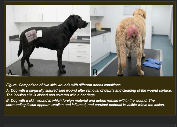
Two dogs sustain similar contaminated skin wounds.
Dog A: Wound is surgically cleaned, all debris removed, and sutured. The wound heals over 10-14 days with diminishing redness and swelling. Histology at day 14 shows restoration of normal tissue architecture with a small fibrotic scar.
Dog B: Wound is cleaned superficially but a fragment of foreign material (plant awn) remains embedded. Redness and swelling persist for weeks. Histology at day 14 shows macrophage and lymphocyte infiltration, progressive connective tissue deposition, and loss of normal tissue architecture in the affected region.
Which interpretation best explains the difference in inflammatory outcome between Dog A and Dog B?
a. Dog A's immune system is more effective than Dog B's, which is why inflammation resolved faster
b. Removal of debris in Dog A eliminated the inflammatory stimulus, allowing vascular permeability to normalize and tissue repair to proceed; retained debris in Dog B provides a persistent foreign body stimulus that maintains mononuclear cell recruitment and progressive tissue remodeling
c. Dog A's wound was sutured, which increased local blood pressure and forced inflammatory fluid out of the tissue faster
d. Dog B developed chronic inflammation because the immune response became self-sustaining and self-amplifying regardless of the original stimulus
b. Correct.
This option correctly identifies the determining variable: stimulus persistence. When the inflammatory stimulus is removed (Dog A), the mediator cascade loses its driving signal, vascular permeability normalizes, leukocyte recruitment decreases, and tissue repair proceeds. The outcome is resolution -- return toward normal tissue architecture. When the stimulus persists (Dog B), mediators continue to be generated, recruiting macrophages and lymphocytes rather than neutrophils (since the acute phase transitions to chronic), and persistent injury drives progressive fibrosis and tissue remodeling. The foreign body cannot be phagocytosed or degraded, so the stimulus never resolves, and neither does the inflammation.
Following tissue injury, the affected area will either undergo complete regeneration or be replaced by a fibrous scar. According to the principles of cellular pathology, which of the following factors primarily determines whether a tissue regenerates perfectly rather than forming a scar?
A) The absolute requirement that all cell types, regardless of their origin, heal exclusively through the deposition of collagen.
B) The presence of "permanent" (terminally differentiated) cells, which rapidly undergo mitosis to replace themselves without scarring.
C) The inherent regenerative capacity of the injured cells (whether they are continuously dividing "labile" cells or quiescent "stable" cells) and the preservation of nearby progenitor cells to fill the gap.
D) The occurrence of extensive liquefactive necrosis, which prevents any scar from forming and guarantees perfect regeneration.
Correct Answer: C.
The innate ability of a tissue to regenerate depends heavily on its cell cycle status. Tissues made of "labile" cells (e.g., epidermis, mucosal epithelium) cycle continuously, and "stable/quiescent" cells (e.g., hepatocytes) can reenter the cell cycle to divide on demand. If the defect is small and these progenitor cells are present, the tissue can regenerate perfectly without a scar.
During the healing of an ulcer or focal tissue defect, a specialized, transient connective tissue known as "granulation tissue" develops at the site of injury. Based on the text, what are the primary structural components that define active granulation tissue?
A) Massive accumulations of amyloid fibrils and dystrophic calcification.
B) Fibroblastic proliferation accompanied by endothelial proliferation (neovascularization).
C) Pools of liquid purulent exudate filled entirely with dead neutrophils and necrotic debris.
D) Aggregates of terminally differentiated neurons and skeletal muscle cells attempting to divide.
Correct Answer: B.
Granulation tissue is a specific hallmark of early healing and repair. It is defined microscopically by the active proliferation of fibroblasts (which lay down new extracellular matrix) occurring simultaneously with the proliferation of endothelial cells, which form new, delicate blood vessels (neovascularization).
In cases where tissue is severely damaged or composed of nondividing cells, healing occurs via replacement with connective tissue (fibrosis). How does early, highly vascular granulation tissue mature into a permanent, firm fibrous scar?
A) Neovascularization subsides, fibroblasts become quiescent, and dense collagen fibers remain to fill the lost space.
B) Fibroblasts undergo squamous metaplasia, transforming into epithelial cells to completely restore the lost organ parenchyma.
C) The tissue undergoes extensive autophagy, enzymatically digesting all the collagen and restoring perfect normal architecture.
D) Endothelial cells continuously multiply without stopping, eventually forming a highly vascularized hemangioma.
Correct Answer: A.
The transition from active granulation tissue to a mature scar is a stabilizing process. The acute need for massive blood supply diminishes, causing the newly formed blood vessels to regress (neovascularization subsides). The active fibroblasts stop dividing (become quiescent), leaving behind a dense, structural matrix of collagen fibers to permanently "fill the lost space" and provide structural continuity.
The success and speed of tissue repair can be heavily influenced by both local conditions at the wound site and the systemic health of the animal. Based on the mechanisms of cellular injury and adaptation, which of the following combinations of factors would most severely impair or delay tissue healing?
A) Intact local blood perfusion, adequate dietary protein, and a youthful population of labile cells.
B) Increased physiological workload combined with adequate hormonal trophic stimulation.
C) A brief, mild thermal stressor that induces temporary, reversible acute cell swelling.
D) Severe local ischemia (hypoxia), systemic nutritional imbalances (such as protein-calorie starvation), and advanced cellular aging (telomere attrition and senescence).
Correct Answer: D.
Healing is an energy- and resource-intensive process. Local ischemia cuts off the essential oxygen and ATP needed for repair cells to function. Systemic malnutrition (protein-calorie starvation) deprives the body of the building blocks needed for collagen and cell division. Advanced cellular aging limits the replicative lifespan of cells (stem cell exhaustion) and creates a pro-inflammatory environment that degrades local tissue, severely impeding repair.
Two wounds occur in a dog on the same day. Wound A is a superficial abrasion affecting only the surface epithelial cells -- the underlying connective tissue framework is completely intact. Wound B is a deep laceration that disrupts the full tissue thickness including the structural connective tissue scaffold. Both wounds are clean with no infection. Which statement best predicts the healing pathway for each wound?
a. Both wounds regenerate because epithelial tissues have high cellular turnover and can always restore normal architecture
b. Wound A heals by fibrosis because superficial injuries trigger stronger inflammation; Wound B regenerates because deep injuries stimulate greater cell proliferation
c. Wound A regenerates because the structural scaffold is intact allowing cells to restore normal architecture; Wound B heals primarily by repair with fibrotic scar because the structural support is disrupted
d. Both wounds heal by fibrotic repair because any tissue injury triggers irreversible scar formation regardless of depth
c. Correct.
The fundamental determinant of regeneration vs repair is whether the structural tissue scaffold is intact. When the scaffold is intact (Wound A), proliferating cells have a framework to organize on and can restore normal tissue architecture -- true regeneration. When the scaffold is disrupted (Wound B), no organizational template exists. Fibroblasts fill the defect with collagen (scar tissue), which restores structural continuity but not the original tissue architecture or full functional capacity.
A horse sustains an open wound on the distal limb that cannot be sutured closed. Over the following week, the wound bed fills with bright red, moist, granular-textured tissue. Histologic examination shows numerous new small blood vessels and abundant fibroblasts within a loose connective tissue matrix. Which interpretation best explains the role of this tissue in wound healing?
a. New blood vessels deliver oxygen and nutrients while fibroblasts produce connective tissue that fills the wound defect and supports subsequent epithelial coverage and scar maturation
b. New blood vessels represent mature scar tissue while fibroblasts prevent collagen formation to keep the wound flexible
c. New blood vessels remove bacteria while fibroblasts eliminate the remaining inflammatory cells from the wound bed
d. New blood vessels block oxygen diffusion to the wound bed while fibroblasts suppress inflammation to prevent further tissue damage
a. Correct.
Granulation tissue is the essential intermediate tissue of open wound repair. Angiogenesis (new vessel formation) restores the oxygen and nutrient delivery that healing tissue requires -- without neovascularization, fibroblasts cannot survive and function in the wound bed. Fibroblasts synthesize collagen and other extracellular matrix components that fill the wound defect, providing structural support for subsequent epithelial migration across the wound surface. The granular texture observed grossly reflects the density of new capillary loops visible at the surface. Granulation tissue is temporary -- it remodels into mature scar as collagen matures and vessels regress.
A dog sustains injury to the lung parenchyma. The inflammatory phase resolves and healing proceeds. Rather than restoring normal alveolar structure, the healing process produces dense collagen deposition that thickens the alveolar walls and partially obliterates the alveolar spaces. Which interpretation best explains the functional consequence of this healing outcome?
a. The collagen deposition strengthens the alveolar wall and improves resistance to future injury with no impact on gas exchange
b. The thickened fibrotic alveolar wall increases the diffusion distance between alveolar air and capillary blood, reducing the efficiency of oxygen transfer from the lung into the bloodstream
c. The collagen deposition restores normal alveolar architecture with full functional equivalence to the original tissue
d. The obliteration of alveolar spaces increases pulmonary blood flow by redirecting blood through patent alveoli
b. Correct.
Gas exchange in the lung depends on the extremely thin alveolar-capillary membrane that normally allows rapid oxygen and carbon dioxide diffusion. This thin interface -- consisting of the alveolar epithelium, basement membrane, and capillary endothelium -- minimizes diffusion distance. When fibrosis replaces this thin specialized structure with dense collagen, the diffusion distance increases substantially. Fick's law of diffusion states that diffusion rate is inversely proportional to membrane thickness -- so thickening reduces gas transfer directly. This is the functional cost of fibrotic repair when it occurs in a tissue specialized for diffusion.
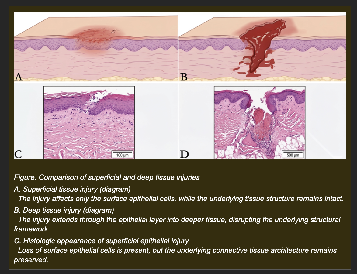
A dog sustains two clean wounds on the same day, both on the skin of the trunk.
Wound A: Superficial abrasion. Only the surface epithelial cells are lost. The underlying dermal connective tissue framework -- collagen, basement membrane, fibroblasts -- is completely intact.
Wound B: Deep full-thickness laceration extending through the epidermis and through the dermis into the subcutaneous tissue. The dermal collagen scaffold and basement membrane are disrupted across the full depth of the wound.
Both wounds are clean with no infection. Both are left uncovered and monitored over 14 days.
Which interpretation best explains the different healing trajectories expected for Wound A and Wound B?
a. Wound A will heal more slowly because superficial injuries have less inflammation to drive repair, while Wound B will heal faster because deep injuries trigger greater proliferative responses
b. Wound A will scar because it is on the trunk where scarring is common, while Wound B will regenerate because the deeper tissue layers are more regenerative
c. Wound A will heal by regeneration because the intact dermal scaffold guides epithelial cells back to their normal position; Wound B will heal by repair with fibrotic scar because the disrupted scaffold cannot be rebuilt and fibroblasts fill the defect with collagen instead
d. Both wounds will heal identically by fibrotic repair regardless of depth because the inflammatory response triggered by any skin injury always produces scar
c. Correct.
The intact dermal scaffold in Wound A provides the organizational template that allows regenerating epithelial cells to migrate across the wound surface and restore normal tissue architecture. When the scaffold is intact, the tissue knows where to put new cells. In Wound B, the scaffold has been destroyed across the full wound depth. Without this template, the wound healing response defaults to repair: fibroblasts fill the defect with collagen, granulation tissue forms, and the wound closes with scar tissue that is structurally continuous but not architecturally equivalent to the original dermis.
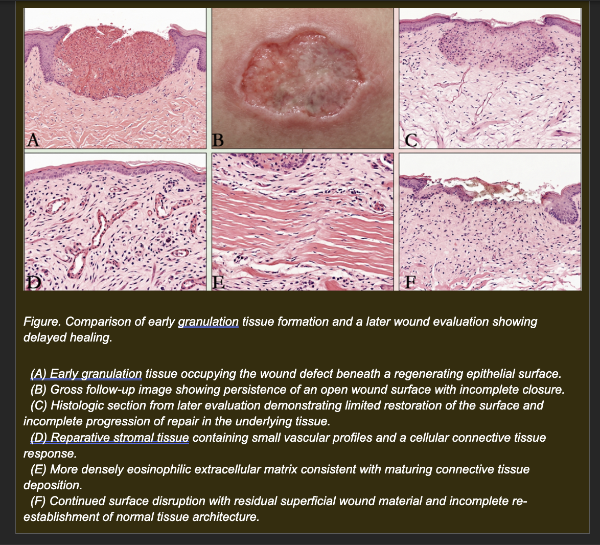
A horse sustains a large open wound on the distal forelimb that cannot be sutured closed due to tissue loss. The wound is managed conservatively. Over the following 7-10 days, the wound bed fills progressively with bright red, moist, granular tissue. Histologic examination at day 10 shows:
- Numerous small new blood vessels (neovascular channels) in a loose connective tissue matrix
- Abundant fibroblasts actively depositing collagen
- Scattered macrophages phagocytosing debris
- The granulation tissue surface begins to be covered by migrating epithelial cells at the wound margins
Which interpretation best explains the role of the granulation tissue in this wound?
a. Angiogenesis restores oxygen and nutrient delivery to the wound bed while fibroblasts produce the collagen matrix that fills the defect and supports epithelial migration and subsequent scar maturation
b. New blood vessels prevent leukocyte entry into the wound while fibroblasts produce inflammatory mediators that maintain the acute inflammatory response
c. Fibroblasts produce scar tissue that permanently replaces the wound defect while new blood vessels are a transient byproduct of the inflammatory response with no role in repair
d. Macrophages produce the collagen scaffold while fibroblasts remove the inflammatory exudate by phagocytosis
a. Correct.
Granulation tissue serves two essential sequential functions in open wound repair. First, angiogenesis: new blood vessel formation restores perfusion to the avascular wound bed, delivering oxygen and nutrients that fibroblasts require to survive and function. Without adequate oxygenation, fibroblasts cannot synthesize collagen effectively. Second, matrix production: fibroblasts produce collagen and other extracellular matrix components that fill the physical wound defect, providing the structural platform on which epithelial cells can migrate to close the wound surface. The macrophages present are clearing cellular debris and secreting growth factors that drive both angiogenesis and fibroblast activity. The granulation tissue is temporary -- as it matures, collagen reorganizes and vessels regress, forming the permanent scar.
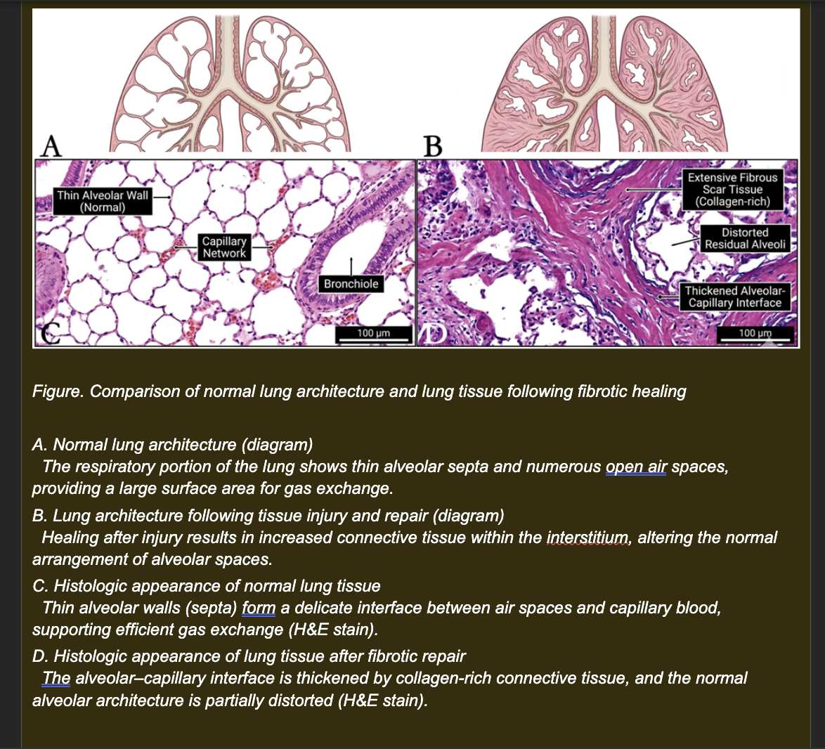
A dog is presented with progressive respiratory signs -- tachypnea, exercise intolerance, and hypoxemia -- 8 weeks after recovering from severe pneumonia. Chest radiographs show increased density throughout the lung fields. Lung biopsy shows:
- Thickened alveolar walls with dense collagen deposition replacing the normal thin alveolar epithelium
- Partial obliteration of alveolar air spaces by the fibrous tissue
- The remaining open alveoli have normal-appearing type I pneumocytes and intact capillaries
- No active inflammation or infection identified
Which interpretation best explains why this dog is hypoxemic despite having no active infection?
a. The fibrous tissue increases alveolar wall strength, which paradoxically traps oxygen within alveolar spaces and prevents its release into the blood
b. The thickened fibrotic alveolar walls increase the diffusion distance between alveolar air and capillary blood, reducing the rate of oxygen transfer and producing hypoxemia -- despite intact capillaries and remaining open alveoli
c. The fibrotic tissue replaces capillaries, eliminating the blood supply needed to carry oxygen from the lung to the rest of the body
d. Fibrosis activates macrophages that consume oxygen in the lung tissue before it can reach capillary blood
b. Correct.
Gas exchange efficiency depends critically on the diffusion distance across the alveolar-capillary membrane. This membrane is normally extremely thin -- approximately 0.5 micrometers in healthy lung -- allowing rapid equilibration of oxygen and carbon dioxide between alveolar air and capillary blood. When fibrosis replaces the thin normal epithelium with dense collagen, the diffusion distance increases substantially. Fick's law states that diffusion rate is inversely proportional to membrane thickness: double the thickness, halve the diffusion rate. In severe fibrosis, oxygen cannot equilibrate fully across the thickened membrane during the transit time of red cells through alveolar capillaries, producing hypoxemia even though alveoli are open and capillaries are patent.
A photograph shows an abnormal area of tissue with a visible change in color, shape, and surface appearance compared with the surrounding normal tissue.
What is the general term for this abnormal finding?
A. Lesion
B. Pathology
C. Histopathology
D. Prognosis
Correct answer: A. Lesion
A cell exposed to severe ischemic injury becomes swollen because it can no longer maintain normal ion and water balance. If the injury continues, the plasma membrane ruptures and the cell dies.
Which mechanism most directly causes these changes?
A. Excessive collagen production
B. Increased potassium retention in the nucleus
C. Depletion of ATP
D. Increased lysosomal storage of glycogen
Correct answer: C. Depletion of ATP
During necropsy, areas of adipose tissue appear firm, chalky-white, and gritty. Histologic examination confirms mineral deposition within damaged fat tissue.
Which tissue change best explains this finding?
A. Acute cellular swelling
B. Fatty change
C. Myxomatous degeneration
D. Fat necrosis
Correct answer: D. Fat necrosis
Pathology Report
History: A 4-year-old male mixed-breed dog is presented with fever, lethargy, and painful swelling on the neck after a bite wound.
Gross Findings: The subcutaneous tissue contains a soft, fluctuant swelling filled with thick yellow-white exudate.
Histopathologic Findings: Severe, focal, suppurative inflammation with abundant neutrophils, necrotic cellular debris, and bacterial colonies.
Comment: The lesion is consistent with a bacterial abscess secondary to a penetrating bite wound.
Use the pathology report to answer this question.
What term describes the following sequence?
Bite wound introduces bacteria → bacterial multiplication in tissue → neutrophilic inflammation → abscess formation
A. Morphologic diagnosis
B. Gross lesions
C. Pathogenesis
D. Disease
Correct answer: C. Pathogenesis
During chronic inflammation, which inflammatory cell is expected to predominate in the tissue?
A. Macrophage
B. Red blood cell
C. Platelet
D. Basophil
Correct answer: A. Macrophage
Session 4A
Hemodynamic disturbances can alter blood volume in tissues or lead to the accumulation of fluid in the interstitial space (edema). Which of the following statements correctly differentiates hyperemia from congestion, or accurately describes a primary mechanism of edema?
A) Hyperemia is a passive process resulting from impaired venous outflow, whereas congestion is an active process caused by increased arterial inflow.
B) Edema is exclusively caused by an abnormal increase in plasma oncotic pressure, which forcefully pushes water out of the vasculature.
C) Congestion is a passive process resulting from impaired venous drainage (e.g., chronic passive congestion of the lungs leading to "heart failure cells"), and edema can result from an increase in intravascular hydrostatic pressure pushing fluid into the interstitium.
D) A severe decrease in plasma oncotic pressure (e.g., due to protein loss in renal failure) prevents edema by trapping water inside the blood vessels.
Correct Answer: C.
Congestion is a passive accumulation of blood caused by impaired venous drainage (e.g., left-sided heart failure causing blood to pool in the lungs, leading to hemosiderin-laden macrophages or "heart failure cells"). Edema occurs when fluid is pushed out of the vessels into the tissue, which is driven by increased hydrostatic pressure (the physical pressure of blood against the vessel wall) or decreased oncotic pressure.
Session 4A
The pathological formation of a blood clot within an intact vessel (thrombosis) is heavily influenced by endothelial injury and altered hemodynamics. Which of the following best explains how these factors contribute to thrombus formation?
A) Altered blood flow (such as stasis) prevents platelets from contacting the vessel wall, making thrombosis impossible.
B) Endothelial injury exposes highly thrombogenic subendothelial extracellular matrix (e.g., collagen) and tissue factor, while altered blood flow (turbulence or stasis) disrupts laminar flow, bringing platelets into direct contact with the damaged endothelium.
C) Normal, healthy endothelial cells continuously produce pro-coagulant factors; therefore, injuring them halts coagulation and leads exclusively to severe hemorrhage.
D) Turbulence within an artery physically washes away fibrin and platelets, serving strictly as a mechanism to dissolve existing emboli.
Correct Answer: B.
In a healthy vessel, blood flows in a "laminar" pattern, keeping platelets in the center of the lumen and away from the walls. Altered blood flow (stasis or turbulence) disrupts this flow, pushing platelets against the endothelium. Furthermore, endothelial injury strips away the protective, anti-thrombotic lining, exposing collagen and tissue factor which immediately triggers the coagulation cascade and platelet aggregation.
Session 4A
An embolus (such as a dislodged fragment from a "saddle thrombus") traveling through the bloodstream can lodge in a narrower distal vessel, completely obstructing the arterial blood supply. In a solid "end-artery" organ like the kidney or spleen, what is the classic macroscopic and microscopic consequence of this sudden occlusion?
A) A pale, wedge-shaped area of coagulative necrosis (an infarct) in which the basic tissue architecture and "ghost cell" outlines are temporarily preserved, often bordered by a red rim of reactive hyperemia.
B) The immediate and diffuse onset of liquefactive necrosis, causing the affected organ to melt into a fluid-filled cystic cavity within minutes.
C) The instant development of wet gangrene, independent of any subsequent invasion by saprophytic putrefactive bacteria.
D) Reversible acute cell swelling (hydropic degeneration) that stabilizes indefinitely without ever progressing to cell death.
Correct Answer: A.
Embolism resulting in arterial occlusion in solid organs (like the kidney or heart) typically causes an infarct. Because the severe initial ischemic injury rapidly denatures both structural proteins and digestive lysosomal enzymes, the tissue undergoes coagulative necrosis. Macroscopically, this appears as a pale, wedge-shaped lesion (conforming to the branching vascular supply) bordered by a red rim of reactive hyperemia and hemorrhage.
Session 4A
Ischemia (reduced vascular perfusion) causes particularly extensive cellular damage because it deprives the tissue of oxygen, glucose, and a means to remove metabolic waste. Which of the following accurately traces the cascade of biochemical and structural events from initial ischemia to irreversible cell death (the "point of no return")?
A) Ischemia → Increased ATP production via glycolysis → Enhanced Na+/K+ pump activity → Cellular shrinkage (apoptosis).
B) Ischemia → Hypoxia → ATP depletion → Failure of Na+/K+ ATPase pumps causing acute cell swelling → Massive intracellular calcium overload and mitochondrial permeability transition (irreversible necrosis).
C) Ischemia → Sudden drop in cytosolic calcium → Inactivation of proteases and phospholipases → Accumulation of intracellular glycogen → Caseous necrosis.
D) Ischemia → Immediate physical rupture of the plasma membrane → Unregulated extracellular fluid influx → Coagulative necrosis without any prior reversible swelling phase.
Correct Answer: B.
This is the classic, fundamental sequence of ischemic injury. The lack of oxygen halts oxidative phosphorylation, causing ATP depletion. Without ATP, the cell cannot run its Na+/K+ membrane pumps, leading to sodium and water influx (reversible acute cell swelling). If ischemia persists, the cell cannot maintain calcium gradients. Massive intracellular calcium overload activates destructive enzymes (phospholipases, proteases) and opens the mitochondrial permeability transition pore, causing irreversible cell death.
Session 4A
During a surgical procedure in a pig, a veterinarian observes two different segments of small intestine. Segment A is bright red, warm, and the small arteries supplying it are visibly dilated. Segment B is dark red-blue, slightly swollen, and the veins draining it are visibly distended with blood. Which statement correctly explains the mechanism producing each appearance?
a. Segment A shows active hyperemia from arteriolar dilation increasing oxygenated blood inflow; Segment B shows passive congestion from impaired venous outflow causing deoxygenated blood to accumulate
b. Segment A shows passive congestion from venous obstruction; Segment B shows active hyperemia from arterial dilation
c. Both segments show hyperemia -- the color difference reflects oxygenation state of the tissue rather than a difference in blood flow mechanism
d. Segment A shows ischemia from arterial spasm; Segment B shows congestion from lymphatic obstruction
a. Correct.
Hyperemia and congestion are both states of increased blood volume in tissue but differ fundamentally in mechanism and blood oxygen content. Active hyperemia (Segment A) results from arteriolar dilation -- an active process driven by neural, metabolic, or inflammatory signals -- increasing inflow of oxygenated blood. The tissue appears bright red because it is filled with oxygenated hemoglobin. Passive congestion (Segment B) results from impaired venous outflow -- a passive process caused by obstruction, compression, or cardiac failure -- causing deoxygenated blood to accumulate. The tissue appears dark red-blue because it is filled with deoxygenated hemoglobin. The dilated arteries in Segment A and the distended veins in Segment B are the clinical indicators of the respective mechanisms.
Session 4A
A horse presents with swelling of the distal forelimbs. Clinical examination shows: venous pressure elevated on central venous measurement, plasma albumin concentration within normal limits, no evidence of infection or inflammation in the limbs, and pitting edema that is bilaterally symmetrical. Which mechanism best explains the edema?
a. Increased vascular permeability from subclinical inflammatory mediator release
b. Decreased plasma albumin reducing plasma oncotic pressure
c. Elevated venous pressure increasing capillary hydrostatic pressure, driving net fluid filtration into the interstitium
d. Lymphatic obstruction preventing clearance of normal interstitial fluid
c. Correct.
Elevated venous pressure is directly confirmed by the central venous pressure measurement. When venous pressure rises, it increases pressure at the venous end of the capillary bed, reducing or reversing the normal fluid reabsorption that occurs there. The net effect is increased filtration from capillary to interstitium -- producing edema. In horses, venous hypertension from cardiac disease or venous obstruction classically produces bilateral dependent limb edema. The pitting quality indicates the edema is fluid-rich, and the bilateral symmetry indicates a systemic or bilateral vascular cause rather than a unilateral local process. Normal albumin and absence of inflammation rule out the other three mechanisms.
Session 4A
A cow presents with ventral subcutaneous edema and abdominal distension from free abdominal fluid. Clinical findings: plasma albumin markedly decreased, venous pressure within normal range, no fever or signs of infection. The abdominal fluid, when sampled, is clear and watery with very few cells and low protein concentration. Which mechanism best explains the fluid accumulation?
a. Inflammatory vascular permeability from subclinical peritonitis
b. Decreased plasma oncotic pressure from hypoalbuminemia allowing net fluid movement from vessels into tissues and body cavities
c. Thrombosis of the portal vein increasing hydrostatic pressure selectively in the abdominal vasculature
d. Lymphatic obstruction within the abdominal cavity preventing fluid clearan
b. Correct.
Albumin is the primary determinant of plasma oncotic pressure. When albumin falls significantly, the oncotic force that normally retains fluid within the vascular space is reduced. Net fluid movement shifts from vessels into the interstitium and body cavities throughout the body, following gravity to dependent and low-resistance regions. The distribution pattern -- ventral subcutaneous edema and abdominal fluid -- is consistent with oncotic edema, which distributes systemically and gravitationally. The transudate profile of the fluid (clear, low protein, low cells) is the defining feature of oncotic or hydrostatic edema, and the confirmed hypoalbuminemia identifies the mechanism. Normal venous pressure rules out hydrostatic edema.
Session 4A
During post-mortem examination of a horse, two vascular obstructions are discovered. Obstruction A is found firmly attached to the vessel wall at a site of endothelial disruption -- the vessel wall at that location shows roughened, irregular endothelium. Obstruction B is found lodged at a branch point in a smaller vessel, is not attached to the vessel wall, and the vessel wall at its location appears grossly normal. Which interpretation correctly identifies each obstruction?
a. Both are thrombi -- the difference in attachment reflects different stages of thrombus maturation
b. Both are emboli -- the difference reflects different locations of the same embolus as it traveled through the circulation
c. Obstruction A is an embolus that has secondarily attached to the vessel wall; Obstruction B is a thrombus that has not yet organized
d. Obstruction A is a thrombus that formed in situ at the site of endothelial injury; Obstruction B is an embolus that traveled through the circulation and lodged at a vessel branch point
d. Correct.
The two defining distinctions between thrombus and embolus are: (1) site of formation -- thrombus forms in situ within the vessel at a site of endothelial injury or activation; embolus originates elsewhere and travels; (2) attachment -- thrombus is attached to the vessel wall at the site of injury; embolus is not attached to the wall at its final location. Obstruction A matches the thrombus profile precisely: firm wall attachment, endothelial disruption at the site. Obstruction B matches the embolus profile: non-attached, lodged at a branch point (where vessel diameter decreases and lodgment is common), normal vessel wall at that location.
Session 4A
Two dogs each experience obstruction of a coronary artery supplying the left ventricular myocardium. In Dog A, the obstruction occurs suddenly and completely -- a thrombus acutely occludes the vessel. In Dog B, the same artery narrows progressively over several weeks due to chronic vascular disease, reaching near-complete obstruction by week 8. At week 8, the degree of luminal narrowing is equivalent in both dogs, but histologic examination shows myocardial necrosis in Dog A and viable (though mildly reduced) myocardium in Dog B. Which explanation best accounts for the difference?
a. Dog B's myocardium is less metabolically active and therefore requires less oxygen, reducing sensitivity to ischemia
b. Dog A's thrombus is larger than Dog B's stenotic plaque, causing more complete obstruction
c. Gradual obstruction in Dog B allowed time for collateral circulation to develop, maintaining myocardial perfusion despite near-complete occlusion of the primary vessel
d. Dog B's myocardium underwent adaptive atrophy in response to progressive ischemia, making it more resistant to irreversible injury
c. Correct.
The rate of vascular obstruction is a critical determinant of infarction risk. When obstruction develops slowly, the progressive reduction in flow through the primary vessel creates a pressure gradient that stimulates the growth and enlargement of pre-existing small collateral vessels connecting adjacent vascular territories. These collateral vessels develop over days to weeks and can provide alternative perfusion routes to the tissue at risk. By the time the primary vessel is nearly completely occluded, the collateral supply may be sufficient to maintain tissue viability. Sudden complete obstruction gives no time for collateral development -- the tissue is immediately deprived of oxygen and infarction occurs rapidly.
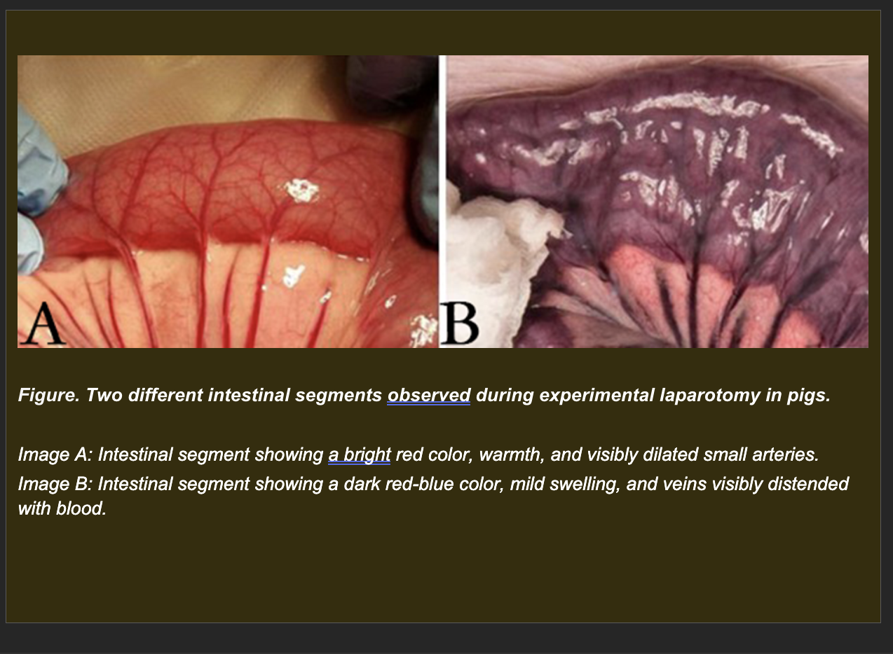
Session 4A
During an experimental laparotomy in pigs, two adjacent intestinal segments are observed simultaneously.
Segment A: Bright red, warm. Small arteries supplying this segment are visibly dilated.
Segment B: Dark red-blue. Slightly swollen. Veins draining this segment are visibly distended with blood.
Which interpretation best explains the difference between Segment A and Segment B?
a. Segment A shows active hyperemia from arteriolar dilation increasing oxygenated blood inflow; Segment B shows passive congestion from impaired venous outflow accumulating deoxygenated blood
b. Segment A shows passive congestion from venous obstruction; Segment B shows active hyperemia from arterial dilation
c. Both segments show hyperemia -- the color difference reflects metabolic rate differences between the two intestinal regions
d. Segment A shows ischemia from reduced perfusion; Segment B shows hyperemia from increased venous flow
a. Correct.
The gross color difference directly reflects the oxygenation state of blood. Bright red indicates oxygenated hemoglobin -- the blood is being actively delivered by arteriolar dilation (active process). Dark red-blue indicates deoxygenated hemoglobin -- blood is passively accumulating because venous outflow is impaired. The dilated arteries in Segment A confirm increased inflow; the distended veins in Segment B confirm outflow obstruction. These two opposite ends of the vascular circuit produce fundamentally different tissue appearances despite both resulting in increased blood volume.
Session 4A
Two animals present with visible fluid accumulation.
Animal A (Horse): Bilateral distal limb swelling. Venous pressure elevated. Plasma albumin normal. No fever or inflammatory signs. Edema is pitting and symmetrical.
Animal B (Cow): Ventral subcutaneous edema and abdominal distension from free abdominal fluid. Venous pressure normal. Plasma albumin markedly decreased. No fever or inflammatory signs. Abdominal fluid is clear and watery with low protein and very few cells.

Which mechanism best explains the edema in Animal A compared with Animal B?
a. Animal A: decreased plasma oncotic pressure from hypoalbuminemia. Animal B: increased hydrostatic pressure from venous hypertension
b. Animal A: increased vascular permeability from subclinical inflammatory disease. Animal B: lymphatic obstruction from abdominal lymph node enlargement
c. Animal A: increased hydrostatic pressure from elevated venous pressure driving net filtration from capillaries into tissue. Animal B: decreased plasma oncotic pressure from hypoalbuminemia reducing the osmotic force retaining fluid within the vascular space
d. Both animals have the same mechanism -- lymphatic obstruction -- but in different anatomic regions
c. Correct.
The clinical data map directly to each mechanism. Animal A's confirmed elevated venous pressure increases capillary hydrostatic pressure, shifting the Starling equilibrium toward net filtration -- fluid moves from the vascular space into the tissue. This produces pitting, symmetrical, dependent edema. Animal B's confirmed hypoalbuminemia reduces plasma oncotic pressure, the primary force retaining fluid within vessels. With reduced oncotic retention, fluid moves freely into the interstitium and body cavities, producing ventral subcutaneous edema and low-protein ascites.
Session 4A
Post-mortem examination of a horse reveals two obstructions in the mesenteric arterial circulation.
Specimen A (gross and microscopic): A firm, organized clot is firmly attached to the vessel wall of the cranial mesenteric artery at a site where the vessel lining is roughened, irregular, and disrupted. Microscopy shows fibrin, platelets, and erythrocytes adherent to a damaged area of endothelium.
Specimen B (gross and microscopic): A firm clot is lodged at a branch point of a smaller mesenteric artery. It is not attached to the vessel wall. The vessel wall at that location appears grossly normal on cut section. Microscopy shows a discrete clot surrounded by normal endothelium.

Which interpretation correctly identifies each obstruction and explains the mechanism of formation?
a. Specimen A is a thrombus that formed in situ at the site of endothelial disruption; Specimen B is an embolus that detached from the Specimen A site and traveled distally to lodge at the branch point
b. Specimen A is an embolus that lodged at the site and caused secondary endothelial disruption; Specimen B is a thrombus that formed in situ at the branch point without endothelial injury
c. Both are thrombi -- Specimen A is a recent thrombus and Specimen B is an older organized thrombus at a different stage of maturation
d. Both are emboli -- they represent two separate emboli that originated from different source sites in the systemic circulation
a. Correct.
Specimen A has the defining features of a thrombus: firm attachment to the vessel wall, associated endothelial disruption at the attachment site, and microscopic evidence of fibrin and platelets organized on the damaged endothelium. Thrombus formation requires endothelial injury or activation to initiate the coagulation cascade. Specimen B has the defining features of an embolus: non-attached position in the vessel lumen, vessel wall appearing normal at the lodgment site, and position at a branch point where luminal narrowing causes lodgment. The embolus most likely detached from the thrombus at Specimen A (or another similar site) and traveled with blood flow until it reached a vessel too small to pass through.
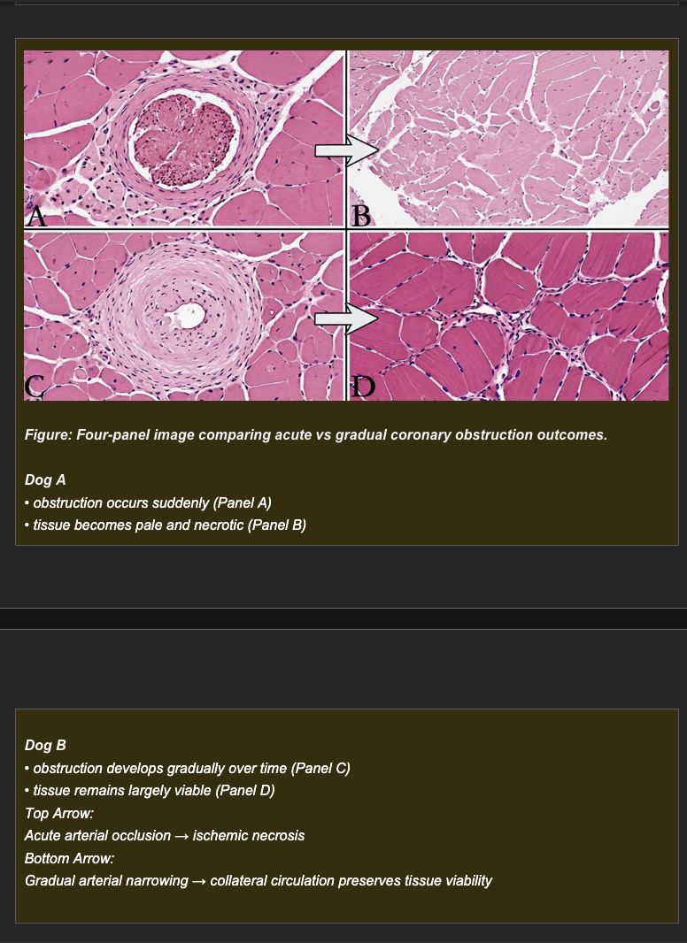
Session 4A
Two dogs each develop obstruction of the left anterior descending coronary artery.
Dog A: A thrombus acutely and completely occludes the vessel. Within 30 minutes, the myocardium supplied by the vessel is pale and begins to show histologic evidence of necrosis.
Dog B: Progressive stenosis from vascular disease narrows the same vessel over 6 weeks, reaching near-complete obstruction by week 6. At week 6, despite equivalent luminal narrowing, the myocardium supplied by the vessel is viable and shows only mild histologic changes.
Which mechanism best explains why Dog B's myocardium remains viable despite equivalent vascular narrowing?
a. Dog B's myocardium has a lower metabolic rate than Dog A's, requiring less oxygen and therefore tolerating the obstruction better
b. Dog B's vascular disease caused fibrous replacement of myocardium that is less metabolically active and less vulnerable to ischemia
c. Dog B received anti-coagulant treatment that prevented thrombus formation over the narrowed area
d. Gradual progression of obstruction in Dog B provided time for collateral vessels to develop and grow, supplying alternative perfusion routes to the at-risk myocardium despite near-complete primary vessel obstruction
d. Correct.
The rate of vascular obstruction is the critical variable. When a vessel narrows slowly, the progressive reduction in flow through the primary vessel creates a pressure gradient that stimulates pre-existing small collateral vessels to enlarge and proliferate. Over 6 weeks, Dog B developed an alternative supply network that maintained myocardial perfusion sufficient to prevent necrosis. Sudden complete occlusion in Dog A gives no time for this compensatory process -- the myocardium is immediately and completely deprived of its blood supply, and irreversible ischemic necrosis begins within minutes.
Session 4A
Hercules | Adult male Labrador | Intraoperative vascular complication
During orthopedic surgery on Hercules, a region of skeletal muscle near the surgical site undergoes sequential vascular changes over 90 minutes. Three observation points are recorded.
Stage 1 -- Observation at 15 minutes post-incision
The surgical region appears: bright red, warm. Small arteries in the region are visibly dilated. The tissue is well-perfused.
Which mechanism best explains the Stage 1 appearance?
a. Impaired venous drainage from surgical positioning causing passive congestion
b. Increased arterial inflow from arteriolar dilation producing active hyperemia
c. Sudden arterial obstruction causing ischemia with tissue pallor
d. Decreased plasma oncotic pressure causing edema formation with tissue swelling
b. Correct.
Bright red coloration (oxygenated blood), warmth, and visibly dilated arteries are the defining gross features of active hyperemia. Surgical tissue handling, inflammatory mediator release, and metabolic signals at the operative site drive arteriolar dilation and increased local blood flow. This is the expected early response to surgical tissue manipulation.
Session 4A
Hercules | Adult male Labrador | Intraoperative vascular complication
During orthopedic surgery on Hercules, a region of skeletal muscle near the surgical site undergoes sequential vascular changes over 90 minutes. Three observation points are recorded.
Stage 2 -- Observation at 45 minutes post-incision
A retractor is inadvertently compressing the venous outflow from the region. Now the tissue appears: dark red-blue, slightly swollen. Veins in the region are visibly distended. Temperature is slightly cooler than Stage 1.
Which mechanism best explains the Stage 2 change from Stage 1?
a. Continued arteriolar dilation has increased venous filling, changing the color to dark red
b. Acute arterial thrombosis has obstructed inflow, producing ischemia and early tissue pallor
c. Decreased plasma albumin from dilutional hypoalbuminemia during surgery is producing oncotic edema
d. Retractor compression of venous outflow is producing passive congestion -- deoxygenated blood accumulates as outflow is obstructed while arteriolar inflow continues
d. Correct.
The retractor physically compresses venous outflow. Arterial inflow continues (not obstructed), delivering oxygenated blood to the capillary bed. But venous outflow is impaired, preventing deoxygenated blood from leaving. The result: deoxygenated blood accumulates in the venous microvasculature, producing dark red-blue discoloration and venous distension. The slight cooling reflects impaired heat dissipation as blood flow velocity decreases. This is the transition from active hyperemia (Stage 1) to passive congestion (Stage 2) -- the same tissue, the same timeframe, a different vascular event.
Session 4A
Hercules | Adult male Labrador | Intraoperative vascular complication
During orthopedic surgery on Hercules, a region of skeletal muscle near the surgical site undergoes sequential vascular changes over 90 minutes. Three observation points are recorded.
Stage 3 -- Observation at 90 minutes post-incision
The retractor compression remains. Arterial blood flow has now also ceased -- combined venous and arterial obstruction. The tissue is now: pale, firm, cool. No pulses detectable in the affected vessels.
Which mechanism best explains the Stage 3 appearance -- and what tissue consequence is now inevitable?
a. Increased hydrostatic pressure from venous congestion is producing severe pitting edema with tissue pallor
b. Inflammatory mediator release from surgical trauma has produced severe arteriolar dilation causing hyperemia and tissue warmth
c. Complete cessation of arterial and venous blood flow has produced ischemia, and ischemic necrosis (infarction) is now occurring in the oxygen-deprived tissue
d. Sudden decrease in plasma albumin concentration from blood loss has reduced plasma oncotic pressure, causing fluid to leave the vasculature and producing pale, edematous tissue
c. Correct.
Stage 3 represents infarction: complete cessation of blood flow producing ischemic necrosis. The sequence is: hyperemia (Stage 1) → congestion (Stage 2, venous obstruction) → infarction (Stage 3, combined venous + arterial obstruction = complete ischemia). Pallor reflects absent blood delivery. Firmness reflects early coagulative necrosis as cellular proteins denature without oxygen for energy-dependent processes. Cool temperature confirms absent perfusion. Absent pulses confirm complete vascular obstruction. Ischemic necrosis is now inevitable -- the tissue has crossed the irreversible injury threshold.
Session 4B
A dog with severe, chronic liver disease is unable to synthesize adequate amounts of albumin. Shortly after, the dog develops a massive accumulation of fluid within the peritoneal cavity (ascites). According to the principles of Starling forces, which specific mechanism is responsible for this fluid accumulation?
A) Increased intravascular hydrostatic pressure forcefully pushing water into the abdomen.
B) Decreased intravascular oncotic (osmotic) pressure, leading to an inability to hold fluid within the vascular space.
C) Primary lymphatic obstruction preventing the normal drainage of interstitial fluid.
D) Severe endothelial cell necrosis leading to massive hemorrhage into the peritoneal cavity.
Correct Answer: B.
Albumin is the primary plasma protein responsible for maintaining intravascular oncotic (osmotic) pressure, which is the force that "pulls" or holds water inside the blood vessels. When the liver fails and albumin drops (hypoalbuminemia), the oncotic pressure decreases, and fluid passively leaks out into the interstitial spaces or body cavities, resulting in edema or ascites.
Session 4B
A veterinarian performs a fluid tap (abdominocentesis) on two different patients. Patient A has clear, watery fluid with very low protein and almost no cells. Patient B has thick, cloudy fluid with high protein content, fibrin, and massive numbers of neutrophils. Which of the following statements correctly conceptually distinguishes these two fluids?
A) Patient A's fluid is an exudate caused by inflammation, while Patient B's fluid is a transudate caused by high blood pressure.
B) Both fluids are exudates, but Patient A's fluid lacks protein because the animal is malnourished.
C) Patient A's fluid is a transudate caused by a pure hemodynamic imbalance (altered Starling forces), whereas Patient B's fluid is an exudate caused by increased vascular permeability due to inflammation.
D) Patient A's fluid is a transudate driven strictly by the active migration of leukocytes, whereas Patient B's fluid is an exudate driven passively by decreased oncotic pressure.
Correct Answer: C.
A transudate (Patient A) is an ultrafiltrate of blood plasma (low protein, low cellularity) that leaks out due to a pure hydrodynamic imbalance in Starling forces (e.g., increased hydrostatic pressure or decreased oncotic pressure) without vascular damage. An exudate (Patient B) is a protein-rich, cell-rich fluid that leaks out because inflammatory mediators have actively widened the gaps between endothelial cells (increased vascular permeability).
Session 4B
Fluid accumulation (edema) can range from a benign incidental finding to a rapidly fatal event depending entirely on the anatomical location and tissue compliance. Based on the pathology of cell injury, which of the following scenarios best demonstrates the most critical clinical consequence of edema?
A) Subcutaneous edema in the ventral abdomen causing immediate cessation of gastrointestinal motility.
B) Cytotoxic edema in the central nervous system (brain), where the rigid cranium provides no room for expansion, leading to a catastrophic increase in intracranial pressure and fatal herniation.
C) Interstitial edema in skeletal muscle leading directly to the systemic release of massive amounts of calcium.
D) Edema within the adipose tissue causing irreversible saponification and fat necrosis.
Correct Answer: B.
The clinical relevance of edema is heavily dictated by whether the tissue can safely expand to accommodate the fluid. The brain is encased in a rigid bone box (the cranium). As described in the text regarding cytotoxic edema, even a slight increase in fluid volume in the brain has catastrophic consequences, causing the brain to swell, compress vital vascular supplies, and herniate through the foramen magnum, leading to death.
Session 4B
An animal suffering from acute bacterial pneumonia has lungs that are heavy, wet, and consolidated. Microscopic examination reveals that the alveoli are completely filled with a protein-rich fluid containing fibrin and millions of dead neutrophils. What is the primary pathophysiological mechanism driving the formation of this specific type of fluid?
A) Inflammation-induced increase in endothelial permeability, allowing large plasma proteins and leukocytes to escape the vasculature and form an exudate.
B) A profound drop in systemic oncotic pressure forcing a transudate to passively fill the lung lobes.
C) Left-sided heart failure causing pure hydrostatic pressure to force a protein-poor transudate into the alveolar spaces.
D) A genetic defect in the sodium-potassium (Na+/K+) ATPase pumps resulting in localized cellular shrinkage.
Correct Answer: A.
The presence of high protein, fibrin, and neutrophils definitively identifies this fluid as an exudate. Exudates form during acute inflammation when chemical mediators cause endothelial cells to contract, increasing vascular permeability. This allows large molecules (like fibrinogen) and leukocytes to leave the blood vessels and enter the tissue/alveoli to fight the infection.
Session 4B
A student is reviewing the Starling forces that govern fluid movement across capillary walls to understand why a patient developed edema. Which of the following statements correctly identifies the role of these forces under normal conditions?
A) Hydrostatic pressure retains fluid within the vessels, while oncotic pressure drives fluid into the interstitium.
B) Both hydrostatic and oncotic pressures are outward forces that drive fluid into the interstitial space.
C) Hydrostatic pressure drives fluid out of the vessels into the interstitium, while oncotic pressure retains fluid within the vessels by osmotic attraction.
D) Oncotic pressure and hydrostatic pressure both function to retain fluid inside the vessels, counteracting lymphatic drainage.
Correct Answer: C.
Capillary hydrostatic pressure promotes movement of fluid from the vascular space into the interstitium. Plasma oncotic pressure, maintained primarily by albumin, attracts water and helps retain fluid within the vascular space. Lymphatic drainage returns excess interstitial fluid and proteins to the circulation.
Session 4B
A veterinarian analyzes pleural fluid samples from two different dogs. Dog X's fluid has a specific gravity of 1.032, total protein of 4.5 g/dL, and a highly cellular profile with predominantly neutrophils. Dog Y's fluid has a specific gravity of 1.009, total protein of 0.9 g/dL, and very few cells. Which classification correctly describes these fluids and their underlying mechanisms?
A) Dog X: transudate from oncotic imbalance; Dog Y: exudate from inflammatory permeability.
B) Dog X: exudate from increased vascular permeability; Dog Y: transudate from hydrostatic or oncotic imbalances with intact vascular permeability.
C) Both fluids are exudates, but Dog Y is in the early stages of inflammation.
D) Dog X: modified transudate from lymphatic obstruction; Dog Y: pure transudate from heart failure.
Correct Answer: B.
Dog X has a protein-rich and highly cellular fluid, consistent with an exudate caused by inflammation-induced increases in vascular permeability. Dog Y has a protein-poor and cell-poor transudate, consistent with altered hydrostatic or oncotic forces while vascular permeability remains relatively intact.
Session 4B
A dog initially presents with a pure transudate in its pleural space due to elevated venous pressure from right-sided heart failure. Two weeks later, the heart failure persists, but a repeat fluid analysis shows intermediate protein (2.6 g/dL) and increased cellularity. What is the most likely explanation for this change in fluid character?
A) The right heart failure has completely resolved, and a primary lymphatic obstruction has taken its place.
B) The fluid is now entirely an inflammatory exudate, and the hydrostatic mechanism is no longer contributing.
C) A severe drop in plasma albumin suddenly converted the transudate into an exudate.
D) Both mechanisms are now contributing: ongoing hydrostatic pressure from heart failure combined with pleural irritation from chronic fluid accumulation, which adds a mild inflammatory permeability component.
Correct Answer: D.
Persistent hydrostatic pressure continues to promote fluid accumulation. Over time, chronic fluid accumulation can irritate the pleural lining and produce a mild inflammatory response, increasing the protein concentration and cellularity. The resulting fluid is consistent with a modified transudate.
Session 4B
An acutely dyspneic dog undergoes emergency thoracocentesis, yielding turbid, yellow-orange fluid. The analysis reveals a total protein of 5.8 g/dL and a nucleated cell count of 28,000 cells/uL with a strong predominance of degenerate neutrophils. Which interpretation best explains these findings?
A) The fluid is a transudate caused by worsening systemic hypertension.
B) The fluid is a modified transudate resulting from simultaneous hypoalbuminemia and heart failure.
C) The fluid is an exudate from a bacterial pyothorax (septic infection), characterized by increased permeability and neutrophil death.
D) The fluid is an exudate caused strictly by severe lymphatic obstruction in the thoracic cavity.
Correct Answer: C.
The high protein concentration, high nucleated cell count, turbidity, and predominance of degenerate neutrophils indicate a septic exudate. Bacterial inflammation increases vascular permeability, allowing plasma proteins and leukocytes to enter the pleural space. Bacterial toxins can contribute to neutrophil degeneration and death.
Session 4B
A cow develops severe, firm, non-pitting swelling confined solely to the right hindlimb. Central venous pressure and plasma albumin levels are completely normal. Physical exam reveals a markedly enlarged right inguinal lymph node but no local heat or systemic fever. Which mechanism best explains this unilateral edema?
A) Decreased plasma oncotic pressure due to severe hypoalbuminemia.
B) Increased hydrostatic pressure from systemic right-sided heart failure.
C) Regional lymphatic obstruction preventing the clearance of interstitial fluid and proteins from the affected limb.
D) Increased vascular permeability from an acute, subclinical systemic infection.
Correct Answer: C.
The unilateral distribution and enlarged regional lymph node support obstruction of lymphatic drainage. Impaired lymphatic clearance causes protein-rich interstitial fluid to accumulate in the affected limb. Chronic lymphatic edema can become firm and non-pitting because of persistent protein accumulation and tissue fibrosis.
Session 4B
Two cats experience sudden internal bleeding following trauma, accumulating exactly 60 mL of fluid. Cat A's fluid accumulates in the pleural space, while Cat B's fluid accumulates in the pericardial sac. Which statement best explains why Cat B is in immediate life-threatening cardiovascular collapse while Cat A only shows moderate respiratory signs?
A) Pleural fluid directly enters the systemic circulation, whereas pericardial fluid destroys the heart muscle.
B) The pericardial sac is rigid and non-compliant; the fluid rapidly compresses the heart and severely limits ventricular diastolic filling (cardiac tamponade).
C) The pleural space cannot expand at all, while the pericardial sac is highly compliant and easily stretches.
D) Both cavities are equally affected by 60 mL of fluid, but Cat B has a pre-existing blood clotting disorder.
Correct Answer: B.
The pericardial sac has limited ability to accommodate rapidly accumulating fluid. Increased pericardial pressure compresses the heart and prevents adequate ventricular filling during diastole, producing cardiac tamponade and a rapid decline in cardiac output. The pleural cavity can generally accommodate the same volume with less immediate cardiovascular compromise.
Session 4B
A dog develops fluid accumulation in the pleural space. To understand why fluid is leaving the capillaries and entering the pleural space rather than remaining within the vessels, a student reviews the Starling forces governing transcapillary fluid movement. Which statement correctly identifies the role of each force?
a. Hydrostatic pressure retains fluid in vessels; oncotic pressure drives fluid into the interstitium
b. Hydrostatic pressure drives fluid out of vessels into the interstitium; oncotic pressure retains fluid within vessels by osmotic attraction
c. Both hydrostatic and oncotic pressure drive fluid out of vessels; lymphatics are the only force retaining fluid
d. Oncotic pressure and hydrostatic pressure both retain fluid in vessels under normal conditions
b. Correct.
Under normal conditions, capillary hydrostatic pressure (generated by cardiac output and blood pressure) exceeds interstitial hydrostatic pressure, producing net outward filtration at the arteriolar end of capillaries. Plasma oncotic pressure (generated by albumin and other plasma proteins) exceeds interstitial oncotic pressure and acts as an osmotic retaining force, drawing fluid back into the vessel at the venous end and resisting excessive filtration. Edema and effusion develop when this balance is disrupted -- either by increased hydrostatic pressure, decreased oncotic pressure, increased permeability, or lymphatic failure.
Session 4B
A veterinarian drains fluid from the chest of a dog with unexplained pleural effusion. The fluid characteristics are analyzed: specific gravity 1.030, total protein 4.2 g/dL, white blood cell count 8,500 cells/uL with predominantly neutrophils. A second dog has pleural fluid with specific gravity 1.010, total protein 0.8 g/dL, and white blood cell count 150 cells/uL. Which classification best describes each fluid and what does the difference indicate?
a. Dog 1: transudate from oncotic imbalance. Dog 2: exudate from inflammatory permeability
b. Dog 1: modified transudate from lymphatic obstruction. Dog 2: pure transudate from hydrostatic pressure
c. Both fluids are exudates -- the difference in protein reflects different stages of inflammation
d. Dog 1: exudate from increased vascular permeability allowing protein and leukocytes to escape. Dog 2: transudate from hydrostatic or oncotic imbalance with intact vascular permeability
d. Correct.
Dog 1's fluid has the defining features of an exudate: high protein (>3 g/dL), high cellularity (>1,500 cells/uL), neutrophil predominance, and high specific gravity (>1.018). These features indicate that the vascular permeability barrier has been breached -- plasma proteins and leukocytes are entering the pleural space. This occurs in inflammatory, infectious, or neoplastic conditions. Dog 2's fluid has the defining features of a transudate: low protein (<2.5 g/dL), low cellularity (<300 cells/uL), and low specific gravity (<1.015). These features indicate intact vascular permeability -- only the low-protein filtrate expected from hydrostatic or oncotic imbalance is accumulating.
Session 4B
Two cats each develop 200 mL of fluid accumulation. Cat A has the fluid in the pleural space. Cat B has the fluid in the pericardial space. Both volumes of fluid are equivalent. Which statement best explains why the clinical consequences differ despite equivalent fluid volumes?
a. Pleural fluid impairs respiratory function by compressing lung tissue and limiting expansion; pericardial fluid impairs cardiac function by compressing the heart and limiting ventricular filling
b. Both cavities are equally affected -- 200 mL produces the same degree of organ compression regardless of location
c. Pericardial fluid is always more dangerous because the heart is smaller than the lungs
d. Pleural fluid causes cardiac tamponade; pericardial fluid causes respiratory compromise
a. Correct.
The clinical significance of an effusion is determined by the functional vulnerability of the organ being compressed. Pleural fluid accumulates in the space where the lungs expand during inspiration. As fluid volume increases, it compresses lung parenchyma and limits tidal volume, producing respiratory compromise (dyspnea, tachypnea, reduced exercise tolerance). Pericardial fluid accumulates in the rigid pericardial sac surrounding the heart. As fluid volume increases, it compresses the myocardium and limits ventricular filling during diastole -- a condition called cardiac tamponade -- reducing cardiac output and producing cardiovascular compromise. The pericardial sac is a rigid, relatively non-compliant structure, making even moderate fluid volumes potentially life-threatening.