MIT211 Lec Exam #2
 Call Kai
 Learn
Learn Practice Test
 Spaced Repetition
 Match
Match Flashcards
 Knowt Play
1/155

There's no tags or description
Looks like no tags are added yet.
Name | Mastery | Learn | Test | Matching | Spaced | Call with Kai | Chat |
|---|
No analytics yet
Send a link to your students to track their progress
156 Terms
Health Physics
The study of the protection of people and the environment from the harmful effects of radiation.
5-minute reset timer
A timer used during fluoroscopy exams to remind technologists to reassess exposure and patient positioning.
HVL
Half-Value Layer, the thickness of a specified material required to reduce the intensity of radiation by half.
Tissue Weighting Factor
A factor used to account for the different sensitivities of various tissues to radiation exposure.
Entrance Skin Dose
The amount of radiation absorbed by the skin at the point of entrance of the x-ray beam.
Gonadal Dose
The amount of radiation dose received by the reproductive organs.
Dose Equivalent
A measure of the biological effect of radiation, taking into account the type of radiation and its energy.
Genetically Significant Dose
The dose of radiation that, if received by a population, would be expected to cause a genetic effect.
Maximum Permissible Dose
The maximum dose of radiation that a radiation worker can be exposed to in a given time period.
Cardinal Principles of Radiation Protection
Time, Distance, and Shielding are the three fundamental principles to minimize radiation exposure.
Time
The dose is directly related to the duration of radiation exposure; reducing exposure time reduces dose.
Calculating Exposure
Exposure rate X Exposure time = Exposure.
Distance
As the distance between the source of radiation and the patient increases, the exposure decreases, following the Inverse Square Law.
Shielding
Material placed between the patient and the radiation source to reduce exposure.
Effective Dose
A dose equivalent that accounts for the risk of exposure to different tissues and organs.
Occupational Dose
The radiation dose received by workers in the course of their employment.
Fluoroscopy
An imaging technique that uses X-rays to obtain real-time moving images of the interior of a body.
Patient Dose Management
The practice of monitoring and minimizing the radiation dose received by patients during medical imaging.
X-ray Ordering Increase
The increase in the ordering of X-rays by 18% each year.
Medical X-ray Exposure Concern
The growing concern regarding the risks associated with medical x-ray exposure.
Entrance Skin Dose (ESD)
Often referred to as the 'Patient Dose'; easy to measure and reasonably accurate; estimates can be made in absence of measurements.
Mammography
A special examination type used in imaging.
Computed Tomography
A special examination type used in imaging.
Unnecessary Exams
Exams not under control, such as screenings, hospital admissions, pre-employment physicals, health exams, ER visits, and whole-body CT screenings.
Repeat Examinations
Examinations that are under control; digital imaging is meant to help maintain the repeat rate within limits; QC testing done every quarter should stay between 4-6%.
Pregnant Patient
The most critical time for a pregnant patient to be irradiated is within the first 2 weeks of conception; patients are asked for the 'first day of the last menstrual cycle' and must sign paperwork stating they are not pregnant before proceeding.
Occupational Radiation Exposure
Refers to the risk associated with radiation exposure for personnel.
Dose Limits
The recommended dose limit for radiologist personnel is .5 Sv/yr (5000 mrem/yr).
Highest Occupational Exposure
Occurs in fluoroscopy, mobile, surgery, and interventional areas.
Maximum Permissible Dose (MPD)
Older term replaced with dose limit (DL); refers to the dose expected not to produce a significant radiation effect.
Whole body DL
50 mSv/yr (5000 mrem/yr).
Skin DL
500 mSv/yr (50 mrem/yr).
Extremities DL
500 mSv/yr (50 mrem/yr).
Lens of the eye DL
150 mSv/yr (15 mrem/yr).
General Public DL
1 mSv/yr (100 mrem/yr).
Film Badges
Worn at the collar to measure thyroid and lens dose; must be worn on the outside of the lead apron at the collar level during fluoroscopy or surgery.
Protective Apparel
Includes protective gloves and aprons; required thickness is .25mm Pb, normal is .5mm Pb.
Holding Patients
Refers to the practice of holding patients during examinations.
Imaging Plate
IP = imaging plate
Work Load
Work Load
Direct Digital Radiography
Direct Digital Radiography
Indirect Digital Radiography
Indirect Digital Radiography
Computed Radiography
Process was first introduced in the United States by Fuji Medical Systems of Japan in 1983.
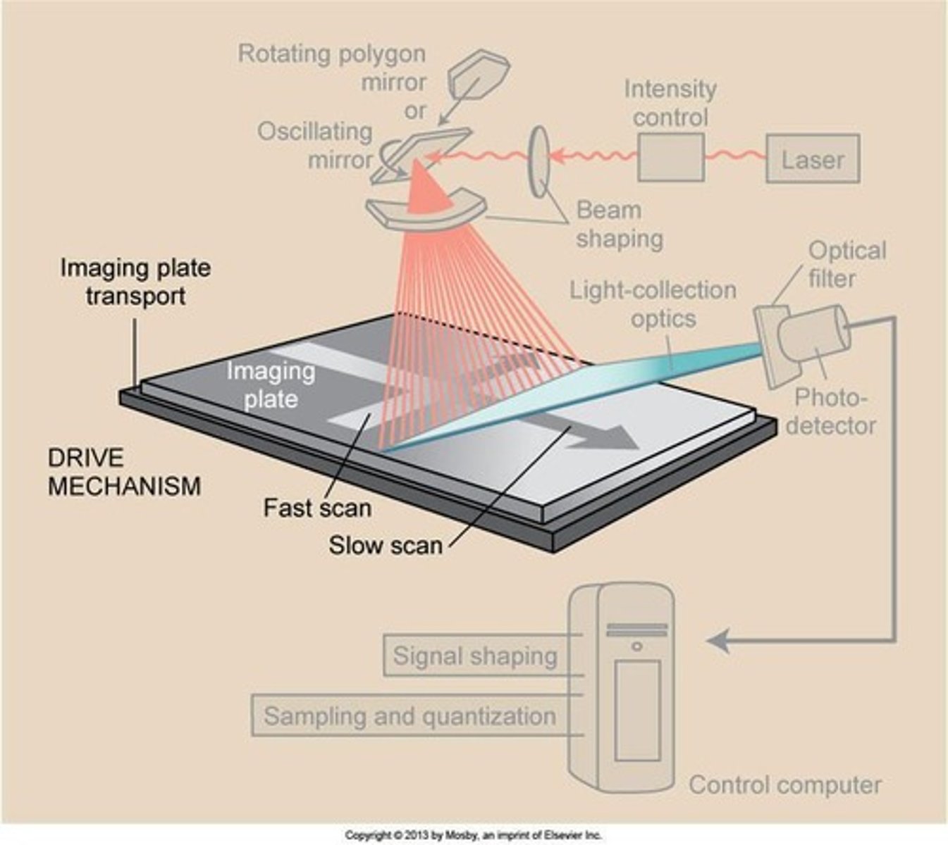
Storage phosphor plates
Similar to intensifying screens. Stores x-ray energy for an extended time.
Erasure cycle
What happens? Computed Radiography Cassettes
Photostimulable Luminescence
A process where materials emit light some time later when exposed to a different light source.
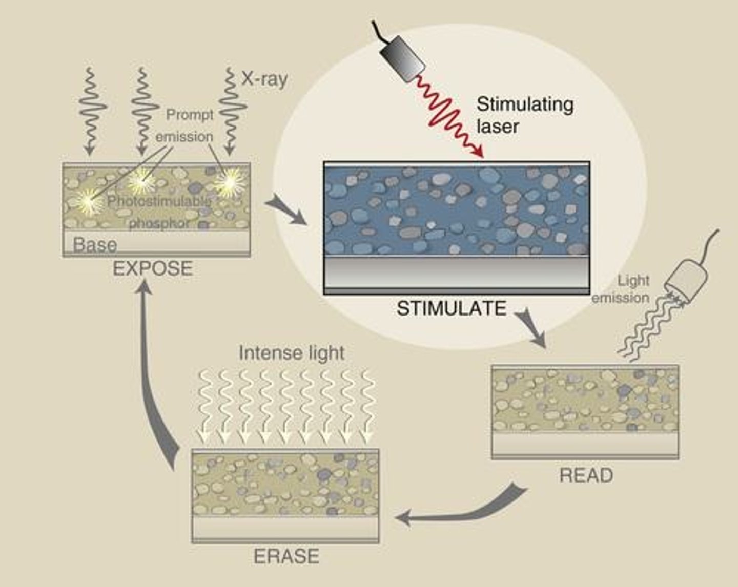
Photostimulable Phosphor
PSP = photostimulable phosphor
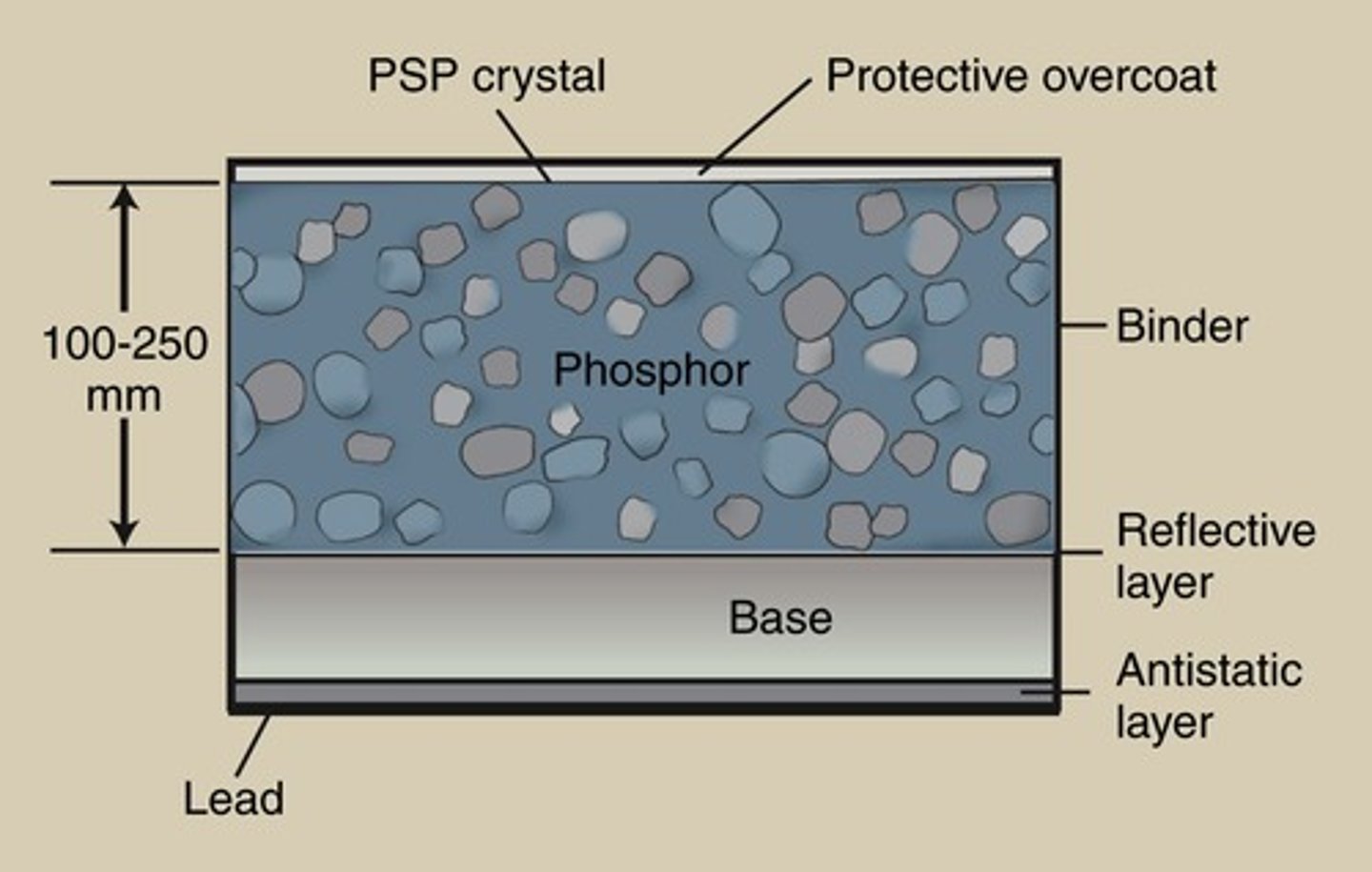
Storage Phosphor
SP = storage phosphor
Storage Phosphor Screen
SPS = storage phosphor screen
Cassetteless system
Uses a flat panel detector or charge-coupled device (CCD) hard-wired to computer.
Digital Radiography
Two types of digital radiography: Indirect capture DR and Direct capture DR.
Indirect capture DR
X-rays absorbed and converted to light. CCD or thin-film transistor (TFT) converts light to electric signals.
Computed Radiography vs. Digital Radiography
Which is more efficient? Which more costly?
Equipment for Computed Radiography
Traditional x-ray room with a table and wall Bucky required.
Equipment for Digital Radiography
A detector replaces the Bucky apparatus in the table and wall stand.
DR
Digital Radiography, which is much more efficient and provides immediate image availability.
CR
Computed Radiography, where the computer is located near the readers.
Image Processing
The process that takes place in a computer for both CR and DR systems.
Scatter
Both acquisition systems are sensitive to scatter.
Noise
Can be seen with low mAs values on both CR and DR acquisition systems.
Post-Processing
Techniques that allow manipulation of gray scale and sharpness of the image to enhance diagnostic interpretation.
Image Enhancement
Techniques used to improve the quality of images.
Window Level
A post-processing technique that adjusts the brightness of an image.
Window Width
A post-processing technique that adjusts the contrast of an image.
Image Restoration
A post-processing technique that aims to recover lost image details.
Image Compression
A technique used to reduce the file size of images.
PACS
Picture Archiving and Communication System, a networked group of computers, servers, and archives to store digital images.
DICOM format
A standard format that PACS can accept for images.
HIS
Hospital Information System, which PACS must interface with.
RIS
Radiology Information System, which PACS must interface with.
PACS Components
Includes reading stations, physician review stations, specialty workstations, web access, technologist quality control stations, administrative stations, and archive systems.
PACS Uses
Facilitates the display of digital images for diagnostic interpretation and review purposes.
Radiologist Monitor
A high-resolution monitor used for diagnostic interpretation.
Technologist Monitor
A desktop workstation used by technologists.
PACS Administrator
Personnel trained in IT/Computer Technology to ensure the integrity of digital imaging and communication systems.
QC
Quality Control, performed by the PACS Administrator.
Ongoing Education
Provided by the PACS Administrator to system users.
Beam restrictors
Devices meant to restrict the beam as the x-rays leave the tube.
Positive Beam Limiting
A device that automatically collimate the beam to the exact size of the cassette when it is locked into place.
Radiographic Grid
A device located between the patient and the image receptor that reduces the amount of scatter reaching the image receptor and improves contrast.
Grid cut off
A phenomenon where the grid absorbs too many primary x-rays, leading to a decrease in image receptor exposure.
Grid Ratio
The height of the grid divided by the interspace width; the higher the ratio, the more effective it is in reducing scatter.
Field Size/Collimation
The size of the area being imaged, which influences the amount of scatter produced.
kVp
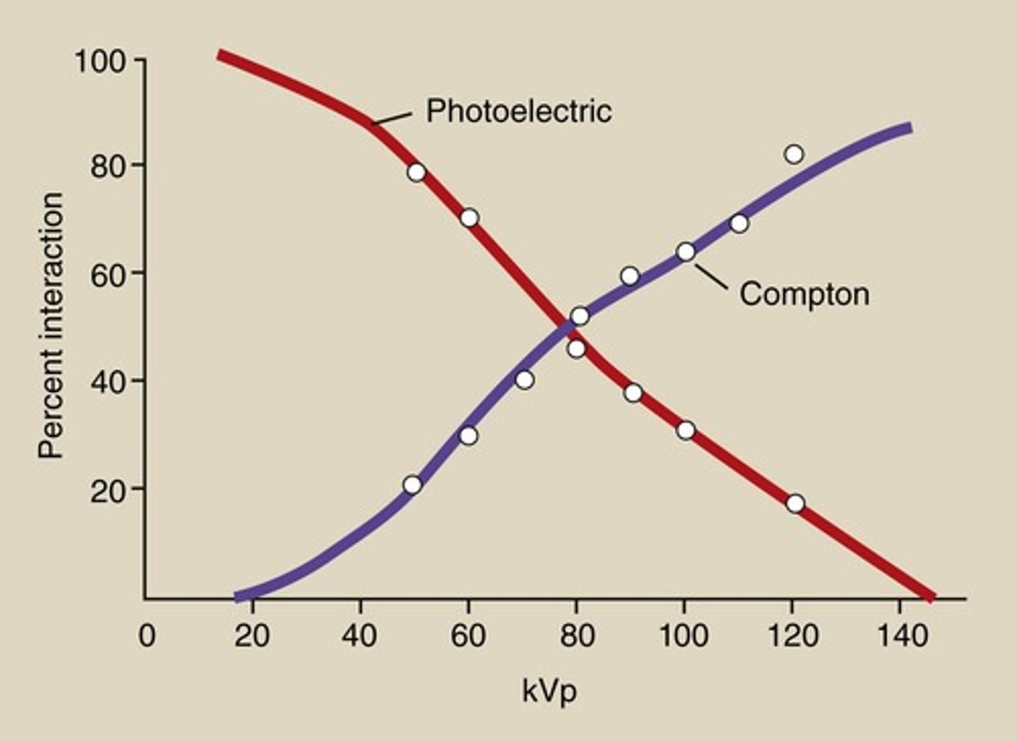
Kilovolt peak; as x-ray energy increases, the number of Compton (scatter) interactions decreases, but the number of Photoelectric interactions decreases much more rapidly.
Patient Thickness
Thicker body parts result in more scatter production than thinner body parts; increased thickness leads to greater average angles of scatter.
Aperture Diaphragm
The simplest type of beam restrictor.
Cones/Cylinders
An extension of the diaphragm that provides more restriction but is heavy to use.
Variable Aperture Diaphragm
A type of beam restrictor that offers a variety of sizes.
Collimators
Devices that allow for a variety of sizes and are commonly used in practice today.
Contrast
The degree of difference between the light and dark areas of an image.
Bucky
A device invented to reduce scatter radiation reaching the image receptor.
Radiopaque material
Material that does not allow x-rays to pass through, used in grid strips.
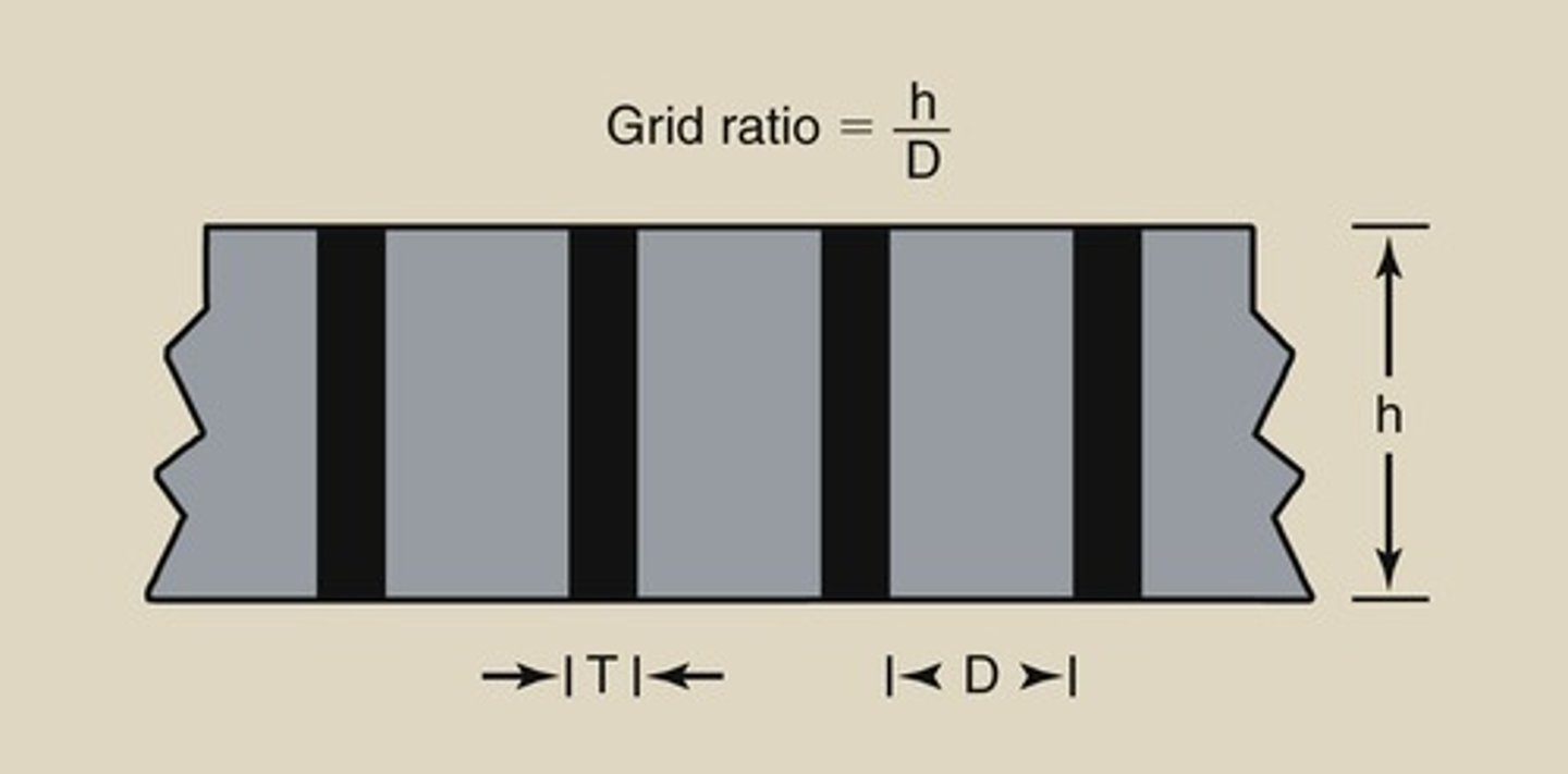
Radiolucent material
Material that allows x-rays to pass through, used as interspace material in grids.
Scatter Radiation
Radiation that is deflected from its original path due to interactions with matter.
Receptor Exposure
The amount of radiation that reaches the image receptor.
Image Receptor
The device that captures the x-ray image, such as film or digital sensors.
Field of View
The area that is visible in the imaging process, which can be affected by collimation.
Postprocessing
Techniques applied after image acquisition to enhance image quality without increasing patient radiation dose.
Grid Frequency
The number of grid strips per centimeter.
Higher Frequency Grids
The higher the frequency = more lead = increased ability to absorb scatter.