abd 3 prep
 Call Kai
 Learn
Learn Practice Test
 Spaced Repetition
 Match
Match Flashcards
 Knowt Play
1/206

There's no tags or description
Looks like no tags are added yet.
Name | Mastery | Learn | Test | Matching | Spaced | Call with Kai | Chat |
|---|
No analytics yet
Send a link to your students to track their progress
207 Terms
Abscess
Irregular shape
Variable echogenicities
May contain gas
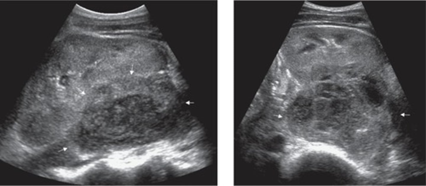
Abdominal Wall Tear/Trauma
Area of tear = loss of muscle striations, complex appearance with hematoma and muscle swelling/thickness
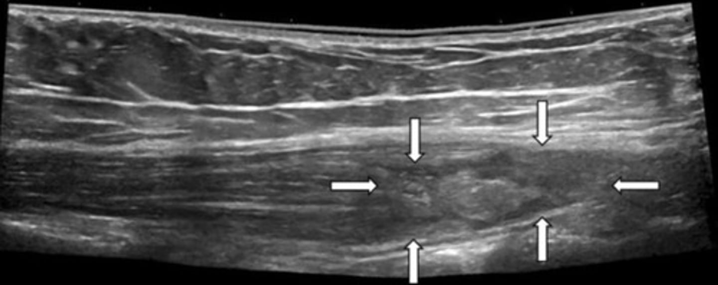
Abdominal Wall Penetration Trauma
Hypoechoic penetration tract where muscles were separated -> will heal hyperechoic
Hematoma "Life" Cycle
Anechoic
Increased size and echogenicity
Solid, mid-level grey
Decreased size, complex
Seroma - cystic
Reabsorption and/or scar tissue
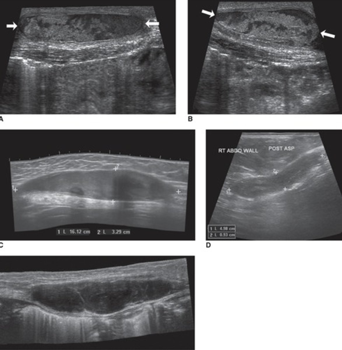
Abdominal Hernia
Opening in abdominal wall with contents protruding through
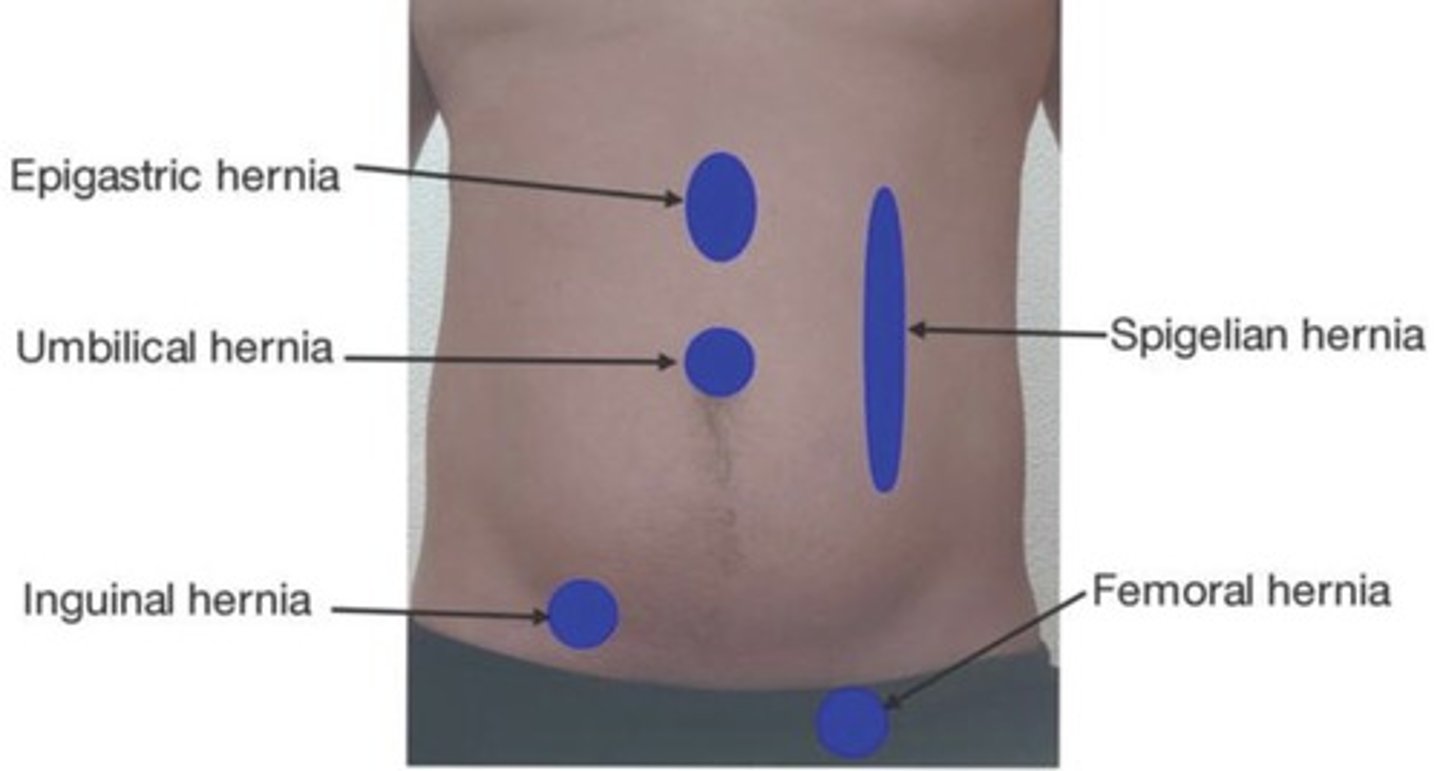
Ventral/Anterior Abdominal Wall Hernia
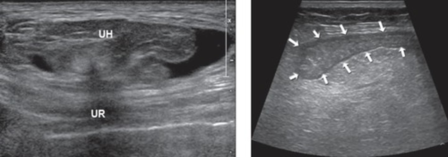
Spogelian Hernia
Along Spigelian fascia
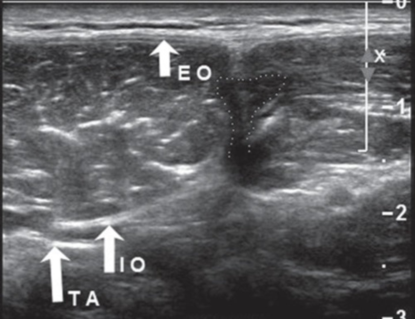
Inguinal Hernia
Direct = contents do not pass through inguinal ring
Indirect = contents pass through inguinal ring
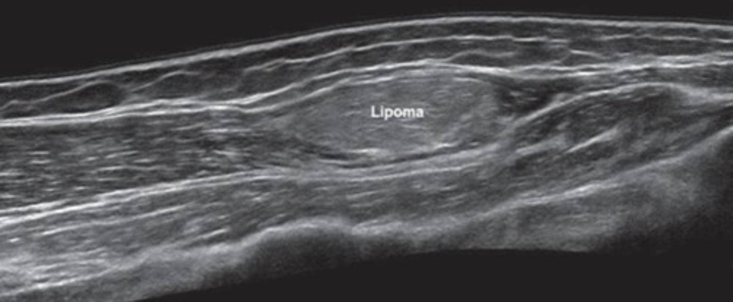
Lipoma
Benign, fatty tumor
Isoechoic (mostly) to hyperechoic
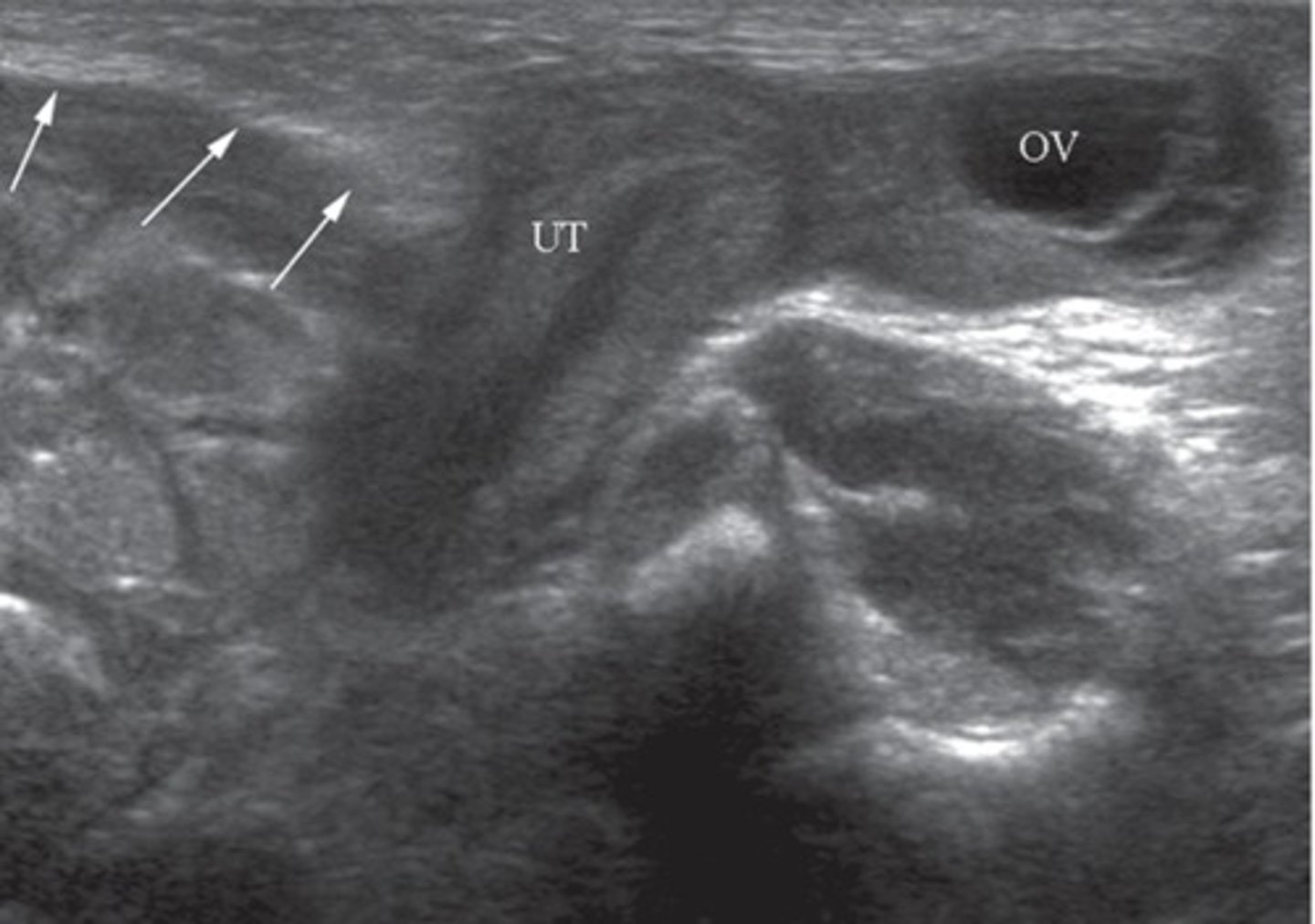
Malignant abdominal wall neoplasms
Irregular shape
Hyperechoic to complex
Inhomogenous
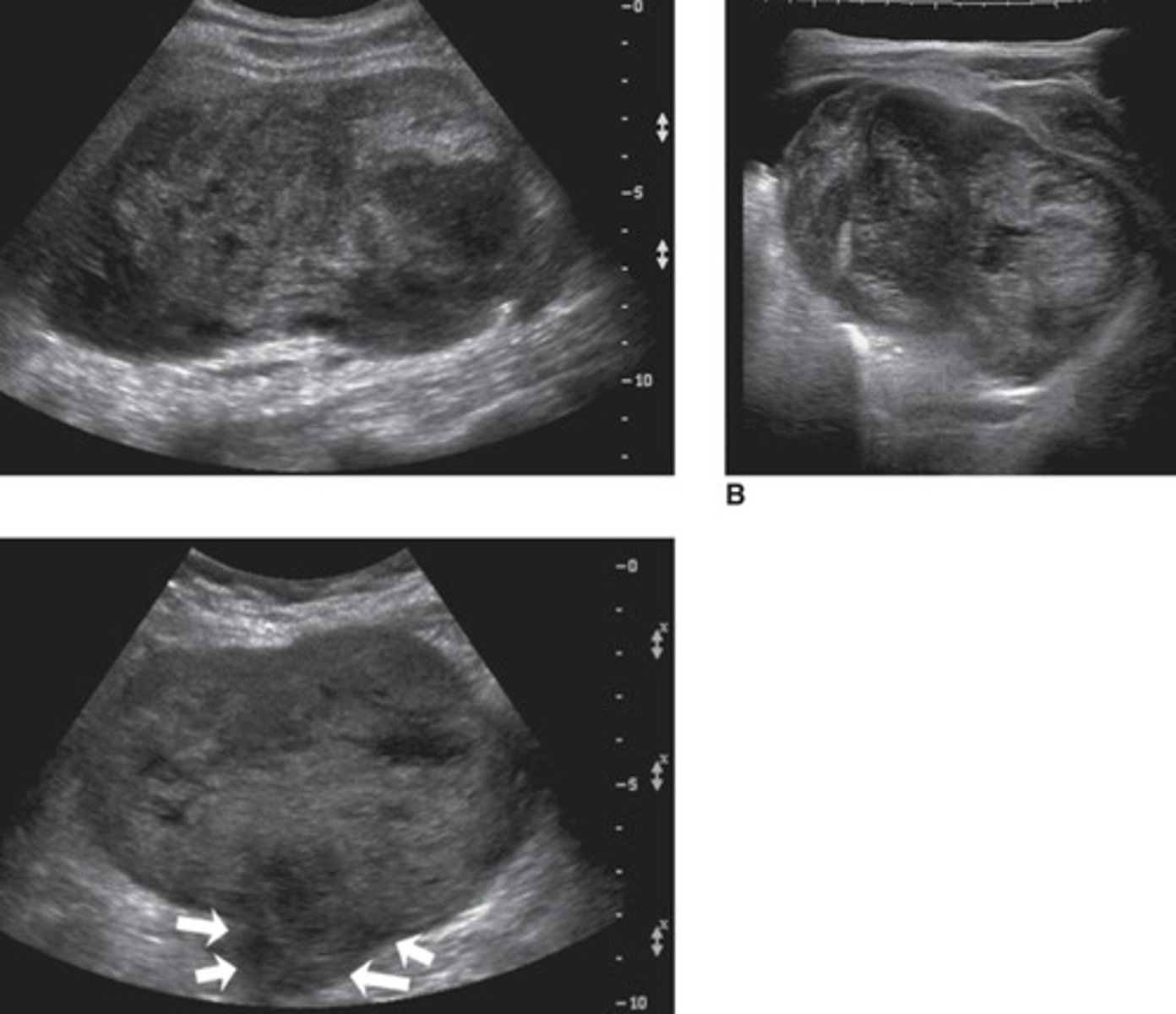
Diaphragm Paralysis
Damaged phrenic nerve
Can be unilateral or bilateral
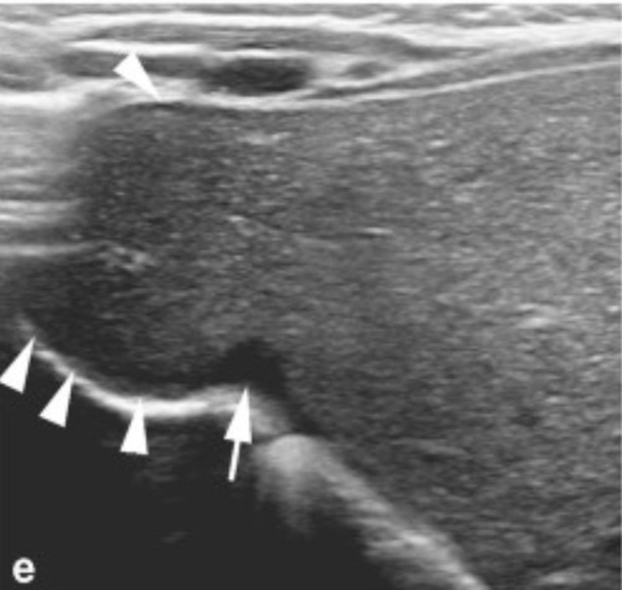
Diaphragmatic Eventration
Denting of diaphragm
Diaphragm does not function properly = breathing issues
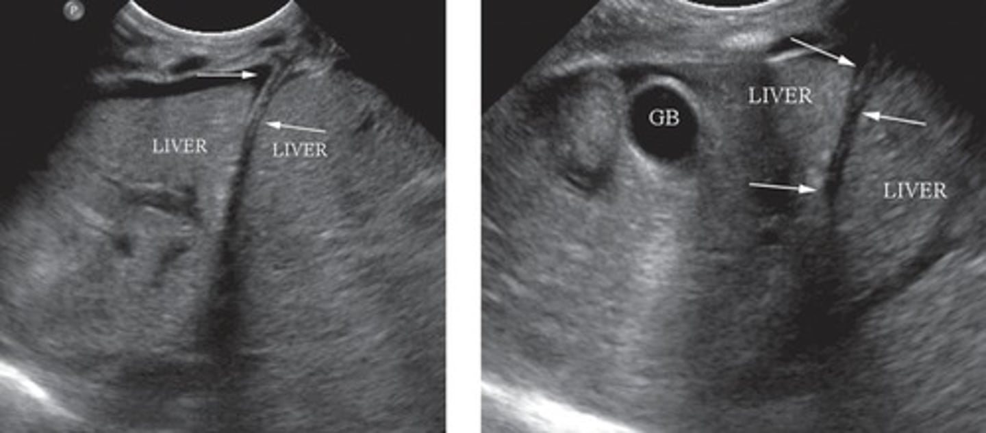
Diaphragmatic Hernia
Abdominal contents enter thorax
Congenital issue
Diaphragmatic Inversion
Little movement of diaphragm with respiration
Sonographic Appearance of Fatty Infiltration
Increased echogenicity
Decreased penetration
Focal Fatty Sparing
Entire liver is fatty with small area of normal tissue
Focal Fatty Infiltration
Entire liver is normal with small area of fatty tissue
Causes of Fatty Infiltration
Alcoholism
Diabetes
Obesity
Hepatitis
Steroids
Chronic illness
Chemotherapy
Grade 1 Fatty Infiltration
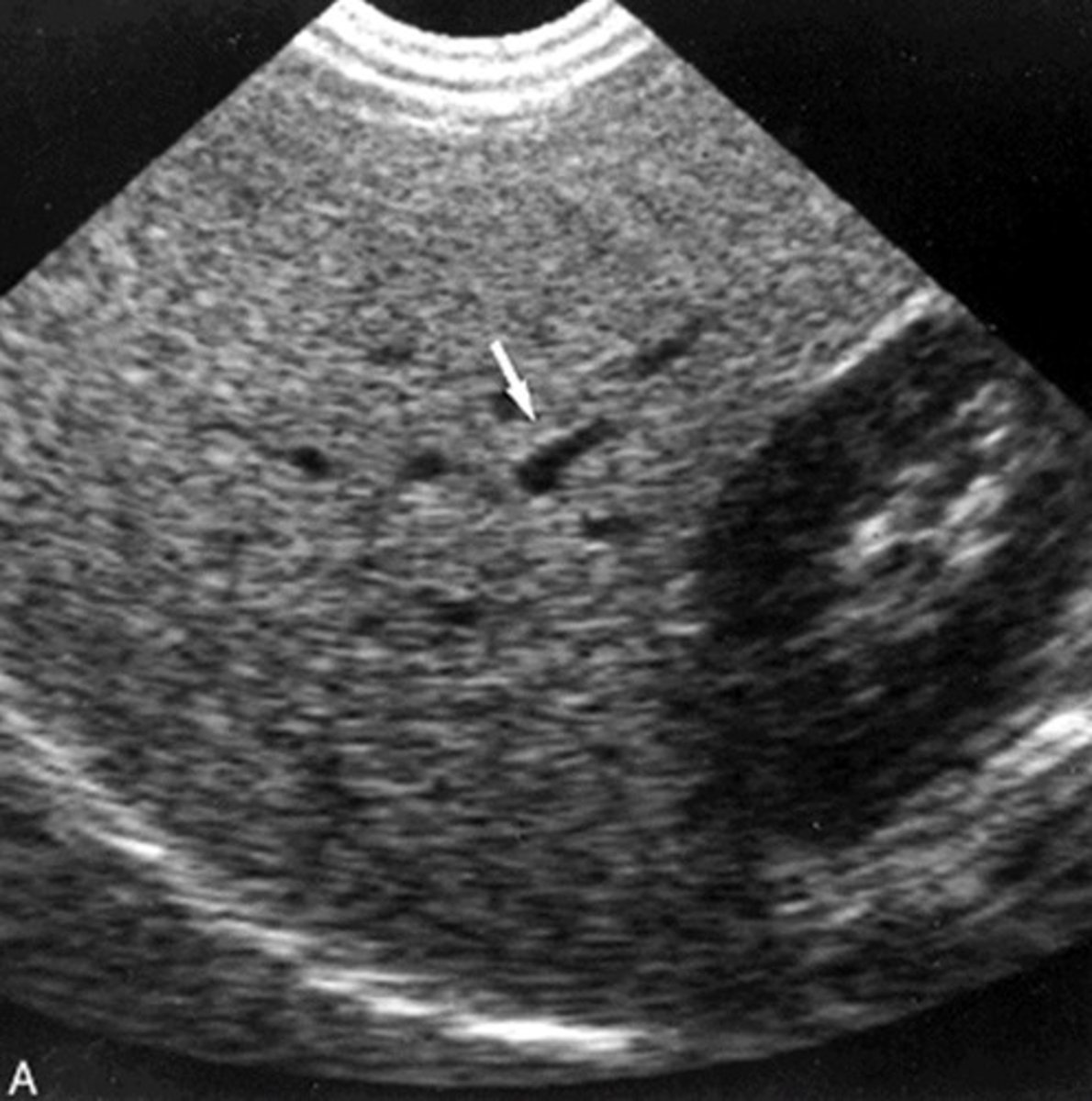
Grade 2 Fatty Infiltration
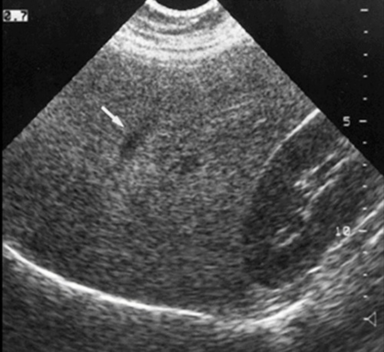
Grade 3 Fatty Infiltration
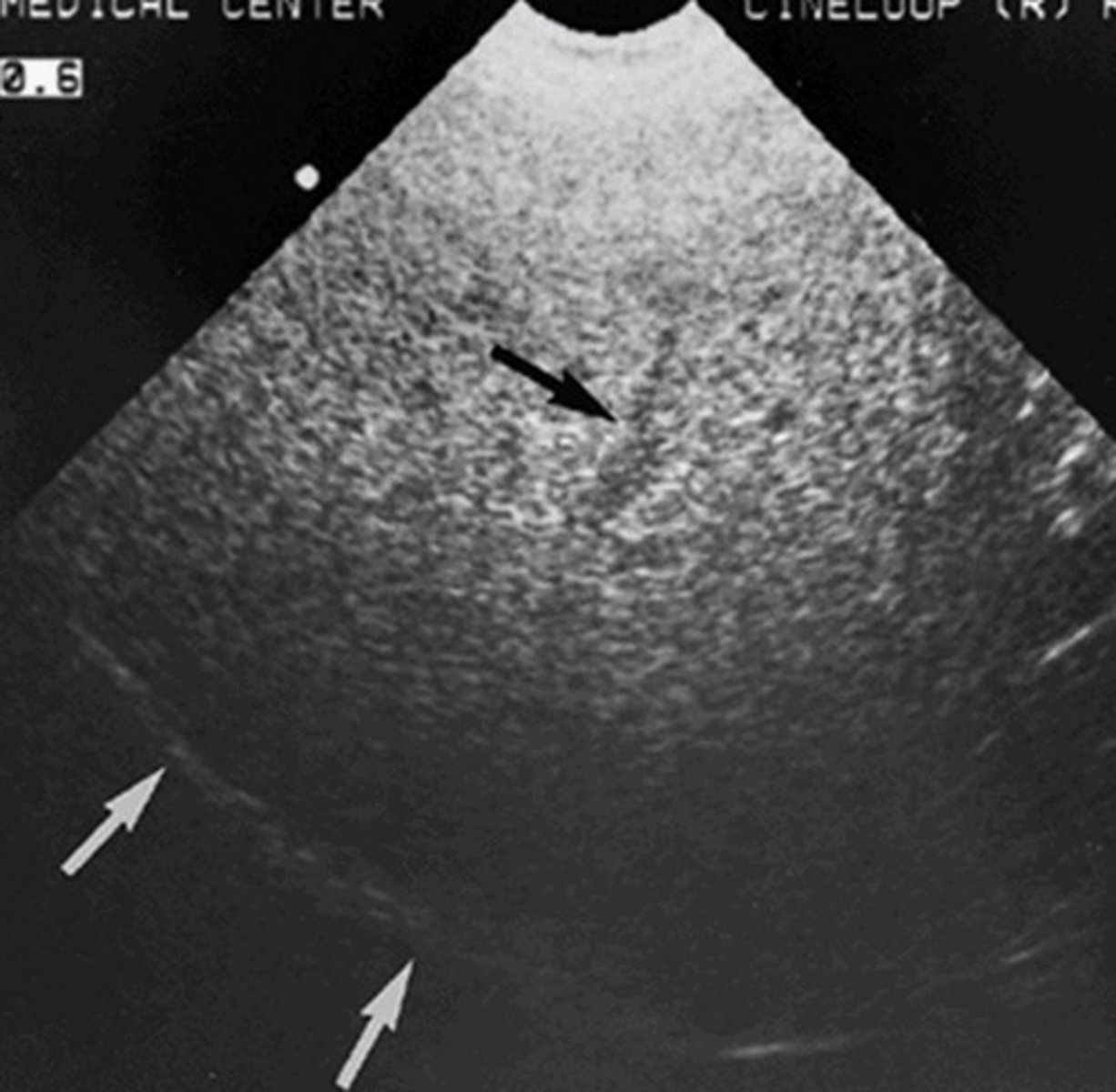
Hepatitis
Inflammation of liver
Causes of Hepatitis
Viruses
Toxins
Symptoms of Hepatitis
Fever/chills
N/V
Hepatomegaly
Jaundice
Lab Values for Hepatitis
Elevated:
ALT
AST
Conjugated bilirubin
Unconjugated bilirubin
Sonographic Appearance of Acute Hepatitis
Normal to hypoechoic parenchyma
Portal vein walls are very visible -> starry sky
Splotchy/inhomogenous
Hepatomegaly
Splenomegaly
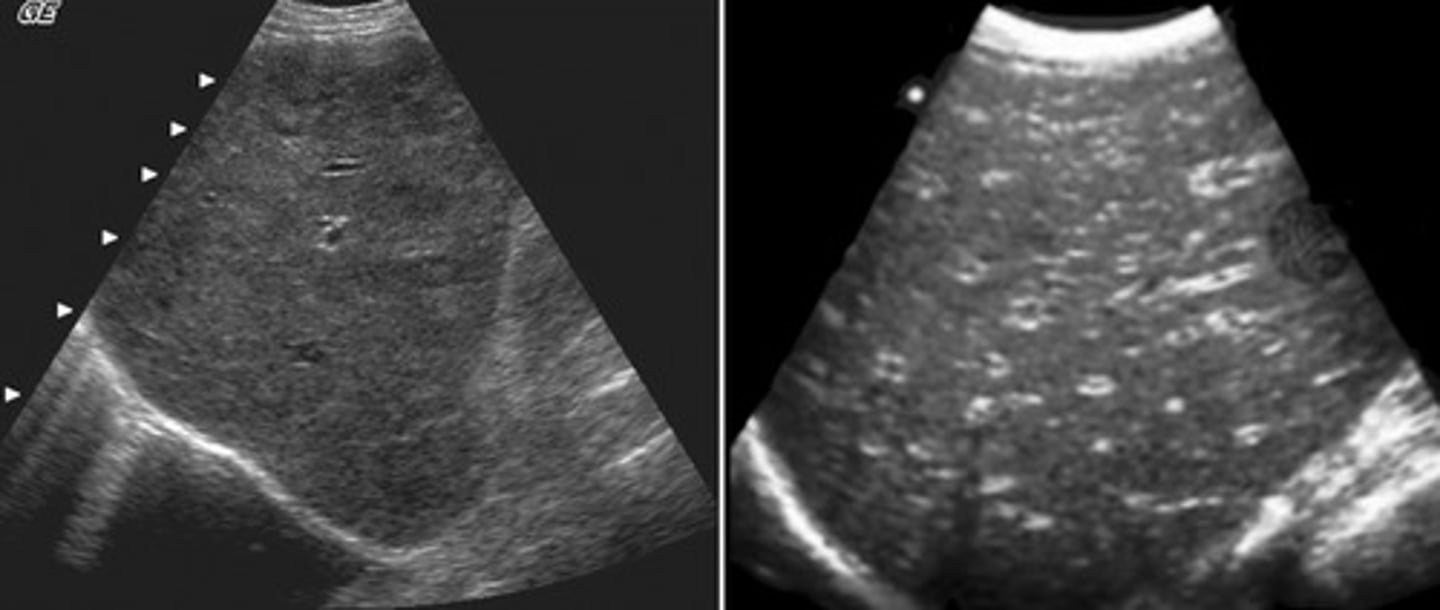
Sonographic Appearance of Chronic Hepatitis
Hyperechoic parenchyma
Decreased liver size
Granular and course texture
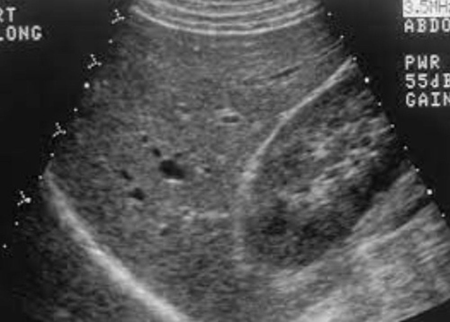
Sonographic Appearance of Cirrhosis
Decreased liver size
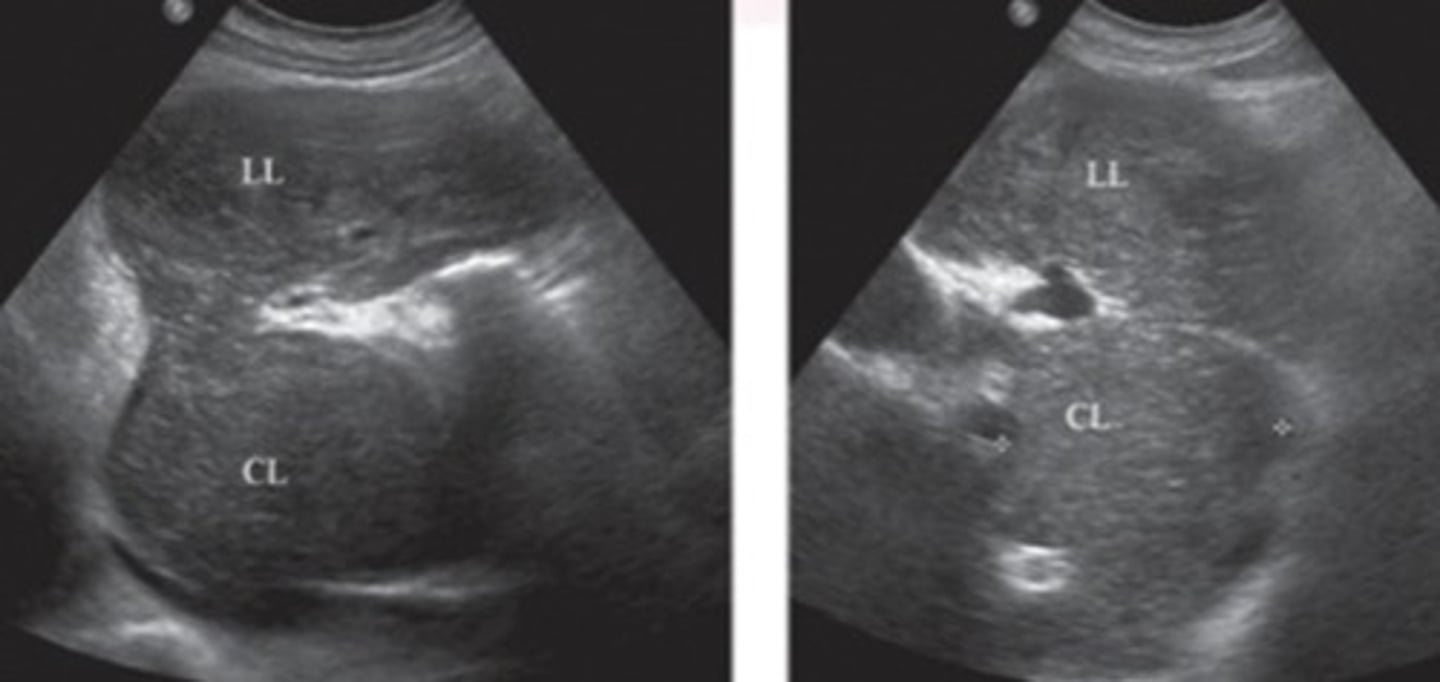
Hypertrophied caudate lobe
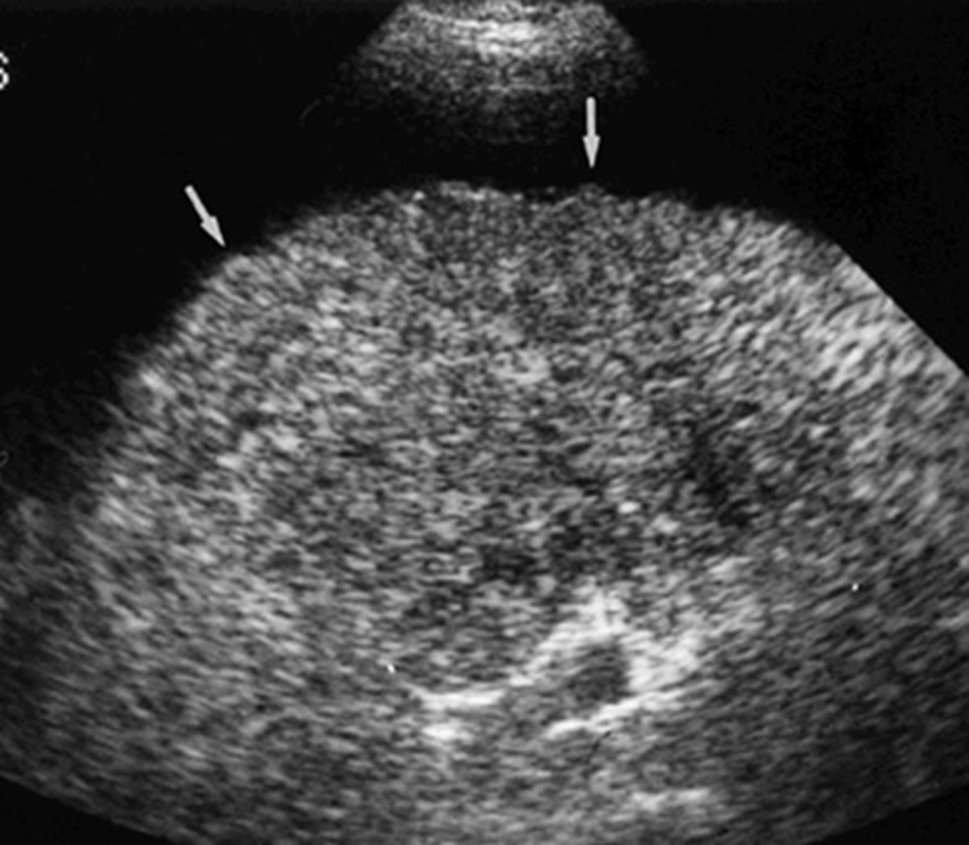
Surface nodularity
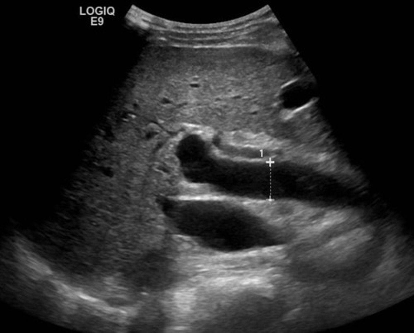
Portal HTN
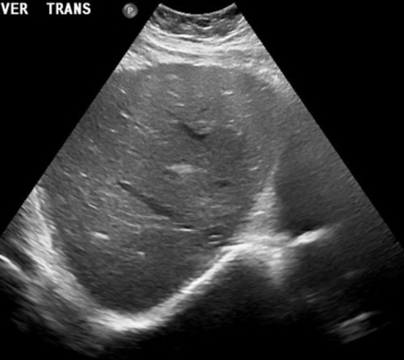
Hypertrophied Caudate Lobe
Liver Surface Nodularity
Portal HTN
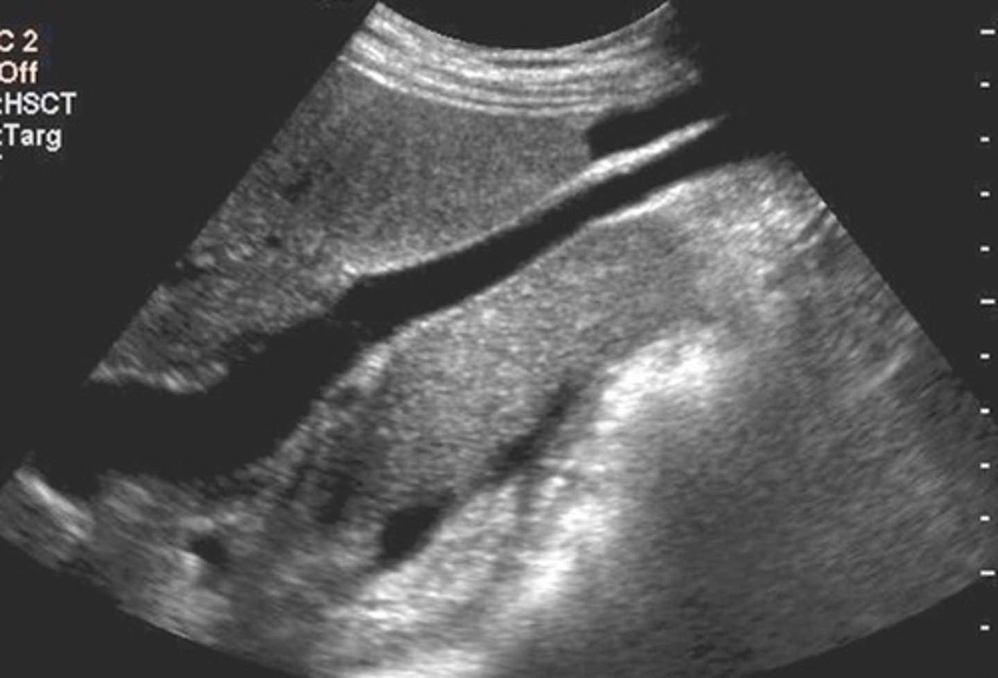
Advanced Cirrhosis Collaterals
Recanalized umbilical vein
Symptoms of Cirrhosis
Asymptomatic
Fatigue
Weight loss
Diarrhea
Jaundice
Ascites
Portal HTN
Lab Values for Cirrhosis
Elevated:
ALT
AST
Lactase dehydrogenase
Serum and fecal conjugated bilirubin
Serum alkaline phosphatase
Urine uribilinogen
Decreased:
Serum albumin
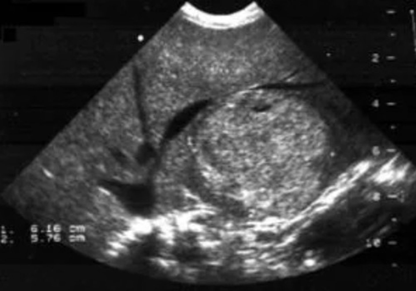
Glycogen Storage Disease
Genetic disorder
Might see solid masses
Lymphoma
Cancer of lymphatic system
Hepatomegaly
Splenomegaly
Lymphadenopathy
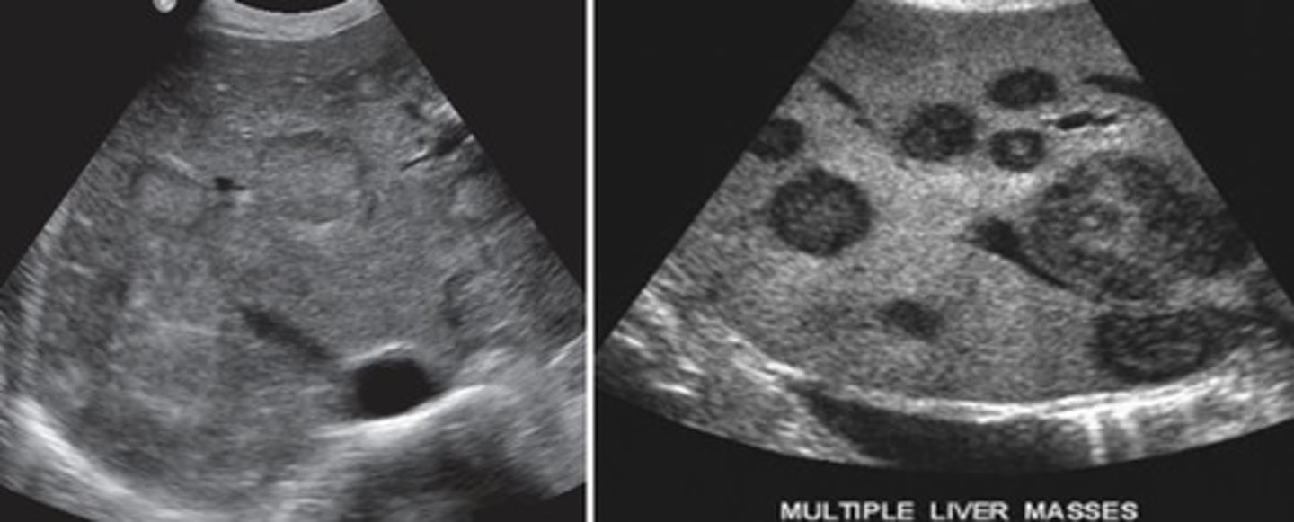
Multiple masses
Bullseye pattern
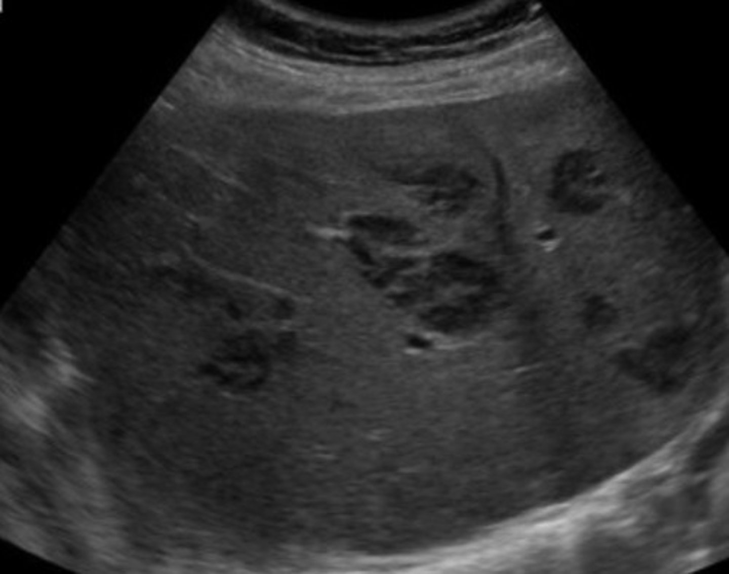
Focal Lymphoma
Stellate (star) pattern
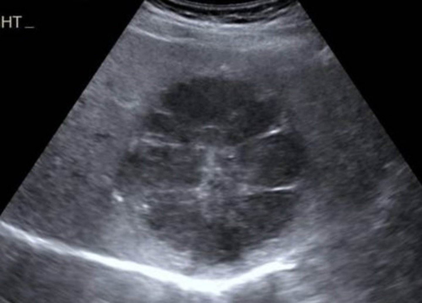
Diffuse Lymphoma
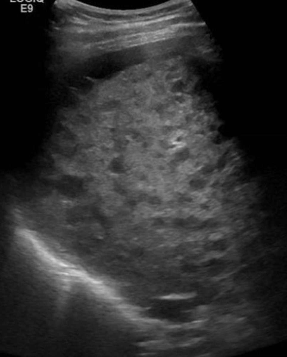
Simple Cysts
Oval to round
Smooth margins
No internal echos
Posterior enhancement
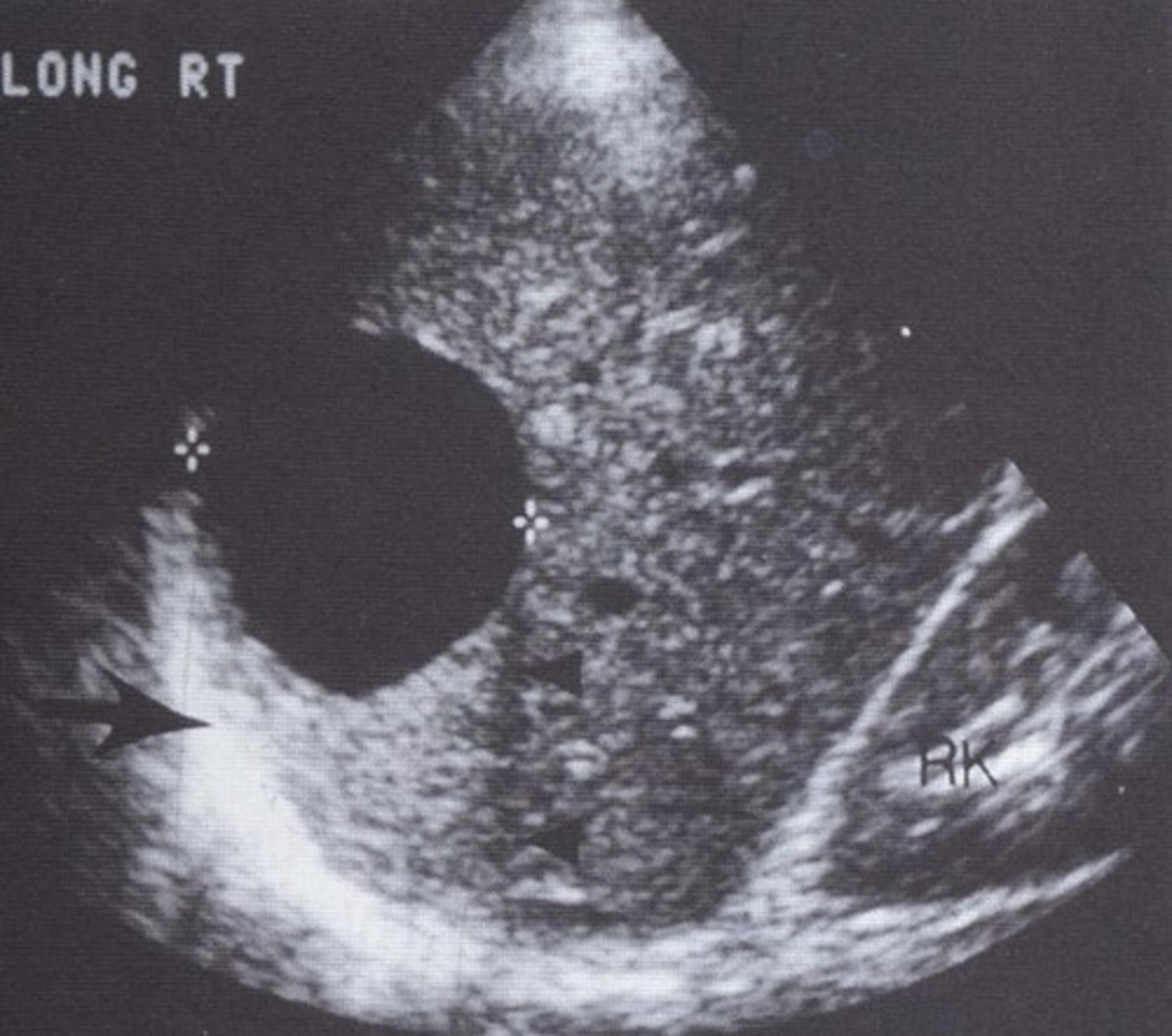
Multicystic Liver
Multiple simple cysts
No genetic component
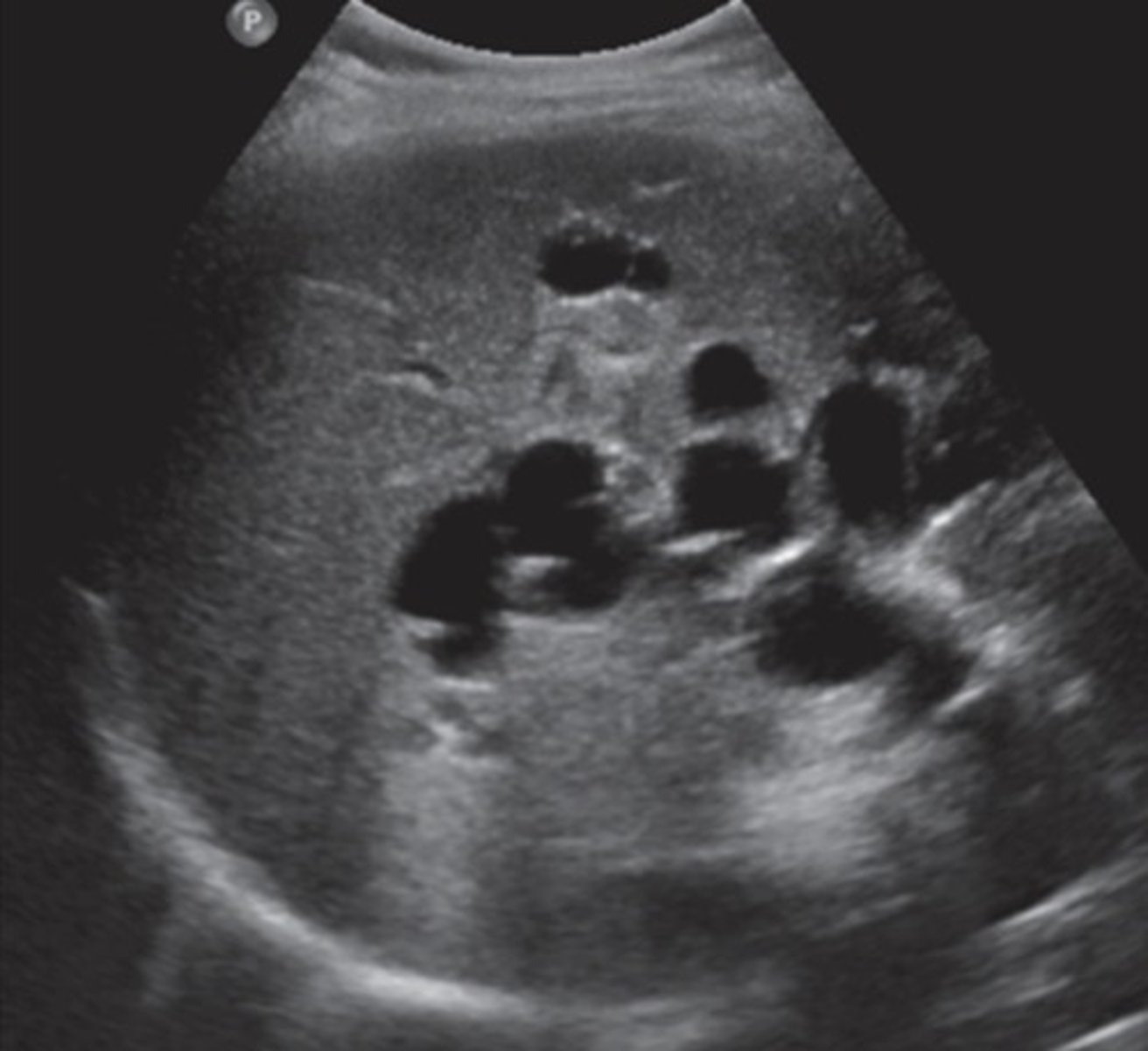
Polycystic Liver Disease
Genetic
Biliary duct anomalies
Will have polycystic kidney disease and pancreatic cysts at the same time
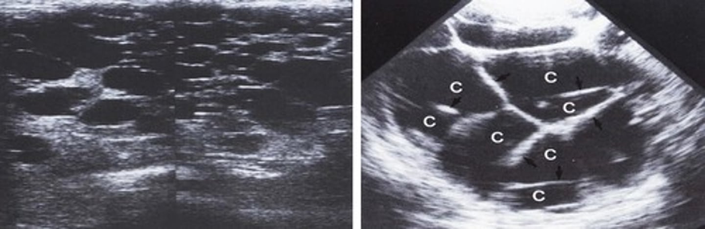
Hematoma
Fresh - hyperechoic
Older - cystic/anechoic and possibly complex
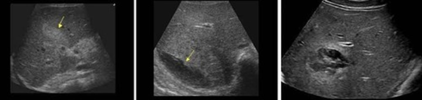
Echinococcal Cyst
Parasitic/hydatid cyst
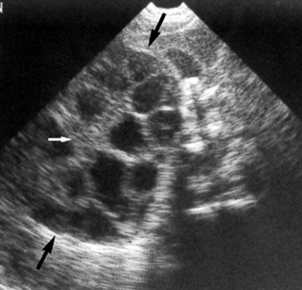
Abscess
Localized infection with pus
Complex appearance
Will have fever/chills, pain, N/V, fatigue, hepatomegaly
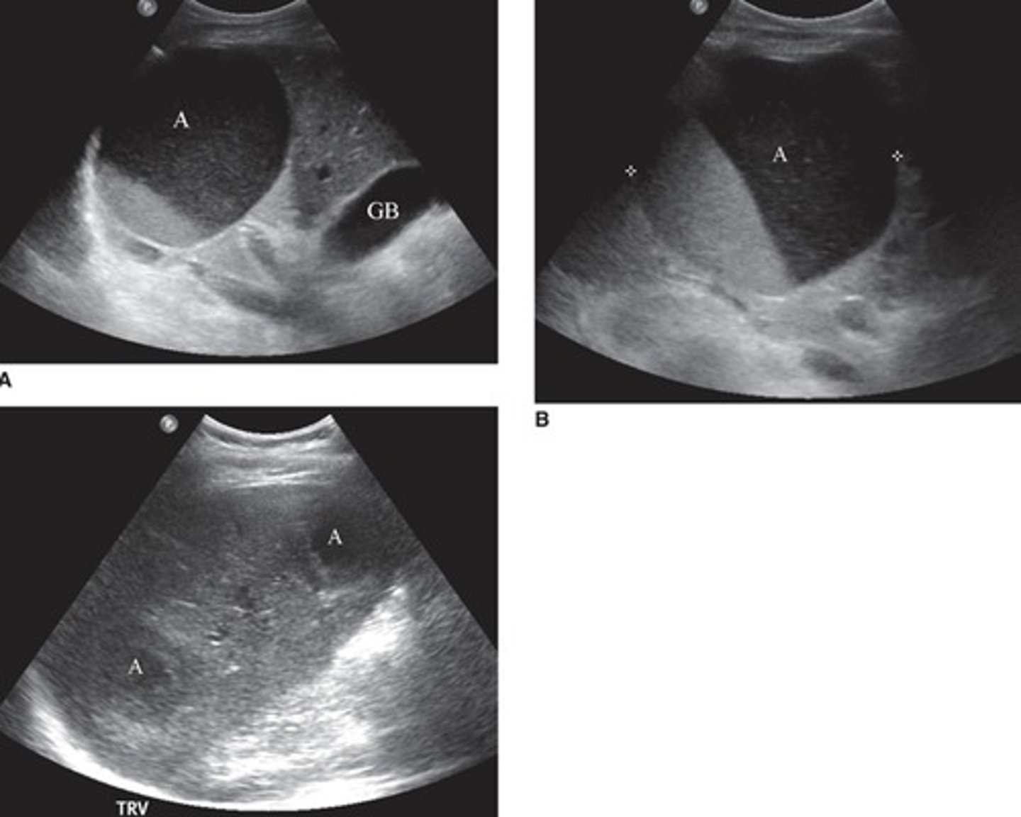
Granuloma
Byproducts from inflammation present as hyperechoic masses throughout the liver
Vary in size, shadowing depends on size
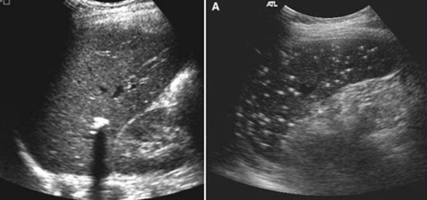
Schistosomiasis
Parasitic infection
Eggs are lodged in liver tissue
Immune response produces lesions
Leads to liver fibrosis and portal HTN
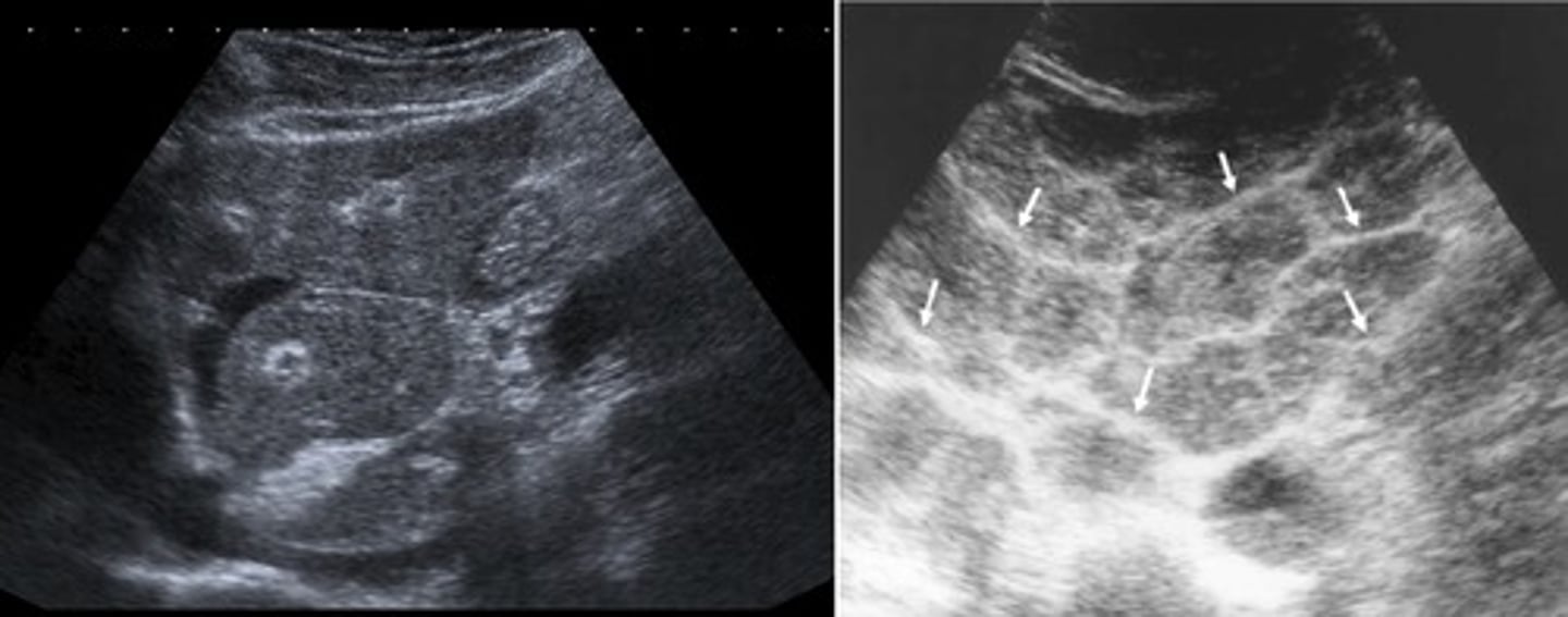
Pneumocystis Carinii
Fungal infection
Seen with HIV-AIDS
Presents as multiple, small, echogenic foci throughout the liver
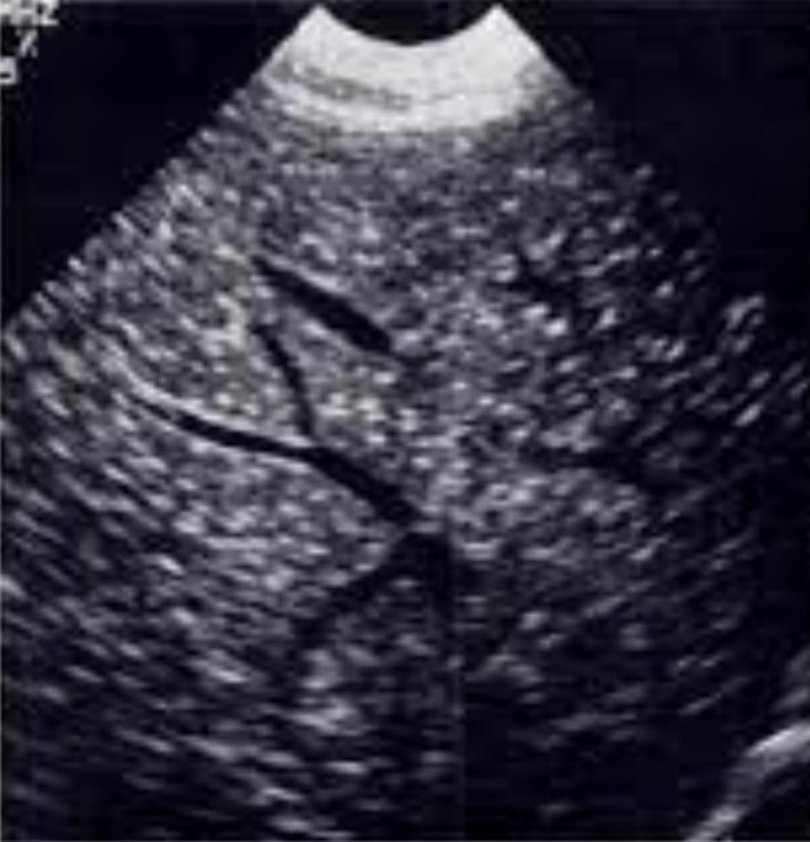
Hemangioma
Benign collection of small vessels
Capillary Hemangioma
Small - 1-3 cm
Homogenous
Posterior enhancement
Well defined margins
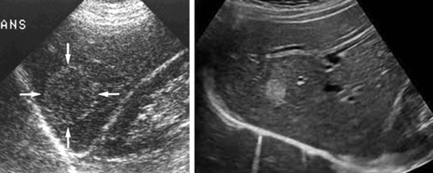
Cavernous Hemangioma
Larger 3-10 cm
Complex appearance
Irregular
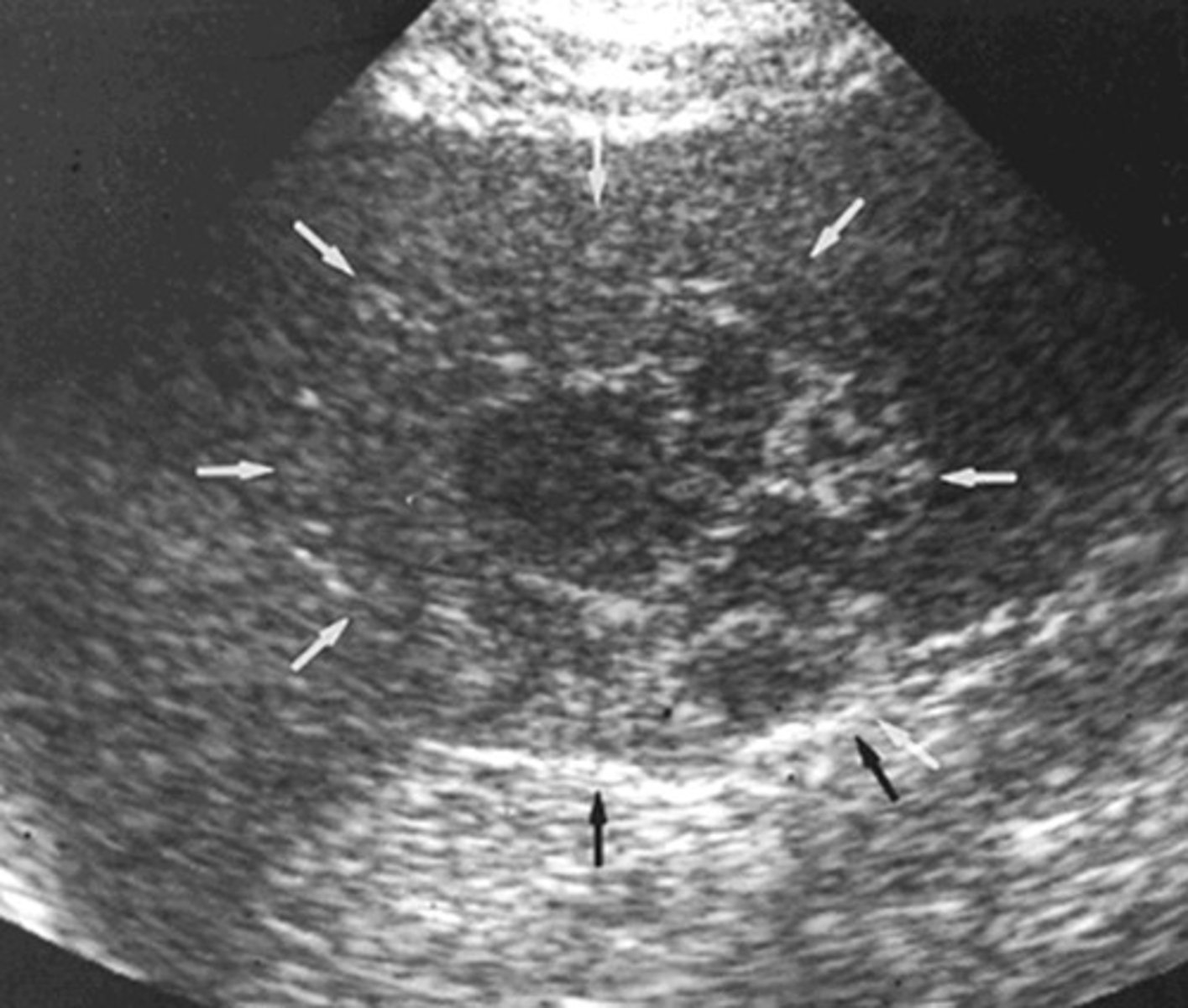
Liver Cell Adenoma
Linked to oral contraceptives
Can actively bleed - halo appearance and posterior enhancement
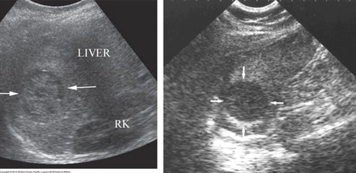
Focal Nodular Hyperplasia
Isoechoic
Area where cells are not put together correctly, do not function well but not cancerous
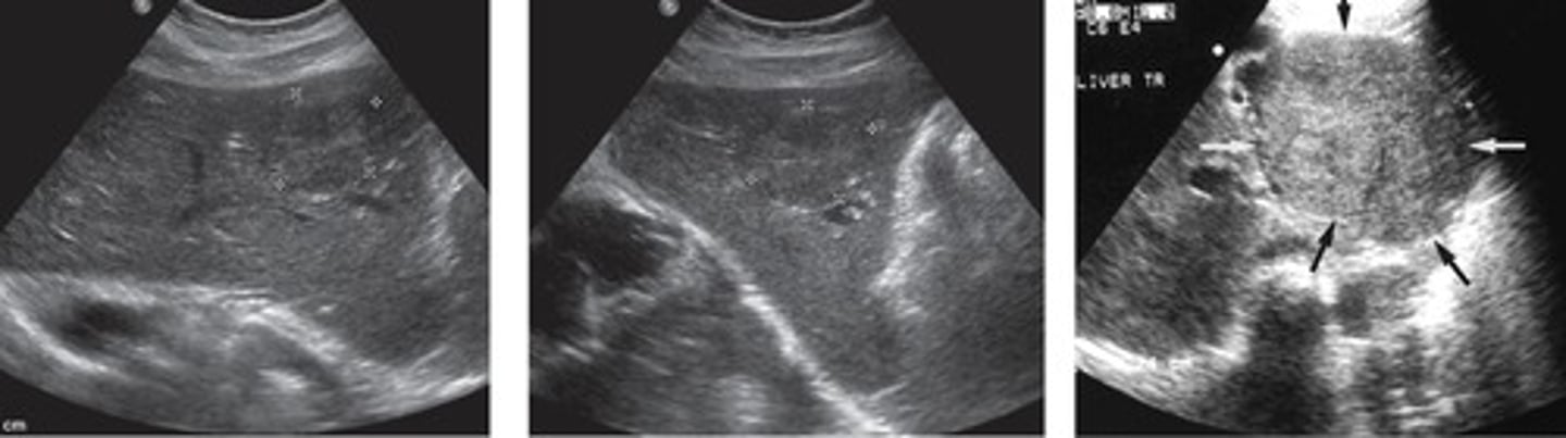
Lipoma
Fatty tumor
Hyperechoic, circumscribed, good margins
cause velocity artifact - diaphragm does not seem complete
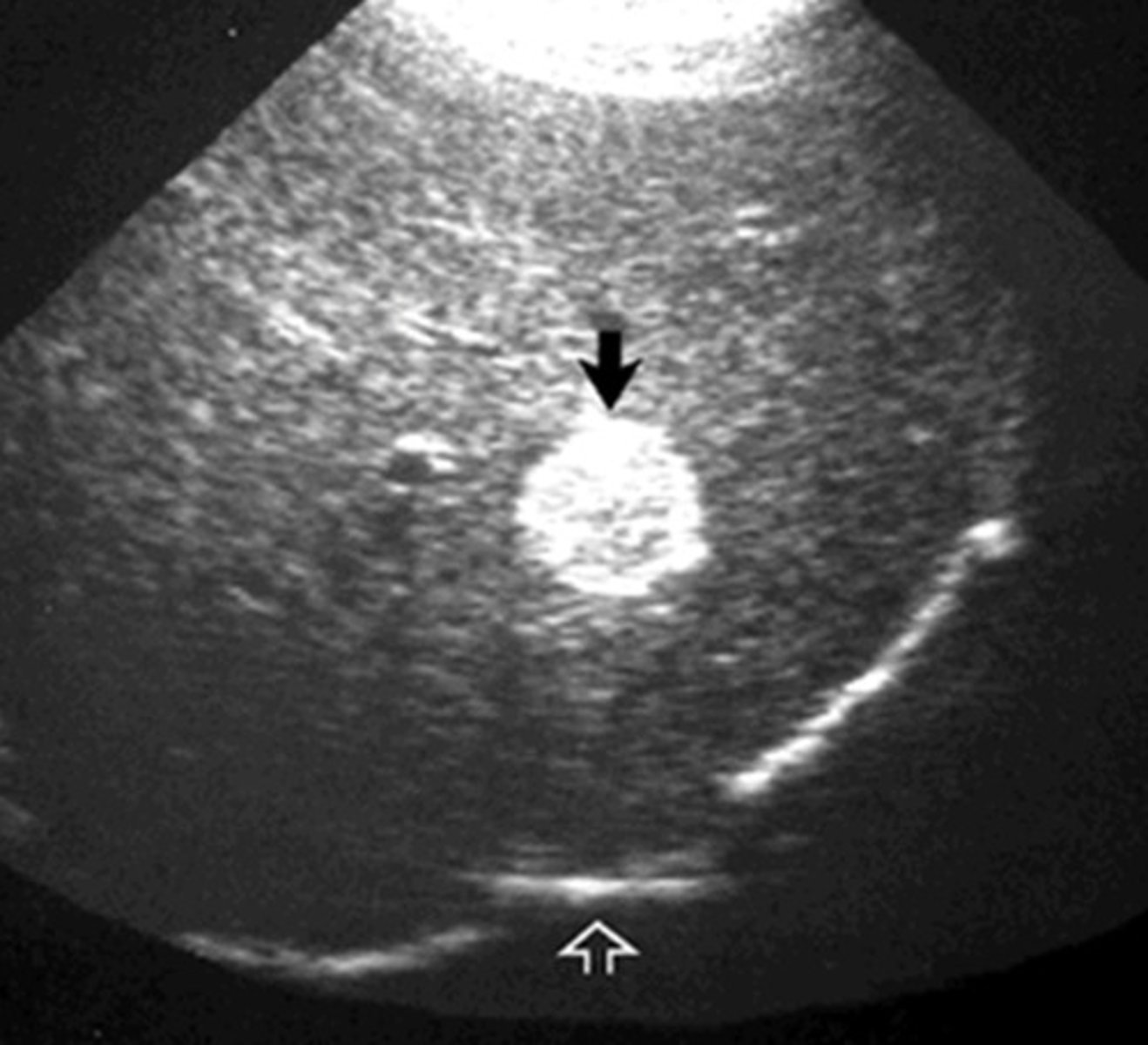
Hepatocellular Carcinoma
Most common primary liver malignancy (but more rare than liver metastasis)
Cirrhosis from Hepatitis B or C leads to increased risk
Solid, inhomogenous, irregular mass
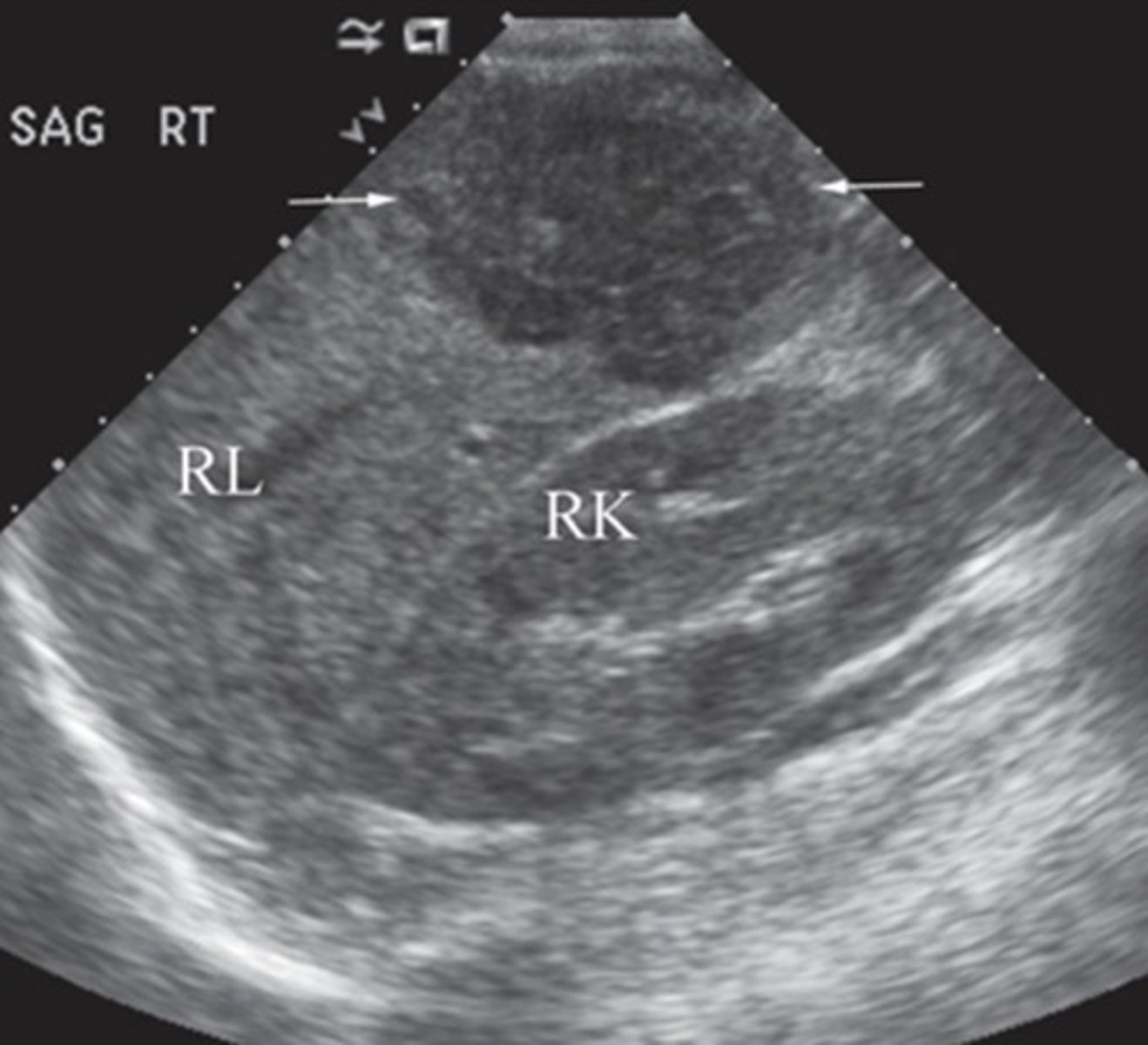
Hepatic Metastasis
Most common secondary liver cancer (more common than HCC)
Presents as multiple lesions througout the liver
Can have bullseye or halo sign
Achalangic Biliary Cirrhosis
Due to partial or complete agenesis of intrahepatic biliary ducts
Alcoholic Cirrhosis
Due to excessive alcohol intake
Atrophic Cirrhosis
Decreased liver size
Biliary Cirrhosis
Due to obstruction or infection of intrahepatic biliary ducts
Cardiac Cirrhosis
Due to congestive heart failure
Fatty Cirrhosis
Due to alchohol abuse
Metabolic Cirrhosis
Due to metabolic storage disorders
Stasis Cirrhosis
Due to obstruction in hepatic veins
Posthepatic Cirrhosis
Due to chronic hepatitis
Postnecrotic Cirrhosis
Due to to viral hepatitis, drugs, toxins, autoimmune destruction
Amylase
Elevated with inflammation or infection
Lipase
Elevated with inflammation or infection
Conjugated/Direct Bilirubin
Elevated with biliary tract obstruction
Urobilinogen
Decreased with biliary tract obstruction
ALT - Alanine Aminotransferase
Elevated with liver cell damage, acute hepatitis, benign hepatic obstruction
AST - Asparate Aminotransferase
Elevated with liver cell damage, cirrhosis, liver mets
ALP - Alkaline Phosphatase
Elevated with everything...
GGT - Gamma Glutamyl Transpeptidase
Elevated with biliary tract obstruction
Globulin
Elevated with inflammation and cancers
Albumin
Decreased with cirrhosis
Cholesterol
Elevated with biliary tract obstruction
Decreased with chronic liver disease
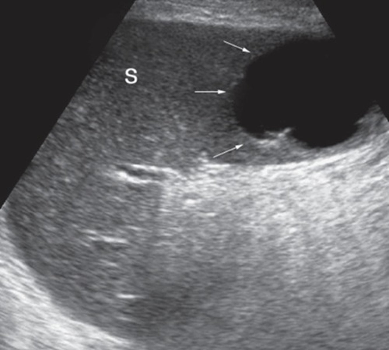
Sonographic appearance of congenital cysts
Anechoic
Well-defined
Posterior enhancement
Singular or multiple
Simple or localated
Causes of congenital cysts
Epithelial origin
Lymphomangiomas in children
Signs and symptoms of congenital cysts
Asymptomatic
Symptomatic when large
Sonographic appearance of acquired cysts
Complex
Ill-defined, thick, irregular borders
Multiple
Dirty shadow if gas is present
Causes of acquired cysts
Post-inflammation -> abscess
Endocarditis
Septicemia
Trauma
Diverticulitis
Other infections
Signs and symptoms of acquired cysts
LUQ pain
High-grade fever
Vomiting
Chest/shoulder pain
Splenic Pseudocysts
Post-trauma
Ex. Hematoma
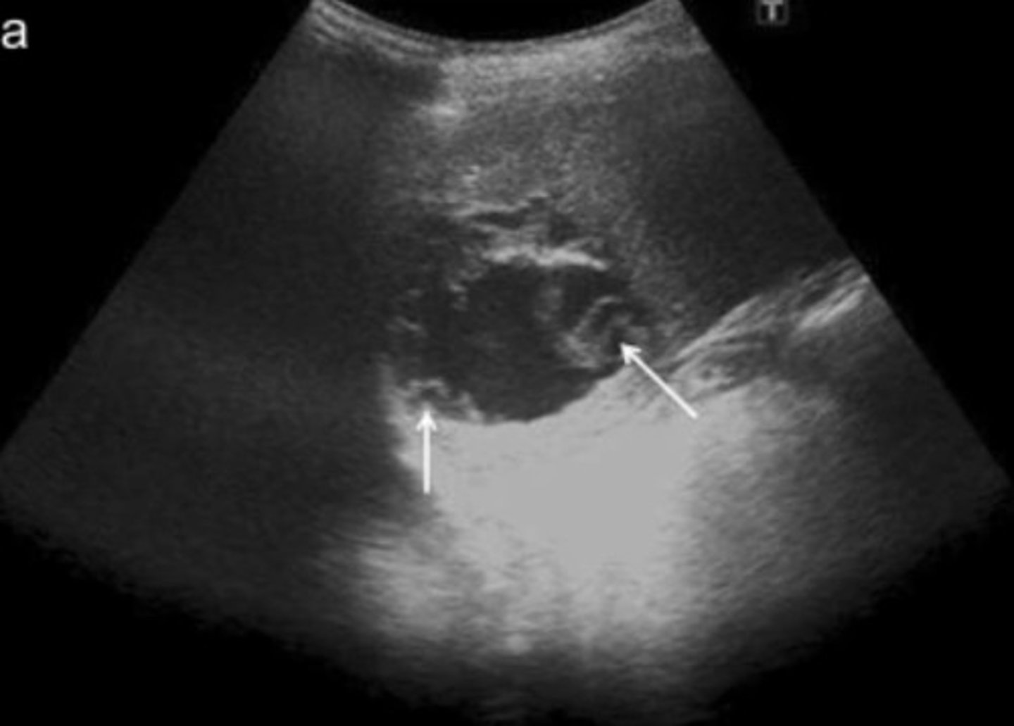
Sonographic appearance of parasitic cysts
Well defined
Daughter cysts (loculations)
Mural calcifications
Posterior enhancement
Simple or complex
Causes of parasitic cysts
Echinococcal parasites
(is considered a hydatid cyst)
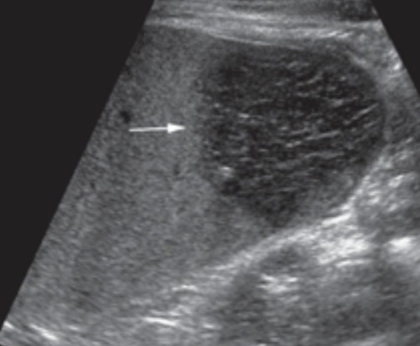
Splenic Rupture or Fracture
Heterogenous appearance
Look for free fluid
Spleen looks enlarged
Splenosis
Sequele of splenic rupture
Splenic tissue transplants into peritoneal cavity
Intrasplenic Splenic Hematoma
Within splenic parenchyma
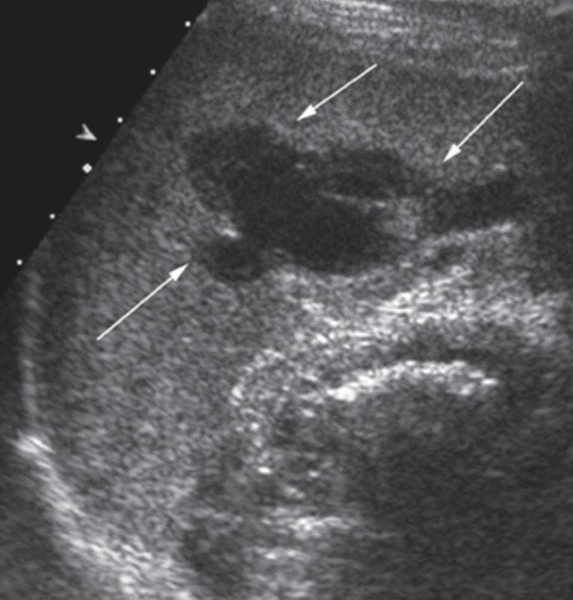
Subcapsular Splenic Hematoma
Blood trapped between splenic capsule and parenchyma
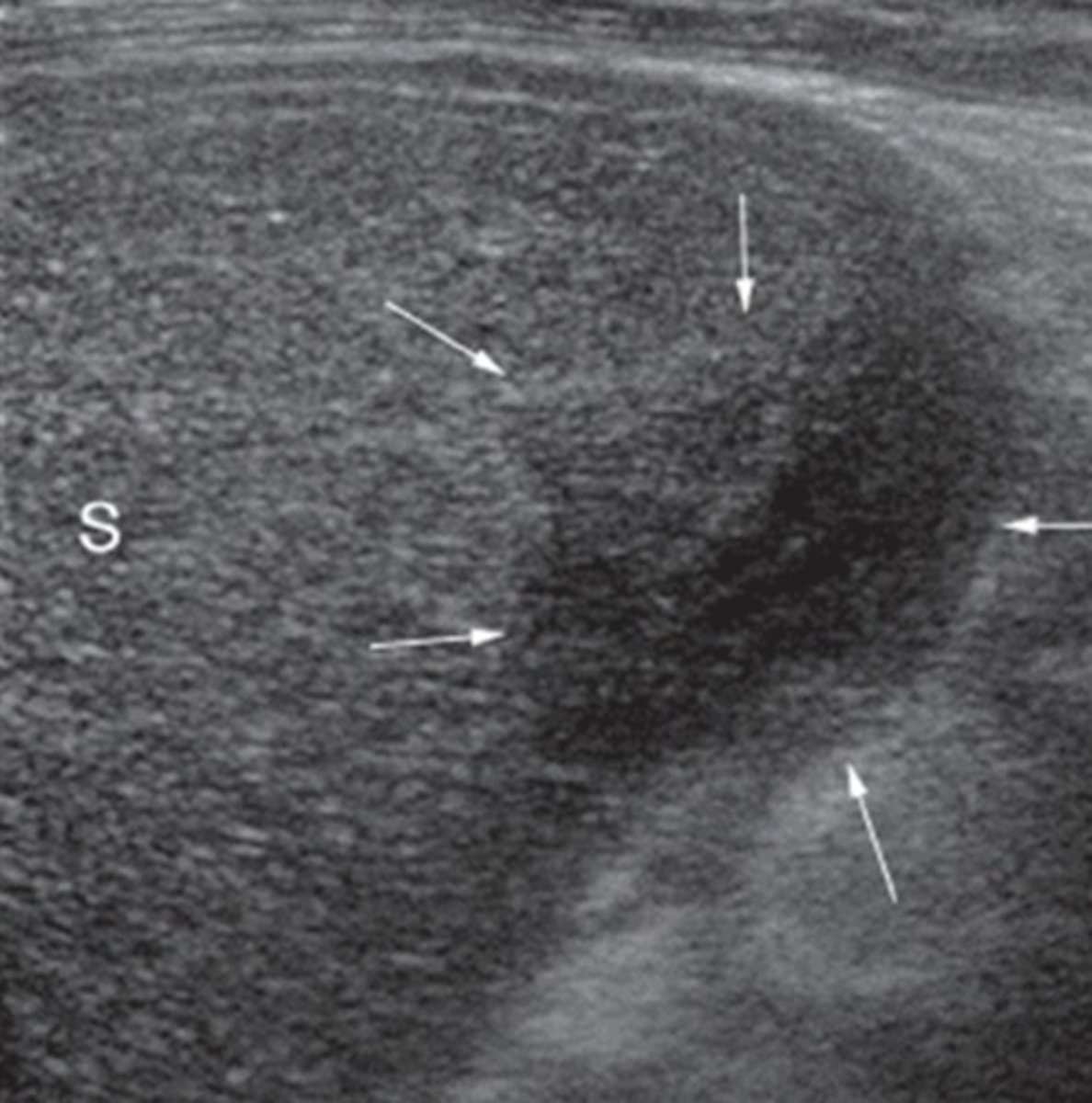
Splenic Infarction
Occlusion of splenic artery - loss of blood supply
Splenic infarctions are secondary to...
Hematologic disorders
Atheromatous disease
Signs and symptoms of splenic infarctions
Asymptomatic
LUQ pain - worsens with inspiration
Sonographic appearance of splenic infarctions
Well-demarcated
Wedge-shaped
Hypoechoic (echogenicity increases with age of infarction)
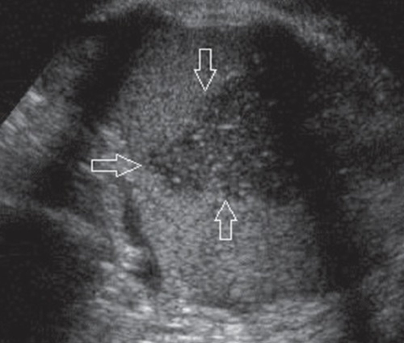
Splenic Calcifications
Hyperechoic
Shadow
Starry Sky appearance
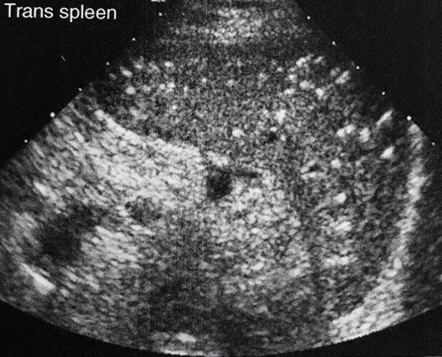
Splenic Hemangioma
Most common benign tumor of the spleen
Made of excess new blood vessels
Variable echogenicities
Similar to liver hemangiomas
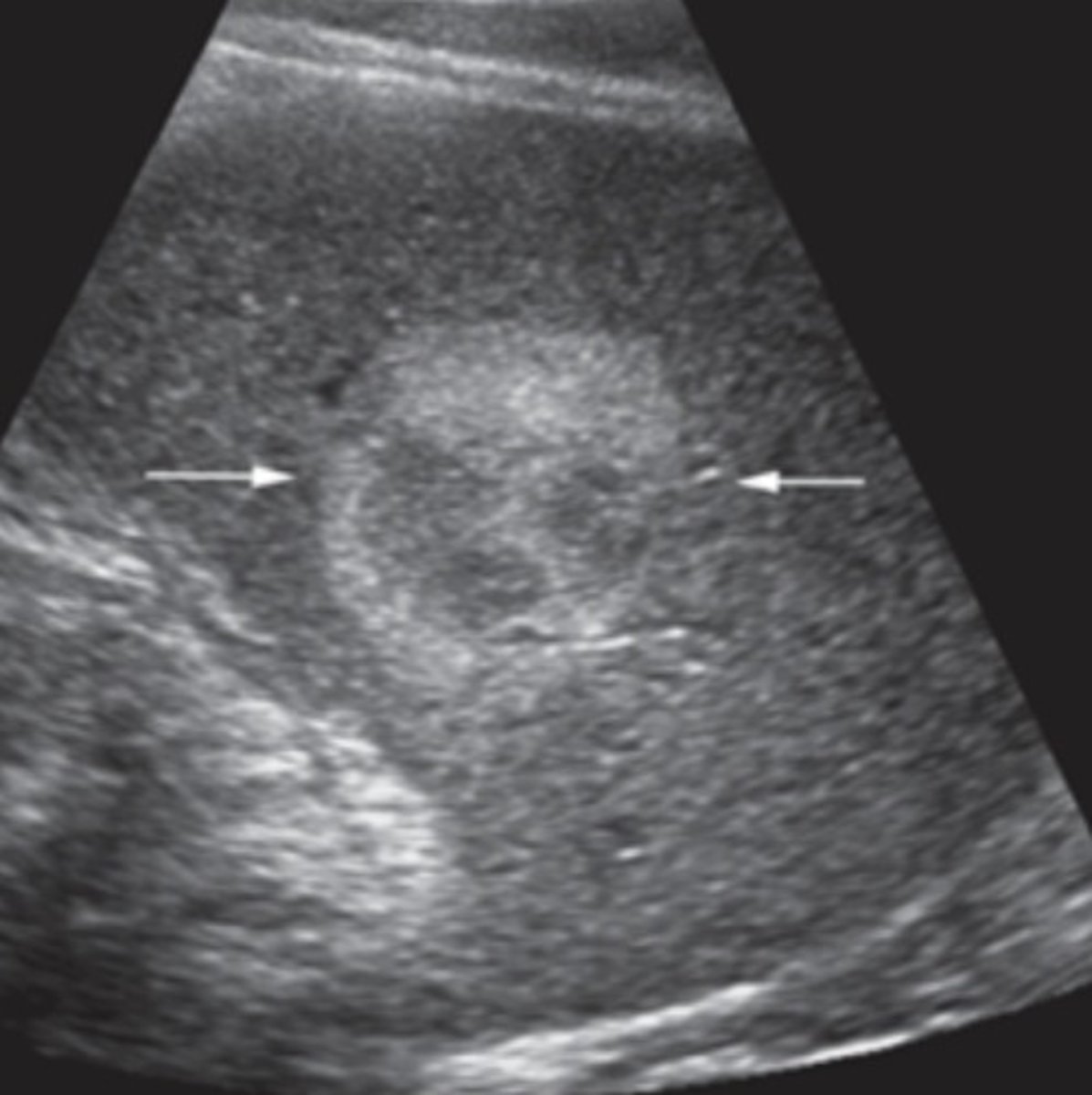
Splenic Hamartoma (splenoma)
Benign splenic tumor
Associated with syndromes
Homogeneous
Solid
Well-defined
Variable echogenicities
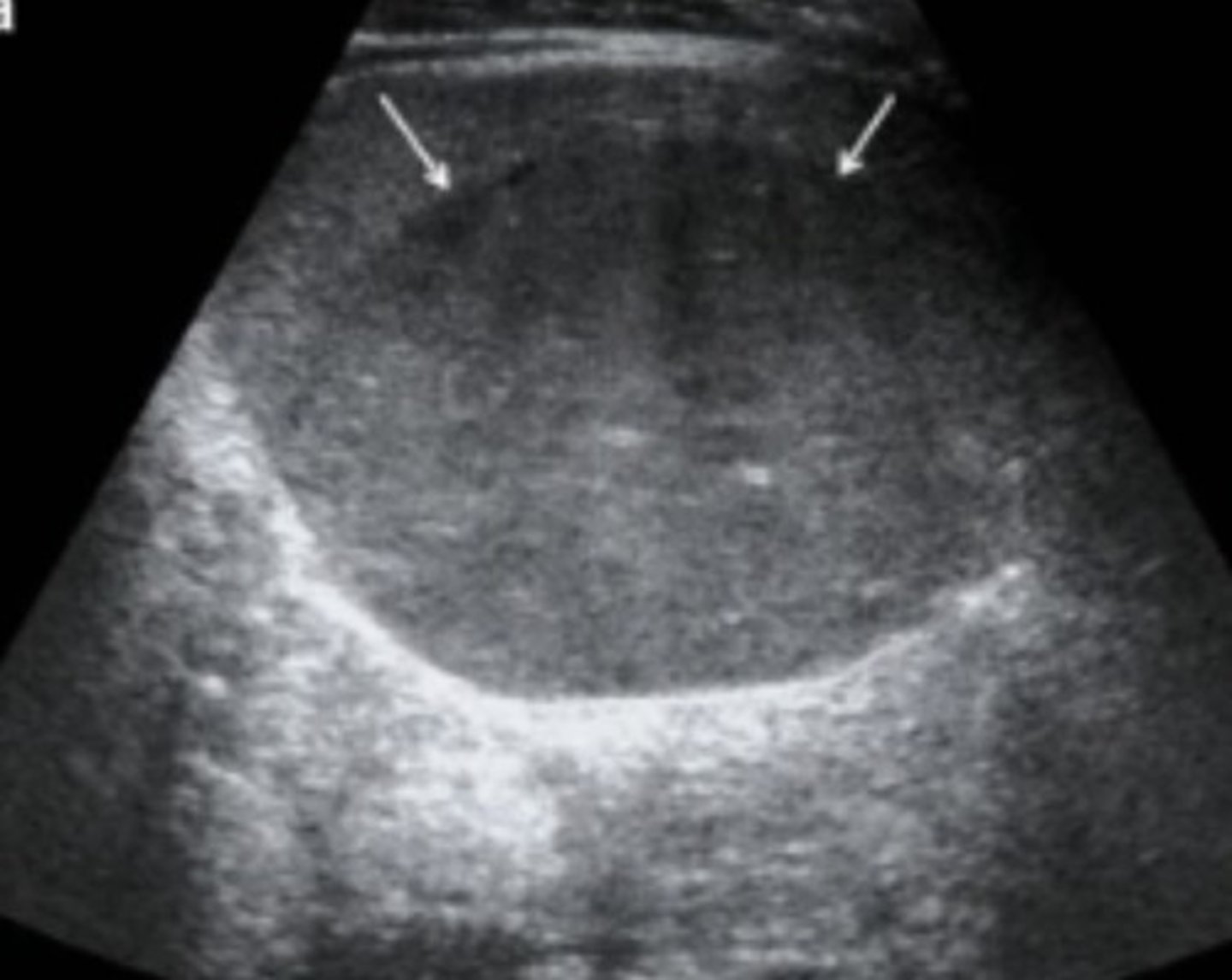
Splenic Lymphoma
Malignant splenic tumor
Hodgkin + Non-Hodgkin lymphomas are most common
Sonographic appearance of focal splenic lymphoma
Hypoechoic lesions with poorly-defined margins
Signs and symptoms of splenic lymphomas
Node enlargement
Fever
Fatigue
Weight loss
Coughing
Pressure
Congestion
Elevated WBC
Anemia
Sonographic appearance of diffuse splenic lymphoma
Modularity and heterogeneity of entire parenchyma