Chapter 7 notes
 Call Kai
 Learn
Learn Practice Test
 Spaced Repetition
 Match
Match Flashcards
 Knowt Play
1/57

There's no tags or description
Looks like no tags are added yet.
Name | Mastery | Learn | Test | Matching | Spaced | Call with Kai | Chat |
|---|
No analytics yet
Send a link to your students to track their progress
58 Terms
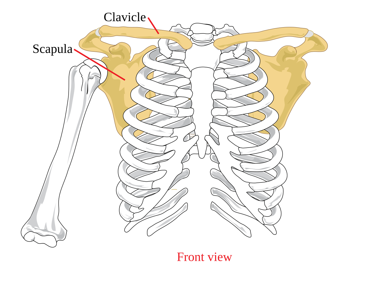
What makes up the pectoral girdle?
Clavicles and scapulae that attach the upper limbs to the trunk
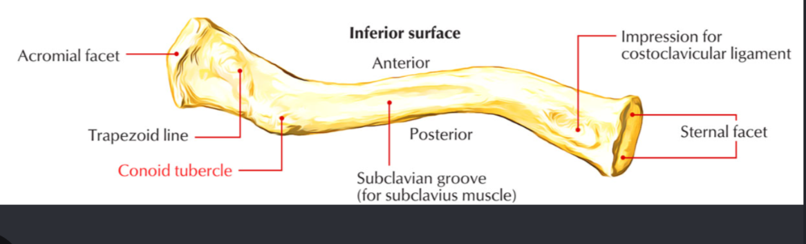
What is the clavicular structure that functions as an attachment site for a ligament?
Conoid tubercle
The pectoral girdle meets the axial skeleton where?
The clavicle attaches to the manubrium


Clavicle
Slender slightly curved long bone, also called the collarbone
Functions:
Provides muscle attachment and braces the scapula and arms away from the thorax while transmitting compression forces

The olecranon is apart of the what?
Ulna

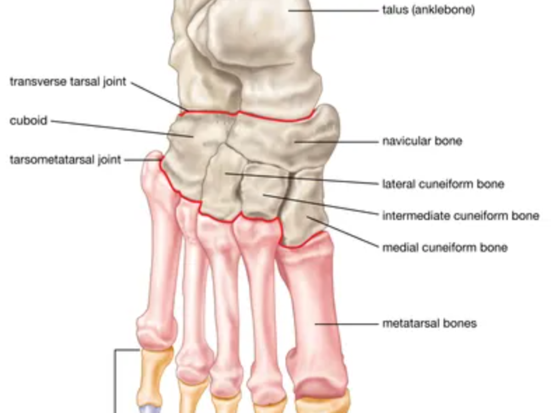
In the ankle bone, the tarsal bone that articulate with both leg bones is the what?
Talus

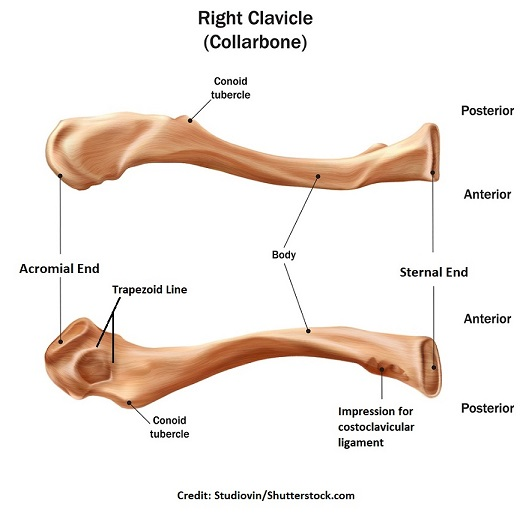
What is the clavicle comprised of?
Sternal end: Medial ice-cream-cone-shaped end that attaches to the manubrium
Acromial end: Flattened lateral end that articulates with the scapula
Trapezoid line: Line on the inferior clavicle that serves as a ligament attachment site
Conoid tubercle: Projection on the clavicle that serves as a ligament attachment site
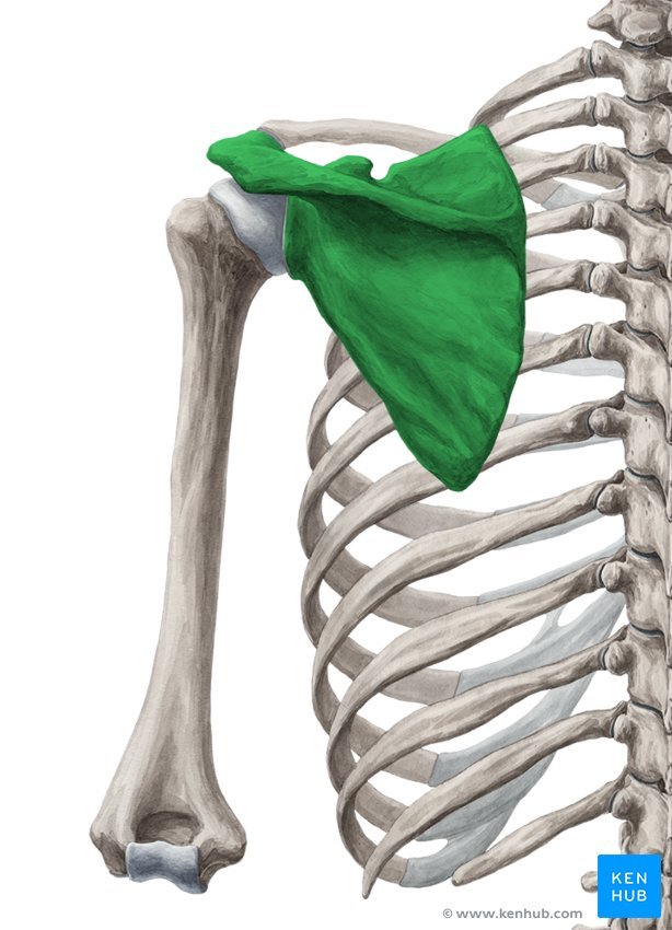
Scapula
Thin triangular flat bone also called the shoulder blade


boarders of Scalpula
superior boarder: Shortest and sharpest border of the scapula
medial boarder: Border of the scapula that parallels the vertebral column
Lateral boarder: Thick axillary border that ends superiorly near the glenoid cavity (socket on the lateral angle of the scapula “shoulder blade” that connects with the head of the humerus “upper arm bone” to form the shoulder joint.)
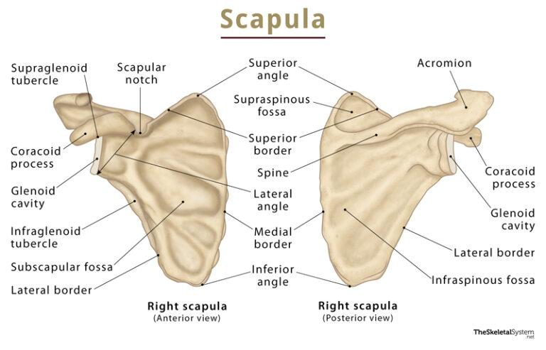

What is the scapula comprised of?
Glenoid cavity: Shallow socket of the scapula that articulates with the head of the humerus. Visible laterally and partially visible anteriorly
Acromion: Scapular process that articulates with the acromial end of the clavicle
Scapular spine: Bony ridge located on the posterior surface of the scapula
Coracoid process: Bent-finger-shaped scapular process serving as an attachment site for the biceps brachii and ligaments
Suprascapular notch: Notch in the scapula that provides a nerve passageway
Infraspinous fossa: Posterior scapular depression below the spine for infraspinatus muscle attachment
Supraspinous fossa: Posterior scapular depression above the spine for supraspinatus muscle attachment
Subscapular fossa: Large depression forming the anterior surface of the scapula for subscapularis attachment
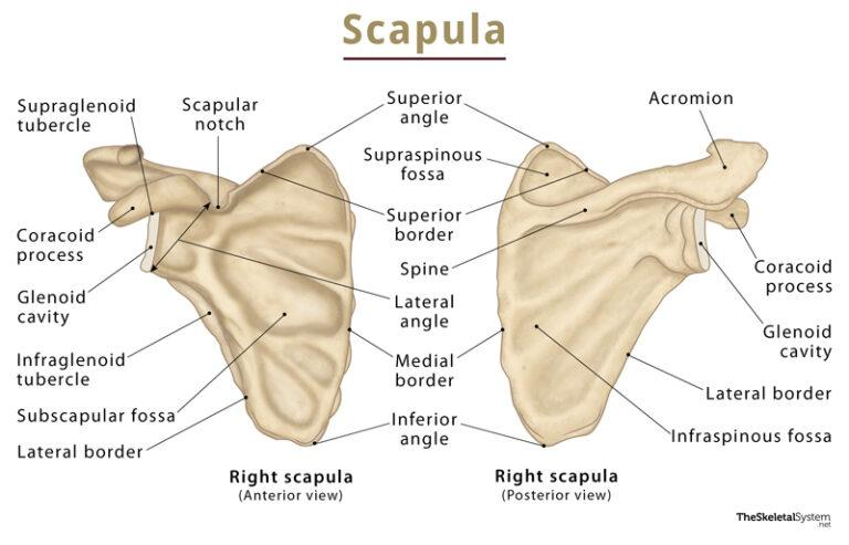

Angles of scapula
Superior angle: Angle located on the superior medial portion of the scapula
lateral angle: Thick angle containing the glenoid cavity
inferior angle: Lowest angle of the scapula that moves as the arm is raised and lowered


The upper limb consists of How many bones?
30 bones total
brachium (1)
Humerus
Antebrachium (2)
Ulna and radius
Hand bones (27)
Carpals (8)
Metacarpals (5) and phalanges (14)


Humerus
Largest and longest bone of the upper extremity and the only bone of the brachium
Articulates with the scapula at the shoulder and the radius and ulna at the elbow


what are the Proximal end features of humerus?
Head: Rounded proximal end that fits into the glenoid cavity of the scapula
Anatomical neck: Constricted region immediately inferior to the head of the humerus
Greater tubercle: Large lateral projection serving as an attachment site for supraspinatus, infraspinatus and teres minor
Lesser tubercle: Anterior and medial projection serving as an insertion site for subscapularis
Intertubercular groove or sulcus: Groove separating the greater and lesser tubercles and guiding the biceps brachii tendon
Surgical neck: Narrowed region distal to the tubercles and a common fracture site
Radial groove: Posterior groove of the humerus that guides the radial nerve
Deltoid tuberosity: Elevated lateral surface of the humerus serving as an attachment site for the deltoid muscle


what are the Distal end features of humerus?
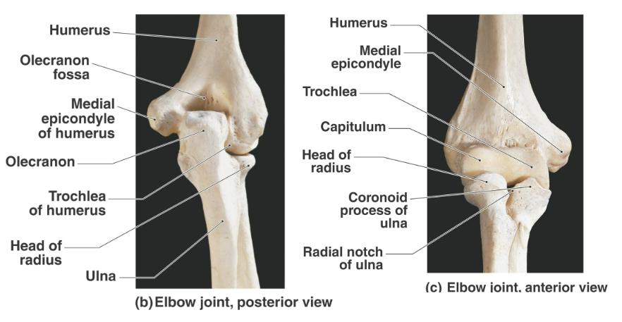
Articular condyle: Distal articular region divided into the trochlea and capitulum
Trochlea: Spool-shaped medial humeral condyle that articulates with the ulna
Capitulum: Rounded lateral humeral condyle that articulates with the radius
Medial epicondyle of humerus: Medial distal projection serving as an attachment site for forearm muscles
Lateral epicondyle of humerus: Lateral distal projection serving as an attachment site for forearm muscles
Medial supracondylar ridge of humerus: Medial ridge superior to the humeral condyle
Lateral supracondylar ridge of humerus: Lateral ridge superior to the humeral condyle and an attachment site for brachioradialis
Olecranon fossa: Posterior depression of the humerus that receives the olecranon process of the ulna
Coronoid fossa: Anterior depression that receives the coronoid process of the ulna during flexion
Radial fossa: Anterior depression superior to the capitulum that accommodates part of the radial head during flexion

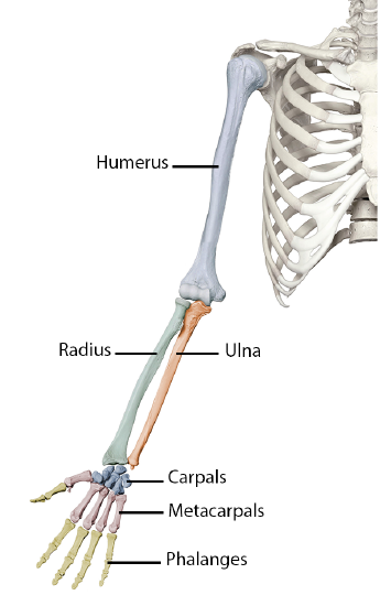
Antebrachium
Anatomical term for the forearm containing the radius and ulna
Radius: Forearm bone located laterally on the thumb side in anatomical position
Ulna: Forearm bone located medially and primarily responsible for forming the elbow
Proximal ends of radius and ulna articulate with the humerus (brachium)
Distal ends articulate with the carpus (wrist)
The radius and ulna articulate with each other both proximally and distally at the proximal, distal and radioulnar joints
Both share a Interosseous membrane connecting the radius and ulna along their lengths
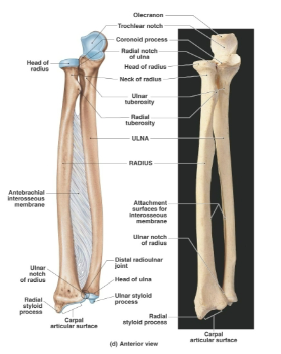
Directional terms for antebrachium
Supination: Position in which the radius and ulna are parallel
Pronation: Movement in which the radius rotates medially over the ulna

Olecranon process
Proximal projection of the ulna that forms the point of the elbow

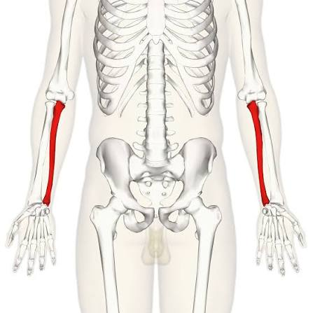
Ulna
Wide at the proximal end, then narrows at distal end.
Slightly longer than the radius
Main function is to form the elbow joint with the humerus
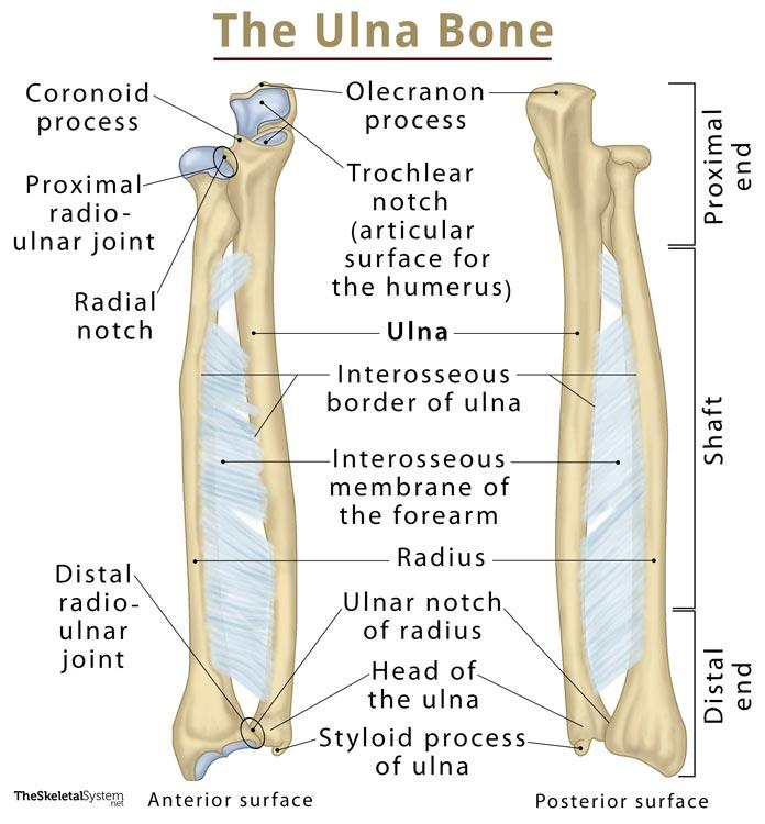

Proximal end features of ulna
Coronoid process: Anterior proximal projection that helps grip the trochlea of the humerus
Trochlear notch: Deep concavity between the olecranon and coronoid processes that grips the humeral trochlea to form a hinge joint, allowing flexion and extension
Radial notch of ulna: Smooth depression where the ulna articulates with the head of the radius
proximal radioulnar joint: a pivot-type synovial joint located just below the elbow that allows the forearm to rotate.

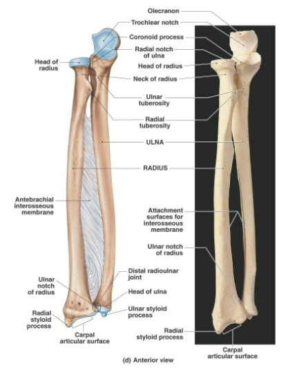
Distal end features of ulna
Head of ulna: Distal end of the ulna separated from the wrist bones by fibrocartilage. little to do with wrist movement
Styloid process: Distal projection serving as an attachment site for a wrist ligament
Distal radioulnar joint: a pivot-type synovial Joint between the ulnar notch of the radius and head of the ulna


Radius
Thin at it’s proximal end and widened at its distal end (aka the opposite of the ulna)
Extreme distal end of radius is concave and articulates (Articulating bones refer to any two or more bones that come together to form a joint.) with the carpal bones of the wrist
The radius is the mobile, rotating bone of the forearm on the thumb side
while the ulna is the stable, supporting anchor on the pinky side.


proximal end features of radius
Head of radius: Proximal spool-shaped end that articulates with the capitulum of the humerus and medially articulates with the radial notch of the ulna, forming the proximal radioulnar joint
Neck of radius: Narrowed region extending from the radial head to the radial tuberosity
Radial tuberosity: Attachment site for the biceps brachii muscle

Distal end features of radius Ulnar notch of radius
Ulnar notch of radius: Medial distal notch that articulates with the head of the ulna, forming the distal radioulnar joint
Styloid process of radius: Lateral distal projection that anchors a ligament to the wrist

Hand bone count
27 bones consisting of:
8 carpals
5 metacarpals
14 phalanges


Carpus
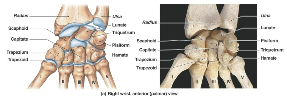
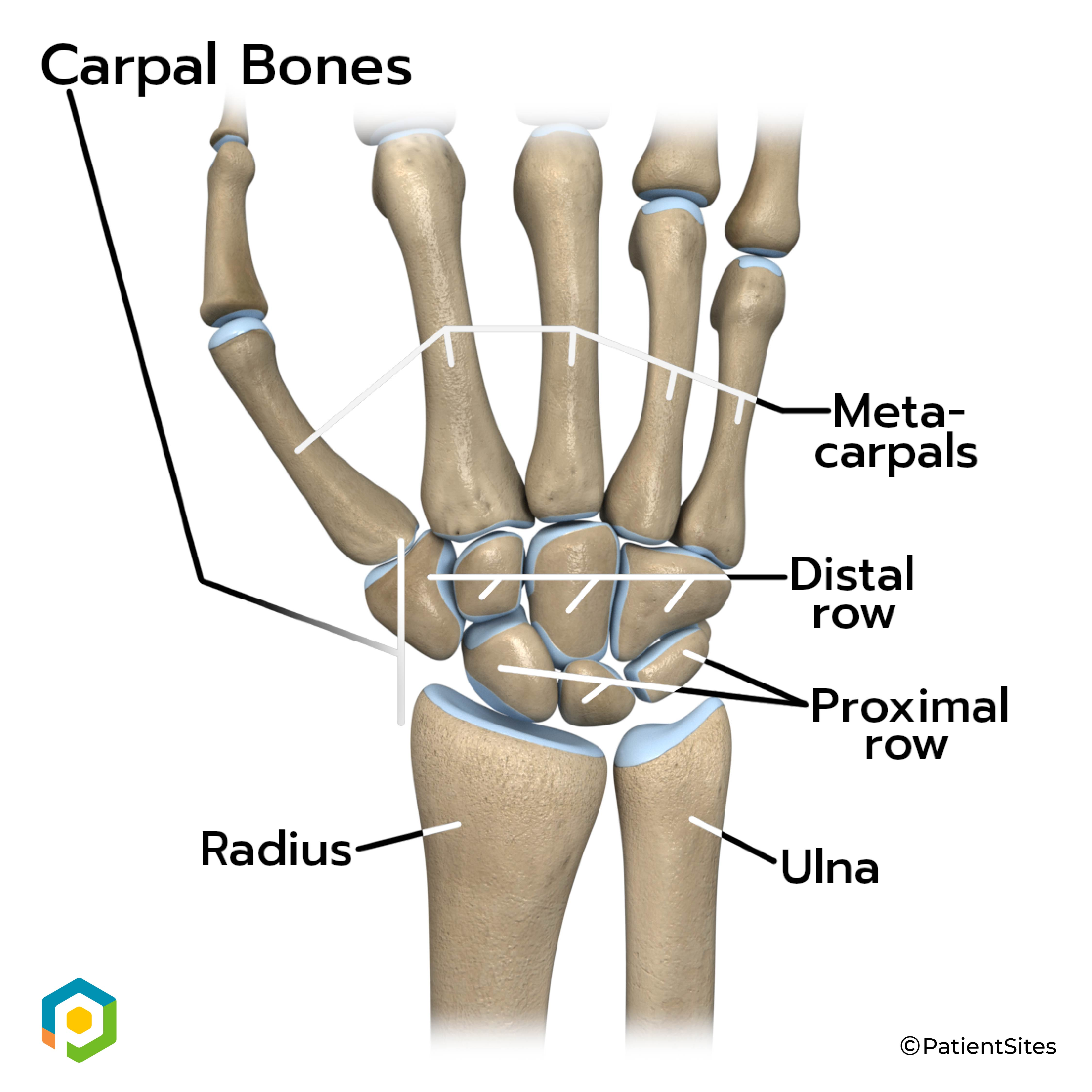
True wrist consisting of eight marble sized carpal bones closely united by ligaments that are arranged in two irregular rows of four
Proximal row of carpals: LATERAL thumb side to MEDIAL
Scaphoid: Proximal lateral carpal that articulates with the radius to form wrist joint
Lunate: Comma-shaped proximal carpal that articulates with the radius to form wrist joint
Triquetrum: Triangular proximal carpal shaped like a small pyramid. articulates with cartilage that separates ulnar head from wrist
Pisiform: Smallest pea-shaped carpal located anterior to the triquetrum and extends farther medially than any other carpal bone
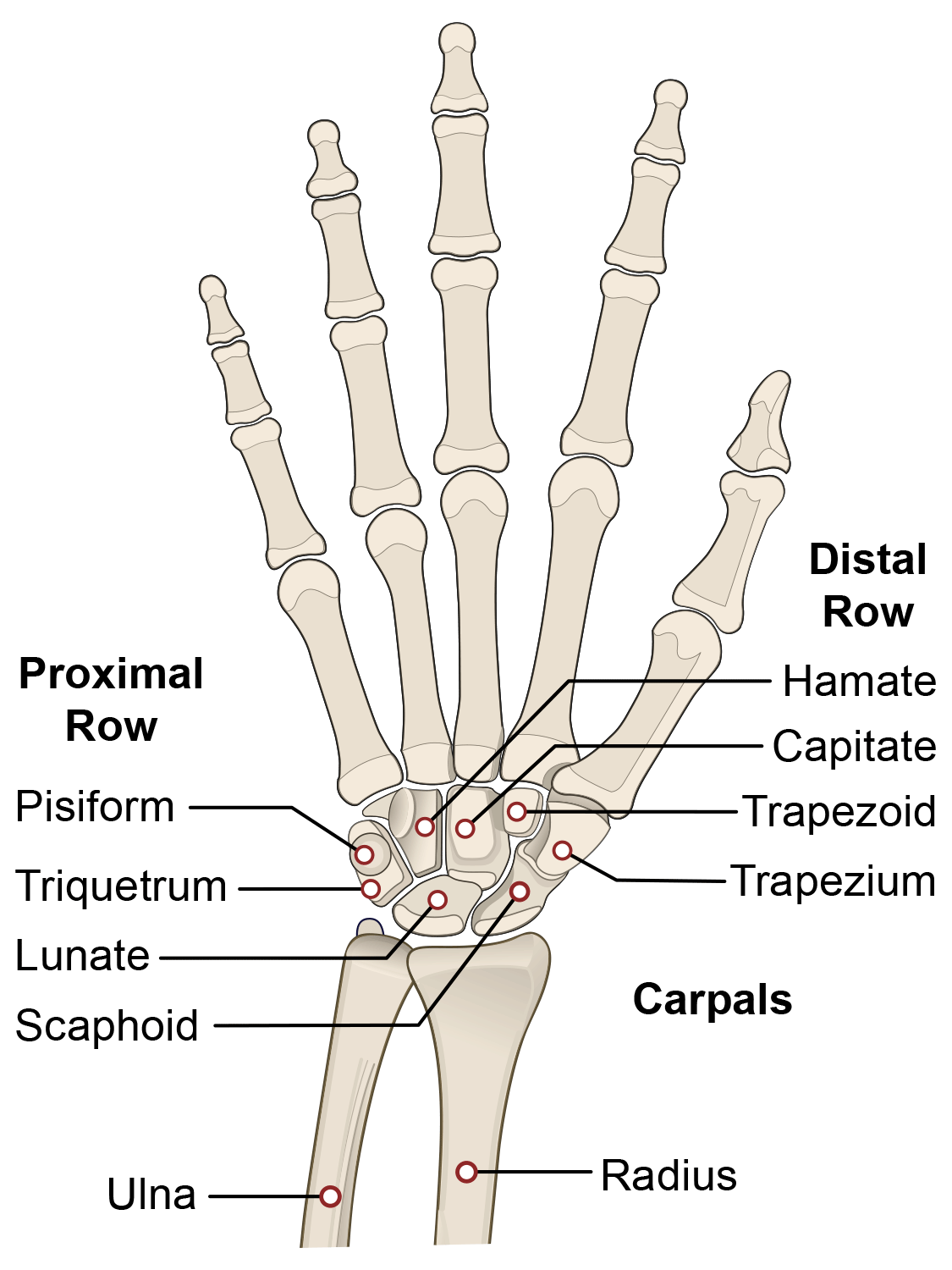
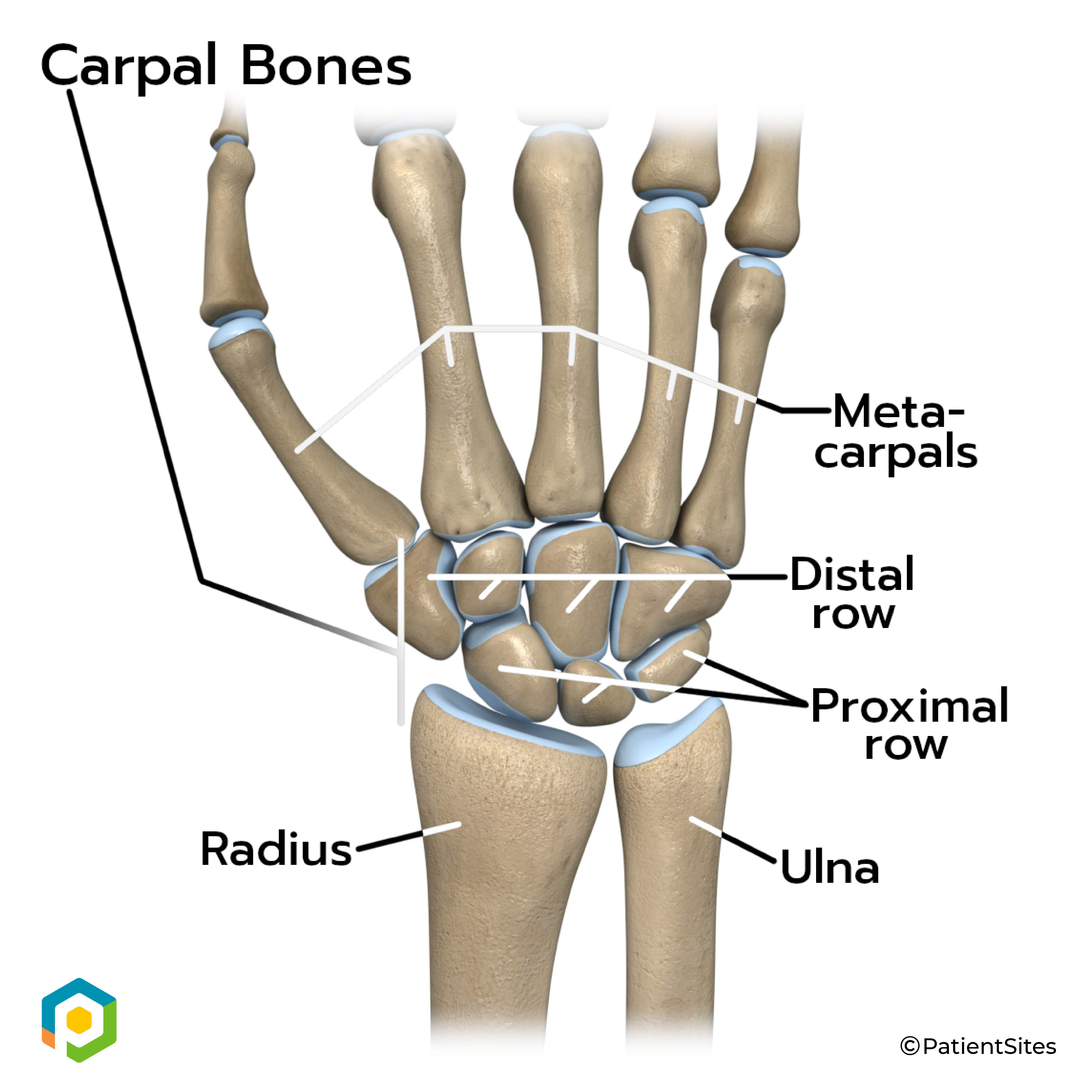
Distal row of carpals: LATERAL thumb side to MEDIAL
Trapezium: Lateral bone of the distal carpal row that articulates proximally with the scaphoid
Trapezoid: Wedge-shaped and smallest bone of the distal carpal row that also articulates proximally with the scaphoid
Capitate: Largest carpal bone and shaped like a head
Hamate: Medial distal carpal containing a hook-like projection

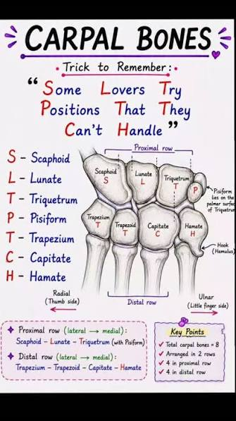
Order of the carpal bones, proximal row to distal row LATERAL to MEDIAL
Scaphoid
lunate
triquetrum
pisiform
trapezium
trapezoid
capitate
hamate
Pneumonic: "Some Lovers Try Positions That They Can't Handle"
Carpal tunnel syndrome
Compression of the median nerve within the carpal tunnel causing pain or numbness

Metacarpus
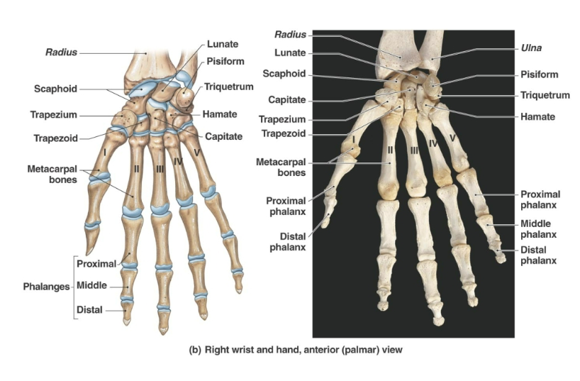
Palm portion of the hand containing five metacarpal bones
Metacarpals are numbered 1 through 5 from the thumb side to the little-finger side
The metacarpal bases articulate with the carpals proximally and with each other on their medial and lateral sides

Metacarpophalangeal joints
Joints between metacarpal heads and proximal phalanges that form the knuckles

Phalanges of hand (digits)
14 finger bones (phalanges) in each hand
Thumb (pollex) (1):
has 2 phalanges each: the proximal phalanx and distal phalanx
The rest of the fingers (2-5):
Have 3 phalanges each: proximal phalanx, middle phalanx, and distal phalanx


Pelvic girdle
Comprised of 3 parts:
ilium
ischium
pubis
Structure that attaches the lower limbs to the axial skeleton with strong ligaments
Supports and protects the lower viscera and developing fetus in females
The pelvic girdle consists of two ossa coxae bones.
The bones of the pelvic girdle and lower limb are much more massive than their homologues of the upper limb.


Coxal bones (os coxae or innominate bones)
Hip bones formed from the Y shaped fused (ilium, ischium and pubis) in adults


Ilium of hip
Superior region of the coxal bone
each consists of an inferior BODY and a superior, wing-like ALA

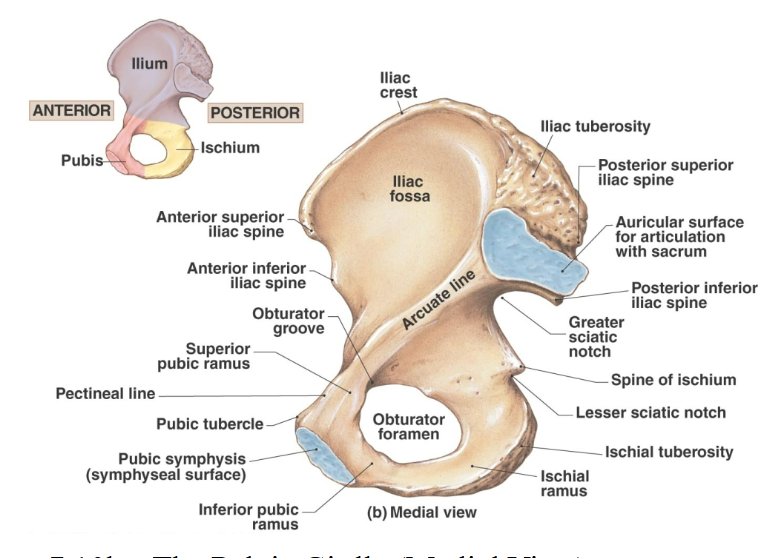
What does the Ilium consist of?
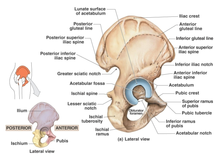
Iliac crest: Thickened superior margin of the ilium serving as a muscle attachment site
Posterior superior iliac spine: Posterior superior projection of the ilium
Anterior superior iliac spine: Prominent (anatomical landmark) anterior superior projection that can be felt at the hip
Posterior inferior iliac spine: Posterior inferior projection of the ilium
Anterior inferior iliac spine: Anterior inferior projection of the ilium
Acetabulum: Deep lateral socket formed by the ilium ischium and pubis that articulates with the femoral head Forming the HIP JOINT
Greater sciatic notch: Deep posterior indentation through which the sciatic nerve passes
Iliac fossa (anteromedial): Concave internal surface of the iliac ala
Auricular surface (posteromedial): Roughened posteromedial surface of the ilium that articulates with the sacrum
Sacroiliac joint: Joint between the auricular surface of the ilium and the sacrum
Arcuate line (medial): Medial line helping define the superior boundary of the true pelvis

Ischium of hip
“L shaped” posteroinferior region of the coxal bone
each consists of a a thicker, superior BODY and a thinner, inferior ischial RAMUS


What does the ischium consist of?
Ischial spine: Triangular medial projection posterior to the acetabulum. Attachment point for SACROSPINOUS LIGAMENT (from the sacrum and coccyx)
Lesser sciatic notch: Notch inferior to the ischial spine that transmits nerves and vessels to the perineum
Ischial tuberosity: Rough thickened inferior region of the ischium


Pubis hip bone
Paired “V shaped” pubic bones within the anterior region of the coxal bone


What does the pubis consist of?
Superior pubic ramus: Superior branch extending from the body of the pubis
Pubic crest: Thickened anterior border of the pubic body
Pubic tubercle: Knob-like lateral end of the pubic crest and attachment site for the inguinal ligament
Obturator foramen: Large opening between the pubis and ischium. Few vessels and nerves pass through this opening, the foramen is almost completely closed by a fibrous obturator membrane (a thin but strong fibrous sheet of connective tissue)
Pubic symphysis (symphysis pubis): Fibrocartilaginous joint connecting the two pubic bones
Pubic arch: Arch formed by the inferior pubic and ischial rami of both coxal bones

What bones are in the lower limbs?
Femur (thigh)
Patella (kneecap)
Tibia (leg)
fibula (leg)
Tarsal bones of the ankle
Metatarsal bones and phalanges of the foot


Femur
Largest longest and strongest bone in the body and the bone of the thigh

Proximal end features of femur
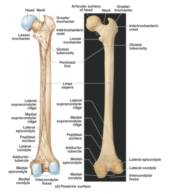
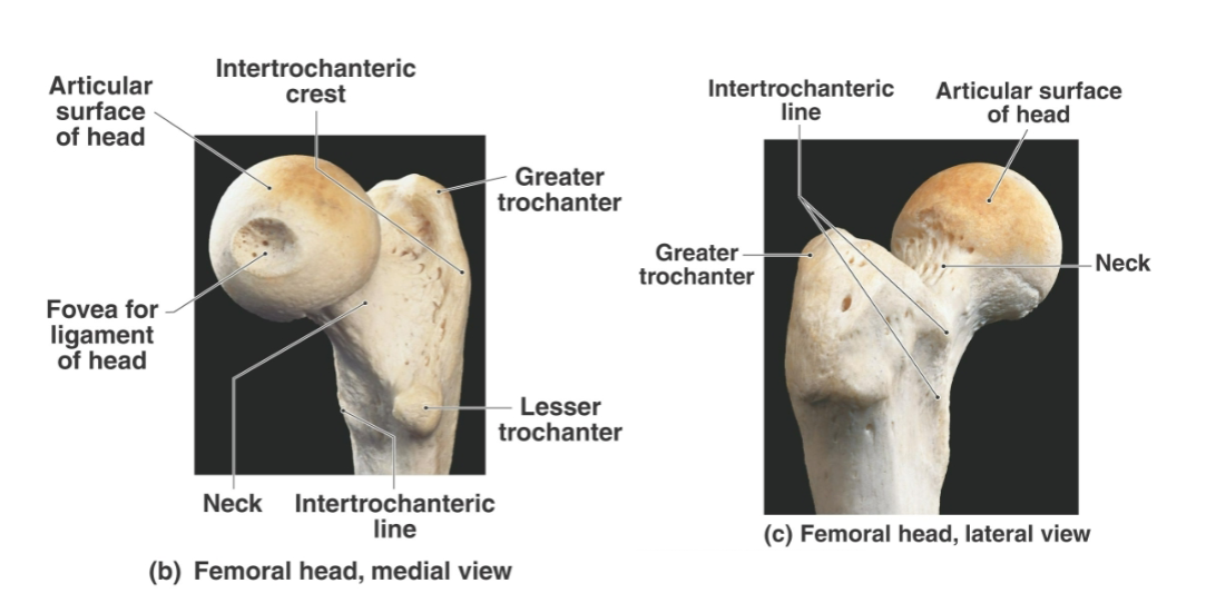
Head of femur: Ball-like proximal end that articulates with the acetabulum
Fovea capitis: Small central pit on the femoral head where the ligament of the head of the femur attaches to acetabulum (hip)
Neck of femur: Narrow region connecting the femoral head to the shaft and a common fracture site
Greater trochanter: Large lateral femoral projection serving as an attachment site for muscles and tendons
Lesser trochanter: Posteromedial femoral projection serving as an attachment site for muscles and tendons
Intertrochanteric line: Anterior line connecting the greater and lesser trochanters
Intertrochanteric crest: Posterior ridge connecting the greater and lesser trochanters
Pectineal line: Medial femoral ridge serving as an attachment site for the pectineus muscle
Gluteal tuberosity: Lateral posterior femoral surface serving as an attachment site for gluteal muscles
Linea aspera: Prominent posterior ridge of the femoral shaft serving as an attachment site for powerful hip muscles
Femoral shaft: Strong, massive lateral bow body of the femur that facilitates weight bearing and balance

Distal end features of femur
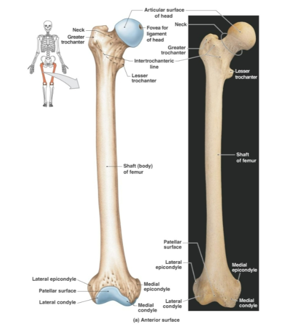
Medial supracondylar ridge of femur: Posterior medial ridge formed as the linea aspera divides distally to triangular area called POPLITEAL SURFACE
Lateral supracondylar ridge of femur: Posterior lateral ridge formed as the linea aspera divides distally to triangular area called POPLITEAL SURFACE
Popliteal surface: Flattened triangular posterior area between the femoral supracondylar ridges
Lateral condyle of femur: Lateral broadened distal articular region of the femur
Medial condyle of femur: Medial broadened distal articular region of the femur
Lateral epicondyle of femur: Raised lateral point of the femoral condyle serving as a ligament attachment site
Medial epicondyle of femur: Raised medial point of the femoral condyle serving as a ligament attachment site
Adductor tubercle: Bump on the upper portion of the medial femoral condyle
Intercondylar fossa: Posterior notch separating the medial and lateral femoral condyles
Patellar surface: Anterior surface separating the femoral condyles and articulating with the patella
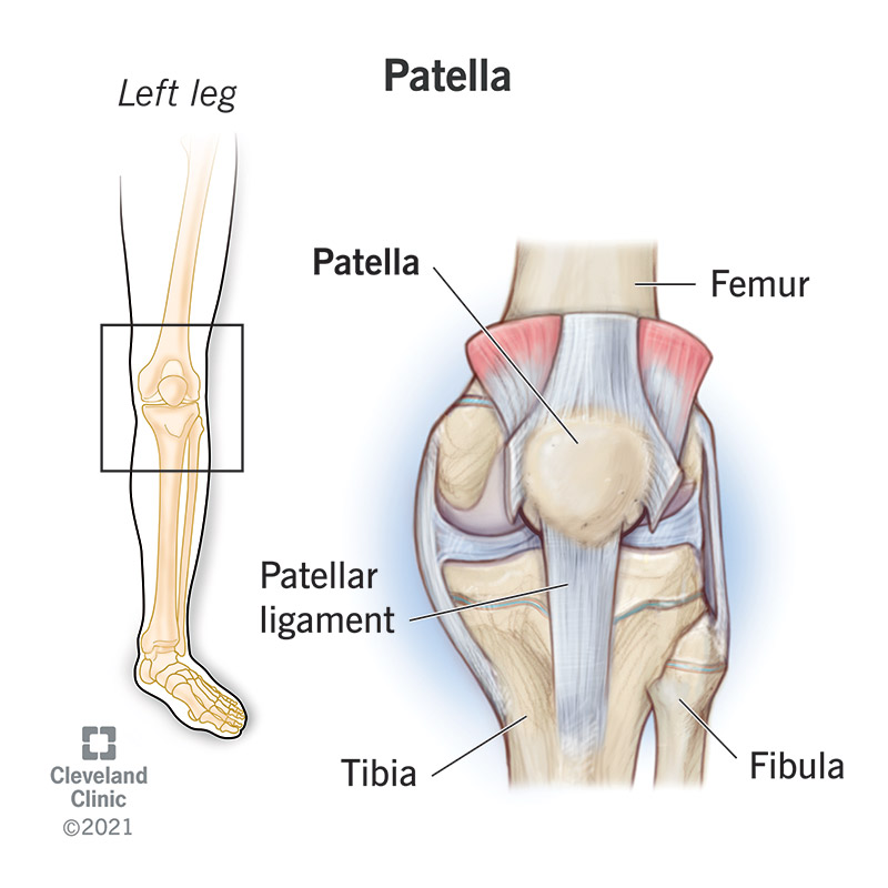
Patella (kneecap)
Triangular sesamoid bone enclosed within the quadriceps tendon and commonly called the kneecap
Rough convex anterior surface.
Strengthens the quadriceps tendon protects the anterior knee and increases quadriceps contraction force
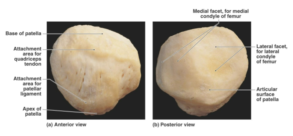

Parts of patella
Base of patella: Broad superior portion of the patella
Apex of patella: Pointed inferior portion of the patella
Patellar facets: Posterior surfaces that articulate with the femoral condyles
Posteroinferior surface: for patellar ligament


Crus (leg)
Anatomical term for the leg between the knee and ankle
Most fractures appear at the medial and lateral malleoli of the tibia and fibula because they are the least massive parts. These fractures are caused by eversion (The sole of the foot faces outward or laterally.) or inversion (The sole of the foot faces inward or medially) of the ankle
Comprised of:
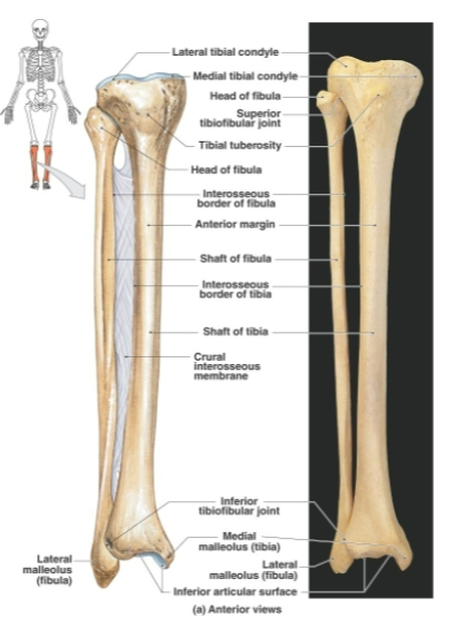
Tibia: Medial weight-bearing bone of the leg and second largest and strongest bone of the body
Fibula: Thin lateral bone of the leg that does not bear significant body weight. located lateral to tibia. origin site of several muscles
What is the tibia comprised of?
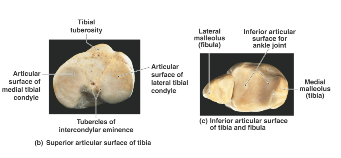
Medial condyle of tibia: Medial portion of the broad proximal tibia that articulates with the femur
Lateral condyle of tibia: Lateral portion of the broad proximal tibia that articulates with the femur
Intercondylar eminence: Irregular projection separating the tibial condyles
Tibial tuberosity: Anterior tibial projection serving as an attachment site for the patellar ligament
Anterior border of tibia: Sharp subcutaneous anterior ridge of the tibial shaft
Medial malleolus: Distal tibial projection forming the medial bulge of the ankle. articulates with the TALUS of foot
Distal articular surface of tibia: Flat distal surface that articulates with the talus
Proximal tibiofibular joint: Facet joint where the lateral tibial condyle articulates with the fibula
Distal tibiofibular joint: Joint where the fibular notch of the distal tibia articulates with the fibula
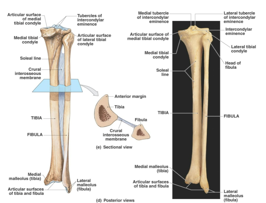

What is the fibula comprised of?
Head of fibula: Expanded superior proximal end of the fibula
Lateral malleolus: Distal fibular projection forming the lateral bulge of the ankle.
Fibular shaft: Heavily ridged shaft that appears twisted about one-quarter turn


Pes (ankle and foot)
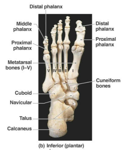
There are 7 tarsal bones that make up the ankle.
The metatarsal bones (5) articulate with the distal tarsal bones and make up the arches of the foot.
The 14 phalanges of the foot make up the toe bones.
Supports body weight and segmentation acts as a lever during movement and adapts to uneven ground
Hallux: Anatomical term for the great toe located on the medial side of the foot (Pollux is lateral to hallux)


Tarsus
Posterior half of the foot containing seven tarsal bones
Types of bones:
Cuboid (lateral): “cubed shaped” tarsal
Navicular (medial): “boat like” tarsal
Medial / intermediate / lateral cuneiform bones (anterior): “wedge shaped” tarsals arranged in a row lateral to the navicular. Named according to their position.


Tarsus consists of what?
Talus: Second largest tarsal that receives and transmits body weight from the tibia anteriorly towards the toes
Trochlea of talus: Smooth superior talar surface that articulates with the medial and lateral malleoli
Calcaneus: Largest tarsal bone and the bone that forms the heel that carries the talus on its superior surface
Calcaneal tuberosity: Part of the calcaneus that contacts the ground
Sustentaculum tali: Medial shelf-like calcaneal projection supporting and articulating with the talus
Achilles tendon: Thick tendon of the calf muscles that attaches to the posterior calcaneus

Metatarsus
Distal portion of the foot containing five metatarsals
Metatarsals I through V are numbered from medial to lateral (starting at Hallux)
Supports body weight and helps form the ball of the foot


Phalanges of toes
14 phalanges in each foot
The great toe has proximal and distal phalanges only while digits 2 through 5 each contains proximal middle and distal phalanges

the sternal end of the clavicle articulates with the ____ while the acromial end of the clavicle articulates with the ____
clavicular notch of the manubrium
acromion of the scapula

The radius articulates with the ___ of the humerus
and the ulna articulates with the ___ of the humerus
capitulum
trochlea

the head of the radius is ___ while the head of the ulna is ___ (directional)
proximal (near the elbow)
distal (near the wrist).
