RHS
 Call Kai
 Learn
Learn Practice Test
 Spaced Repetition
 Match
Match Flashcards
 Knowt Play
1/258

There's no tags or description
Looks like no tags are added yet.
Name | Mastery | Learn | Test | Matching | Spaced | Call with Kai | Chat |
|---|
No analytics yet
Send a link to your students to track their progress
259 Terms
apical pathology
inflammatory conditions around the root tip of tooth
what are caries
cavities
purpose of periapical dental images
to show the entire tooth from crown to root apex and surrounding bone
purpose of bitewing dental images
to detect interproximal caries, recurrent decay, periodontal disease, and evaluate restorations
Purpose of Full Mouth Series (FMX)
full survey of teeth and supporting structures using pariapicals and bitewings
Purpose of occlusal
shows large areas of the maxilla or mandible and helps locate impacted teeth or foreign objects
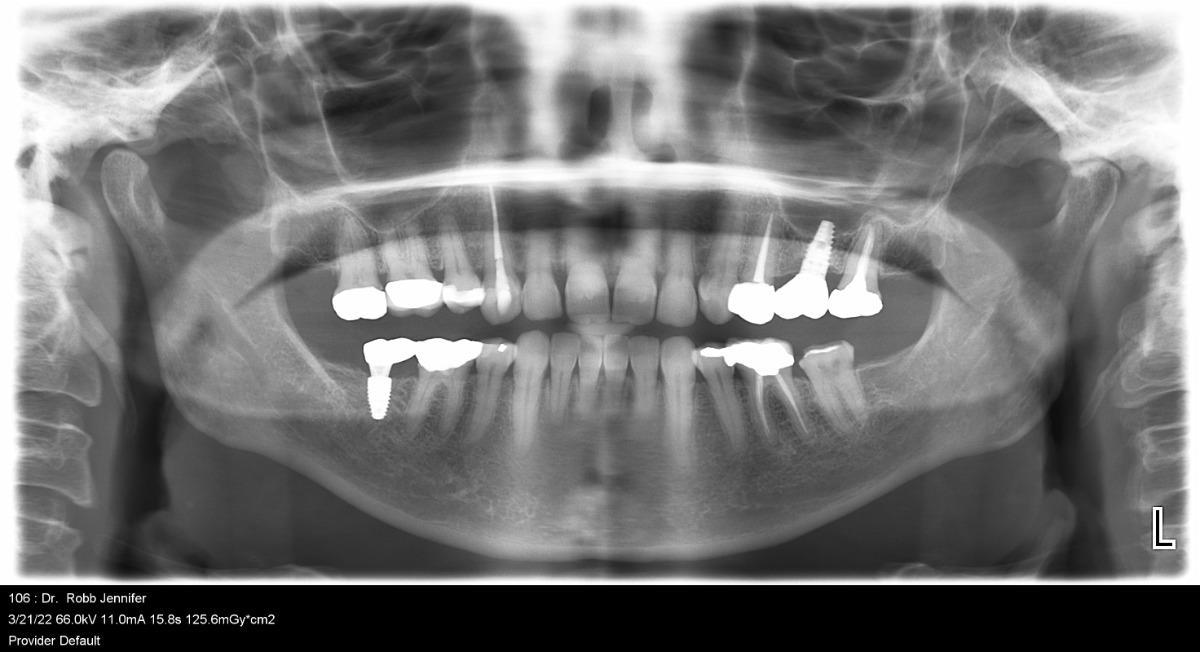
purpose of panoramic
shows a view of both jaws, tmjs, sinuses, and surrounding structures
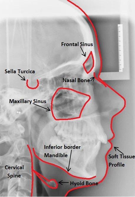
purpose of cephalometric
shows facial profile and to assess the exact relationship between the teeth, jawbones, and skull structure
purpose of cone beam computed tomography (CBCT)
used for implants, impacted teeth, pathology and surgical planning
what are the features of a diagnostically acceptable image? (6)
includes requires anatomy
proper density
proper contrast
sharp details
minimal distortion and magnification
free from errors and retakes
what is density
overall darkness or blackness of the image
what is contrast
difference between shades of black, gray, and white
what is sharpness
ability to reproduce fine details clearly
what is distortion
incorrect size or shape caused by improper angulation
why should you review medical history
to identify contraindications and medications that may affect treatment
How would you prepare patient for a radiographic procedure
you would remove:
glasses
earrings
necklaces
facial piercings
removable appliances
purpose of patient positioning
to prevent distortion and ensure accurate image acquisition
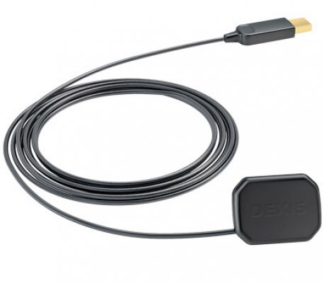
what is an image receptor
the sensor that captures the x-ray

what is the image holder
the device that holds it in position (XCP)
Yellow XCP holder?
posterior x-rays
Green XCP holder
endodontic x-rays
Blue XCP holder
anterior x-rays
Red XCP holder
bitewing x-rays
What is a bite block
plastic piece that patients bite down on to stabilize the receptor holder
What is a bitwing tab
a sticky tab that holdd the biting film/sensor in place
what is positioning indication device (PID)
the cone/tube end that directs x-ray beam
What is the beam alignment and paralleling devices
tools that help align x-ray beam and receptor correctly
what are the 3 components of an x-ray unit
control panel
extension arm
tubehead
what is the control panel
button/settings for exposure
what does the control panel do
sets kVp, mA, and exposure time
what is the extension arm
moves tubehead into postion
what does the extension arm do
positions the x-ray beam towards patients' mouth
what is the tube head
where x-rays are produced
what are the 3 components in a tubehead
anode
cathode
target
what do you do with a tubehead
aim it at receptor using PID
what do you do with the image receptor and holder (3)
place receptor inside the patient's mouth
use holder to stabilize it
ensure that it is flat and correctly positioned against the tooth area
what do you do with the bite block
patient gently bites in it to keep everything still and helps maintain correct alignment
what do you do with bitewing tabs
attach tab to the receptor, and patient bites on it to stabilize.
what do you do with the PID
aim it toward the receptor and tooth; it must be centered correctly to avoid cone cuts
what do you do with beam alignment and paralleling devices
align tubehead→ring→receptor so beam is parallel
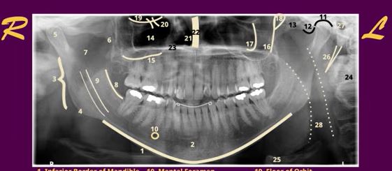
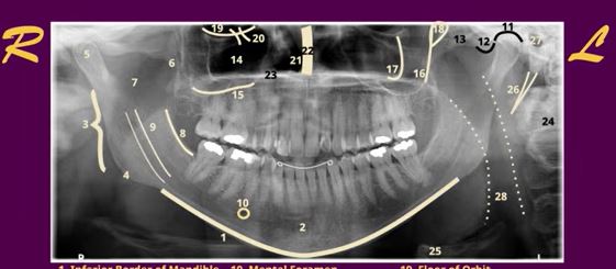
1
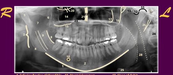
inferior border of mandible
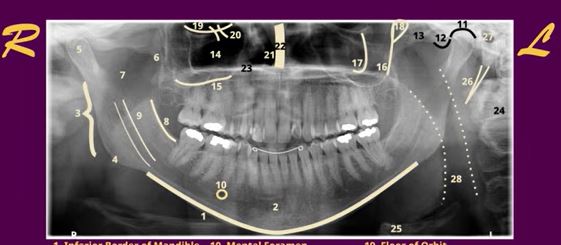
2
body of mandible
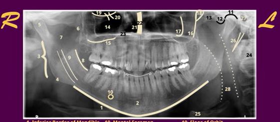
3
ramus of mandible
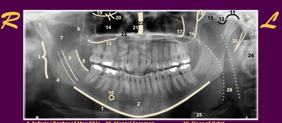
4
angle of mandible
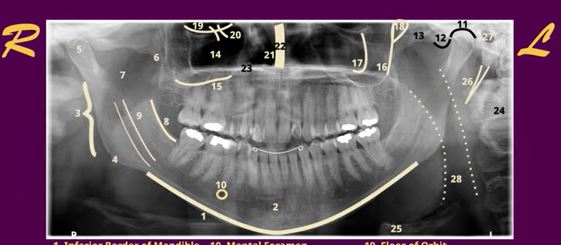
5
condyle
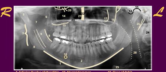
6
coronoid process
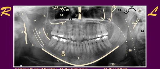
7
sigmoid notch
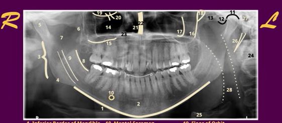
8
external oblique ridge
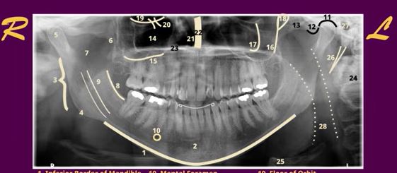
9
manibular canal
10
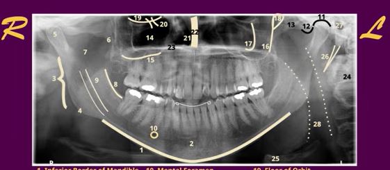
mental foramen
11
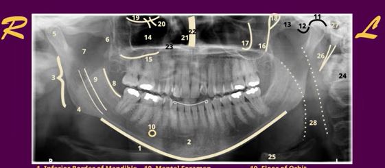
glenoid fossa
12
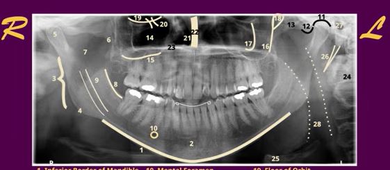
articular eminence
13
zygomatic arch
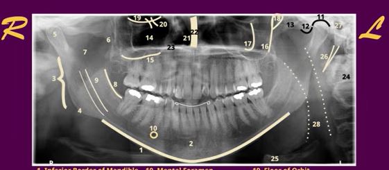
14
maxillary sinus
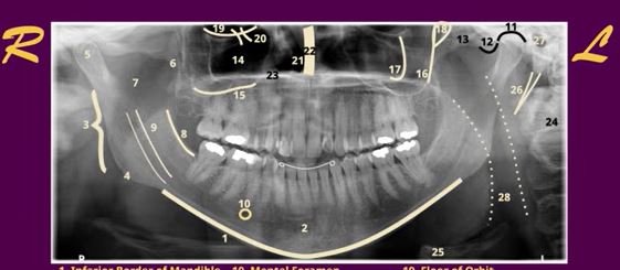
15
floor of maxillary sinus
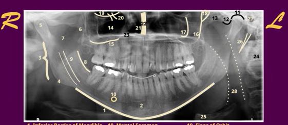
16
posterior wall of max sinus
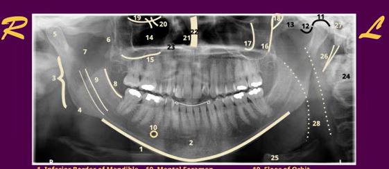
17
zygomatic process of max
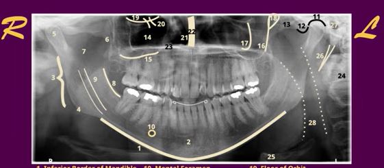
18
pterygomaxillary fissure
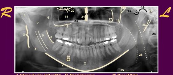
19
floor of orbit
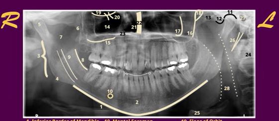
20
infraorbital canal
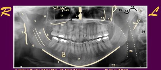
21
nasal cavity
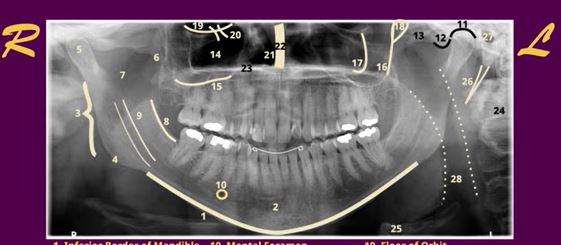
22
nasal septum
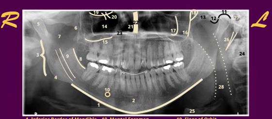
23
hard palate
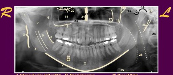
24
vertebrae
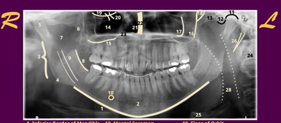
25
hyoid
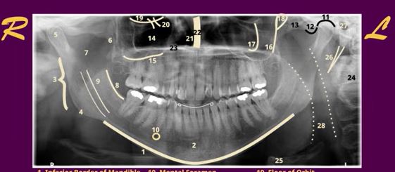
26
styloid process
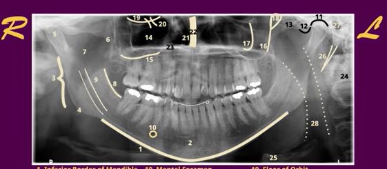
27
external auditory meatus
28
airway
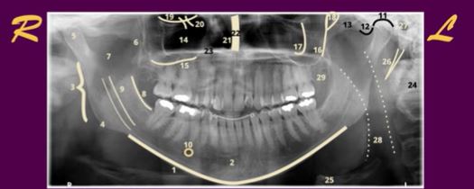
29
maxillary tuberosity
Radiopaque (3)
white light, that is high density and blocks the passage of x-rays
Radiolucent (2)
1 black/gray
low density that lets x-rays pass through
example of radiopaque
bone, metal implants, dental filling
example of Radiolucent
air, fat, soft tissue, and tooth decay
labial surface
towards the lip
buccal surface
towards the cheek
lingual surface
faces the tongue
occlusal surface
top of posterior teeth
palatal surface
faces the palate (roof of mouth)
mesial surface
towards the midline
distal surface
away from the midline
what is ionization
the removal of an electron from an atom
why is ionization important
it is responsible for biological damange caused by radiation
where are x-rays produced
inside the x-ray tube
what is a cathode
the negative side of the x-ray
what is the anode
the positive side of the xray
what is the tungsten filament
a wire inside the cathode that produces electrons when heated
what is thermionic emission
release of electrons from the heated tungsten filament
what is the focal spot
area of the tungsten target where electrons strike and x rays are produced
what percentage of energy becomes x-rays
aprox. 1%
what percentage becomes heat
aprox. 99%
what is primary radiation
useful beam produced at the target
what is secondary radiation
radiation produced after the primary beam interacts with matter
what is scatter radiation
secondary radiation that changes direction after interacting with matter
why is scatter radiation dangerous
it exposes patients and operators to unnecessay radiation
what is leakage radiation
radiation escaping through the tube housing
what does kVp control
beam quality and penetrating power
increasing kVp causes what
greater penetration and lower contrast
what does mA control
quantity of x-rays produced
increasing mA causes what
more x-rays and a darker image
what does exposure time control
length of radiation exposure