PNB 2264: Neurophysiology, skeletal muscle, brain and physiology practice questions
1/137
There's no tags or description
Looks like no tags are added yet.
Name | Mastery | Learn | Test | Matching | Spaced | Call with Kai |
|---|
No analytics yet
Send a link to your students to track their progress
138 Terms
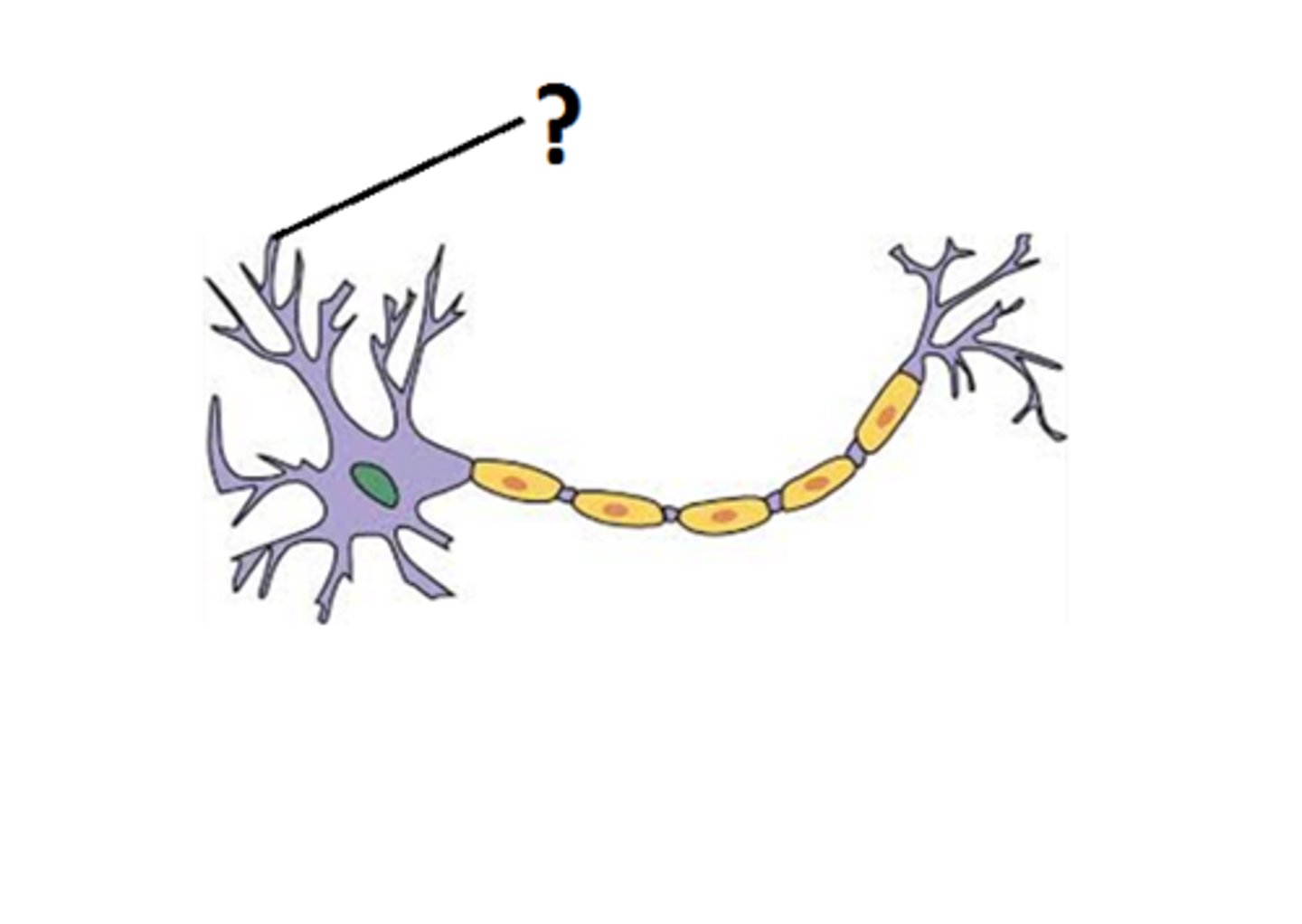
dendrite
What part of the neuron is this?
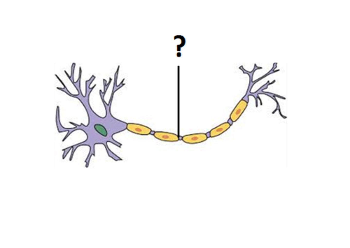
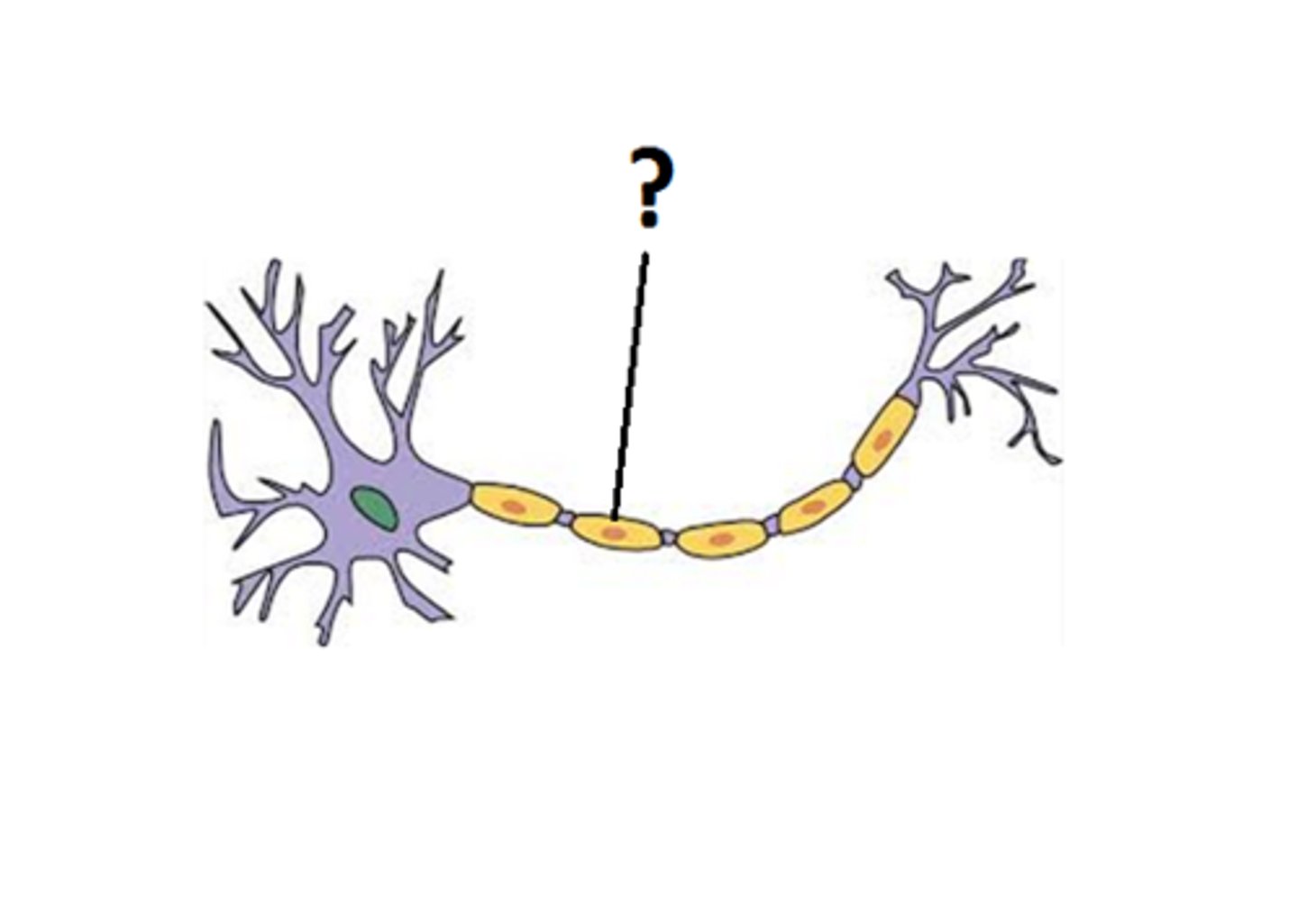
node of ranvier
What part of the neuron is this?
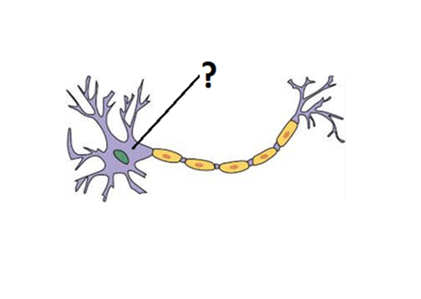
cell body (soma)
What part of the neuron is this?
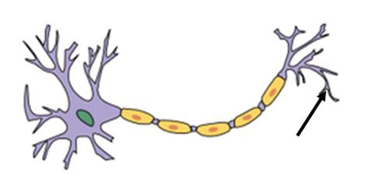
axon terminal
What part of the neuron is this?
myelin sheath
What part of the neuron is this?
What is the function of the dendrite?
The dendrites are a place of signal input to the neuron. They contain high densities of receptors because they are constantly receiving signals from internal and external environment.
What is the function of the soma (cell body)?
Signal integration and other cellular functions
What is the function of the axon hillock?
It is where the action potential is generated and spreads through the axon into the axon terminal
What is the function of the axon terminal?
Forms synapses or connections with other cell types (neurons or non neuronal cell types inside the body)
What is a synapse?
Point of communication between two different cells.
What is the term for the phase when a membrane at rest experiences a positive change in membrane potential?
Depolarization
What is the term for the phase when a membrane returns to rest from depolarization?
Repolarization
What is the term for when a membrane at rest experiences a negative charge in membrane potential?
Hyperpolarization
What type of graded potential describes a positive or depolarizing change from rest? What is an example of this potential?
Excitatory postsynaptic potential (EPSP)
Example is opening a sodium channel.
These push the neuron closer to threshold and make an action potential more likely.
What type of potential describes any negative or hyperpolarizing change from rest? What is an example of this potential?
Inhibitory postsynaptic potential (IPSP)
Example is
opening up a potassium channel
These push the neuron further from threshold and make an action potential harder to achieve.
What is the difference between a graded potential and an action potential?
Action potentials are able to maintain their strength over time and distance, whereas a graded potential decreases over time and distance
Explain what a passive electrical signal is and give an example.
Passive electrical signals are transient changes in the membrane potential that is specific to one region of the cell and only occurs for a short period of time
Examples would be graded potentials and synaptic potentials
Explain what an active electrical signal is and give an example.
Active electrical signals create changes in membrane potential that can spread over a very long distance
An example would be an action potential
Describe a typical axon segment
An axon is separated into segments that are like work stations. Each of them have what they need (coltage gated Na+ channels) to create action potential in that spot. The action potential needs to be able to travel all the way from the axon hillock to the axon terminal. It's easier for the action potential to travel from one segment to the next than all the way through (in the same way we take breaks between sets at the gym).
How does myelin increase the speed of an action potential?
Think of it like an electrical cord. If you have holes in the rubber around the wires your device is going to charge slower or not at all. It acts as a insulating wrapper that keeps local currents from dissipating into external environments. It creates an increase in conduction velocity and efficiency of the overall neuron.
What is a myelinated segment called?
Internode
What is a non myelinated segment called?
Node of ranvier
What are the characteristics of nervous tissue?
High metabolic rate: Nervous system depends on concentration gradients which means neurons must have a very high capacity to generate apt to keep the ions in and out of the cell within homeostatic ranges
Extreme longevity:
they live longer than other cells in the body
Non-mitotic
Terminally differentiated cell that does not go through mitosis.
We can replace neurons but we have to go through the stem cell differentiation process.
We normally can't get the neurons to differentiate in the right spot which makes it hard to treat neurodegenerative diseases.
Where is action potential initiated and why?
Action potential is initiated at the axon hillock.
It starts here because there is a high density of voltage gated sodium channels
How do changes in ion concentration impact action potential?
The depolarization associated with an action potential generated at one point along an axon spreads passively to the adjacent segment, where it triggers opening of voltage-gated Na+ channels and hence another action potential. Propagation of the action potential occurs in one direction only because of the short inactive period of the Na+ channels and the brief hyperpolarization resulting from K+ efflux
How does a change in permeability impact the membrane potential
An increase in sodium permeability correlates with depolarization which will lead to an ever greater number of Na+ channels opening and membrane potential becoming even more depolarized.
What is an inactive state?
think of a house with a screen door and a glass door. One of them is open but the other door is still closed. Sodium still cannot enter the cell because one of the doors is closed.
What is an open state?
An open state is when the activation gate and the inactivation gate are open
What is a closed state?
A closed state is when the activation gate is closed but the inactivation gate is still open.
What are the effects of channelopathies on nervous system function?
What are examples?
Channelopathies are caused by changes to the voltage gated sodium channels. Lack of sodium channels leads to action potentials not firing and no ability to feel pain.
An example of this is CIP or congenital insensitivity to pain
It's also possible to have overactive voltage gated sodium channels which lead to action potentials continued firing. An example of this would be PEPD or paroxysmal extreme pain disorder where even the lightest touch can hurt.
How are action potential propagation and graded potential propagation similar? How are they different?
Graded potentials are brought about by external stimuli (in sensory neurons) or by neurotransmitters released in synapses, where they cause graded potentials in the post-synaptic cell.
Action potentials are triggered by membrane depolarization to threshold. Graded potentials are responsible for the initial membrane depolarization to threshold.
What are the general properties of muscle?
Excitability: muscle generates graded and action potentials through the same mechanisms as neurons
Elasticity: muscles are constantly changing in size
Contractility: Muscles use their action potential to initiate the process of contraction, which generates force.
Extensibility: Muscle doesn't lose its membrane integrity when it experiences large changes in size.
What are anatomical features of muscle at the cellular level?
Multinucleated tissue (3+ nuclei per cell)
Overall rectangular or cylindrical type shape
Vertical lines known as striations
Densely packed tissue with little space between adjacent cells
What are the components of a sarcomere?
Sarcomeres make up myofibrils.
They are the basic unit of muscle contraction
Made up of three structural proteins; myosin (thick filament), actin (thin filament), and titan (tiny elastic protein filaments.
They contract in an oscillatory manner, similar to a spring.
Actin and myosin slide to contract the muscle.
How does the sliding filament model work to regulate contraction?
Actin and myosin repeatedly slide over each other, contracting (similar to how a spring does) the sarcomere and generating tension in the muscle. This is how a muscles work to contract.
What is the role of calcium in muscle tissue?
Muscles require a calcium signal in order to initiate muscle contraction. They also need energy. Binding must happen between actin and myosin in order to allow muscles to contract.
Referred to as a cross-bridge
Cross bridges must be formed in order for a contraction to occur.
At rest (low Ca2+), tropomyosin blocks the myosin binding site on actin, which prevents binding
Impossible for cross bridges to form
When depolarized, (high Ca2+), Ca2+ binds to TnC (subunit of troponin) and causes ejection of tropomyosin so binding can occur
Possible for cross bridges to form
They also require energy
What are the components of the excitatory contraction coupling machinery?
Muscle fiber is densely packed with microfibrils
T-tubules allow rapid exchange between the interstitial fluid and the deepest muscle fibers.
Necessary because muscles have very little cytoplasm
Sarcoplasmic reticulum is well developed in muscle and the main calcium storage site in muscle
T tubule and sarcoplasmic reticulum are so close together that they practically touch each other
What are the steps of excitation contraction coupling?
in response to an action potential a
motor neuron releases acetylcholine
acetylcholine crosses the synaptic cleft
and binds to receptors on the plasma
lemma if enough acetylcholine binds to
the receptors an action potential is
generated and transmitted along the full
length of the muscle fiber the action potential triggers the release of
calcium ions from the sarcoplasmic reticulum into the sarcoplasm in the resting state
tropomyosin molecules cover the myosin
binding sites on the actin molecules of
each sarcomere preventing the binding of
the myosin heads once calcium ions are
released from the sarcoplasmic reticulum
they bind to the troponin on the actin
molecules troponin with its strong
affinity for calcium ions is believed to
then initiate the contraction process by moving the tropomyosin molecules off the
myosin binding sites on the actin
molecules allowing myosin heads to
attach to the actin filaments
What are the components of the neuromuscular junction?
An alpha motor neuron and a skeletal muscle fiber. Presynaptic cell is a neuron. Postsynaptic cell is a muscle fiber. Every single skeletal muscle is provided supply by the axon terminal of a motor unit
How do changes in ion concentration impact muscle tissue?
Once you start neuron action potential it is the same as neuron action potential.
What is curare? How is it treated?
Competitive antagonist that binds to the acetylcholine receptor and blocks the acetylcholine site
which stops all postsynaptic events at the NMJ
It induces muscle paralysis without causing a loss of consciousness.
To fix this you need to take AChE inhibitor, which stops the destruction of acetylcholine.
What does eserine (physostigmine) do?
inhibitor of acetylcholinesterase (enzyme that breaks down acetylcholine)
increases the half life of acetylcholine
This drug can cross the blood brain barrier and can be used for neurological purposes
What does succinylcholine do?
ACh receptor that binds and activates Acl receptor. Cant be broken down by acetylcholinesterase. This results in signals not repeating
What is myasthenia gravis?
Autoimmune disorder where the body produces antibodies that lead to the destruction of aCh receptors.
not be able to produce enough end plate potential to start the action potential because they don't have enough receptors
Causes muscles to get fatigued more easily
What is Lambert-Eaton Myasthenic Syndrome (LEMS)
Mutation in voltage gated calcium channel inside of axon terminal
Decrease calcium influx
Decrease release of aCh
Muscle is not stimulated enough so patients get fatigue and weak muscles
Guillain-Barre Syndrome
Random rapid onset muscle paralysis
Autoimmune disorder where the immune system attacks the glial cells that myelinate the internodes of the motor neuron
When this happens conduction efficiency through the axon slows down and the conduction velocity slows down.
This means that action potentials dont always reach axon terminal
ACh never gets released
Muscle never gets stimulated
Compare and contrast muscle and neurons with respect to excitability and synaptic transmission
There is no response diversity in muscle. the muscle is either contracted or not contracted (all or none) .
Neurons can open and close with activation and inactivation gates. it doesn't follow the all or none concept.
Neurons can depolarize, repolarize and hyperpolarize.
Muscles have one ACh receptor and one response (contraction)
Describe structure function relationships in muscle at the histological and organ level
A system of tubes within a tube
Series of tube-like structures called fascicles. Each fascicle is a bundle of muscle fibers held together by a connective tissue wrapper
Each individual muscle fiber contains myofibrils which are created from the arrangement of the contractile proteins inside the cell
Myofibrils are very abundant because they contain the contractile proteins of the cell
Arrange themselves into repeating units that are called sarcomeres which are the basic units of muscle contraction
One myofibril contains many sarcomeres
Three structural proteins that make up the sarcomere
Myosin: thick filament
Actin: thin filament
Titan: tiny elastic protein filaments
Sarcomeres contract in an oscillatory manner similar to a spring
Sliding filament (actin and myosin) model
On a large scale this contracts the entire muscle cell
Contraction is a really tightly regulated process
Regulatory proteins in the same spots as actin and myosin
What mechanisms do individual fibers and entire muscles use to increase the amount of force generated??
Difference in color is due to an oxygen binding protein called myoglobin
Type 1 fibers are the darkest and have the most myoglobin
They are really good at binding and trapping oxygen in the tissue which allows them to produce a lot of ATP and stay contracted for a long period of time
Postural muscles are an example of this
Type 2b fibers have little if any myoglobin
They rely on anaerobic respiration which is much less efficient at producing ATP
Used for short activities such as sprinting
Type 2a fibers
Have some myoglobin and are considered intermediate
Limited amount of aerobic respiration
How is the blood brain barrier formed?
In the capillaries that form the blood-brain barrier, endothelial cells are wedged extremely close to each other, forming so-called tight junctions. The tight gap allows only small molecules, fat-soluble molecules, and some gases to pass freely through the capillary wall and into brain tissue
What are the components of the neurovascular unit?
endothelial cells form it. tight junctions regulated by astrocytes and pericytes regulate blood flow.
How is CSF produced?
75% of the CSF is produced by the ependymal cells (transporting epithelium) which form a tissue known as the choroid plexus
25% is produced by regions outside of the choroid plexus
How is CSF stored?
It is stored in the ventricles
How is CSF circulated?
It is circulated by ventricles. It moves from the lateral ventricles on either side into the third ventricle and then through the cerebral aqueduct into the fourth ventricle.
It goes through the central canal into the spinal cord
What is the function of CSF
It provides physical and chemical protection
What are the major anatomical features of a spinal cord cross section?
sensory info comes in through the dorsal route and motor leaves through ventral route
What are the major anatomical regions of the brain
Cerebrum
Diencephalon
Brainstem
Cerebellum
What is the function of the cerebrum?
Higher brain function
What is the diencephalon do and what are the three major parts?
Responsible for homeostatic controls
Epithalamus
Thalamus
Hypothalamus
What are the parts and functions of the epithalamus?
Made up of the pineal gland which secretes melatonin, a hormone that regulates day and night cycles, and habenular nuclei which relay signals from the limbic system to the mesencephalon/ midbrain and are involved in emotional responses to odor
What is the function of the thalamus?
Acts as a filter, only a small amount of signals actually make their way up to the primary somatosensory cortex.
What is the function of the hypothalamus?
Regulates homeostatic mechanisms throughout the body.
What is the function of the brainstem and what parts is it made up of?
Bidirectional passageway for all tracts extending between the cerebrum and the spinal cord. Houses nuclei of many cranial nerves. Responsible for involuntary processes and contains many autonomic centers and reflex centers. Made up of the midbrain (mesencephalon), pons, and medulla.
What does the pons do?
Regulates respiratory centers and arousal.
What does the medulla do?
Regulates respiratory system and cardiovascular system
What is the cerebellum responsible for?
Balance and coordination
Compare and contrast blood with CSF
CSF has a lower concentration of protein, glucose, and potassium, and a higher concentration of sodium than blood. It allows for a large gradient to generate action potential. Low extracellular potassium concentration allows us maintain negative resting membrane potential.
How would the blood brain barrier be altered if there were changes in the neurovascular unit??
Changes in the NVU could mean that the composition of different compartments is not being maintained. When tight junctions that limit the paracellular junction are not working properly the toxins that were being kept out will go in. This can cause damage to the structures inside of the BBB
What are the parts of the cerebral cortex?
frontal lobe, parietal lobe, occipital lobe, temporal lobe
What are the parts and functions of the frontal lobe?
Responsible for motor, speech, memory formation, personality, and emotion
It is the largest and most developed region of the brain. Crucial in decision making, hypothesis generation, inhibition/impulse control. Includes broca's area, orbitofrontal cortex, and olfactory bulb.
What is brocas's area responsible for?
Speech
What is the orbitofrontal cortex responsible for?
decision making/ adaptive learning
What is the olfactory bulb responsible for?
odor perception
What is the parietal lobe responsible for?
Contains the primary somatic sensory area, where sensory information is collected. The amount of real estate that different parts of the body get inside of the brain is dependent on how often they are used.
What is the occipital lobe responsible for?
Visual processing and storing visual memories
What is the temporal lobe responsible for?
Hearing, speech and language comprehension, memory formation and retrieval.
Wernicke's area is important for language comprehension.
What are the effects of damage to the cerebral cortex?
Personality changes (depressive disorders), difficulty concentrating and remembering, poor decisions, and trouble controlling impulses.
What are the effects of damage to the basal ganglia?
problems controlling speech, movement, and posture. Difficulty starting, stopping, or sustaining movement. Parkinson's is a good example.
What are the effects of damage to the diencephalon?
Severe and long lasting amnesia.
What are the effects of damage to the cerebellum?
Loss of coordination of motor movement (asynergia), the inability to judge distance and when to stop (dysmetria), the inability to perform rapid alternating movements (adiadochokinesia), movement tremors (intention tremor), staggering, wide based walking (ataxic gait), tendency toward falling, weak muscles (hypotonia), slurred speech (ataxic dysarthria), and abnormal eye movements (nystagmus).
In the brain, how are white matter and gray matter different.
Grey matter contains numerous cell bodies and very few myelinated axons. White matter has few cell bodies and is composed mainly of long range myelinated axons. The color difference is due to the myelin. Whit matter is also deeper.
How do explicit and implicit memories differ?
Information that you have to consciously work to remember is known as explicit memory, while information that you remember unconsciously and effortlessly is known as implicit memory
What cerebral features are responsible for increasing the surface area for exchange of ions with the CSF?
gyrus/gyri: raised portions (hills)
and
Sulcus/sulci: grooves (valleys)
What does the longitudinal fissure separate?
cerebral hemispheres
What does the transverse fissure separate?
the cerebrum and the cerebellum
What does the sylvian/lateral fissure separate?
Frontal, parietal, and temporal lobes
What does the central sulcus separate?
The frontal and parietal lobes
What are the language areas of the brain? Explain each.
Found in the left hemisphere. Wernicke's area is responsible for language comprehension, Broca's area is responsible for speech. Broca's area includes the arcuate fasciculus (connects Broca's area to Wernicke's area and allow us to produce coherent spoken language) and angular gyrus (help us comprehend written language, connect visual cortex to wernicke's area).
Chemical messengers that cross the synapse bind to ______ channels, which create _______ potentials.
a. Ligand gated; action
b. voltage gated; graded
c. Ligand gated; graded
d. voltage gated; action
c. ligand gated; graded
An IPSP could be caused by opening a ________ channel or closing a _________ channel
Potassium, sodium
Place the following events in the order they occur, starting with the depolarization of the axon hillock:
- Sodium enters through a ligand gated channel
- Voltage gated sodium channels open on the sarcolemma
- End plate potential develops
- Voltage gated calcium channels in the axon terminal open
- Cross bridges form
- Voltage gated sodium channels open in an internode segment
- Calcium levels in muscle increase
- Acetylcholine enters the synapse
1. Voltage gated sodium channels open in an internode segment
2. Voltage gated calcium channels in the axon terminal open
3. acetylcholine enters the synapse
4. Sodium enters through a ligand gated channel
5. End plate potential develops
6. Voltage gated sodium channels open on the sarcolemma
7. Calcium levels in muscle increase
8. Crossbridges form
During filament sliding, Z discs move towards the M line and:
a. The size of the A bands become smaller
b. The size of the I-bands become larger
c. The size of the H-zones become smaller
d. All of the above
The size of the H-zones become smaller
The knee jerk reflex requires ____ synapse(s) to activate quadriceps contraction in response to quick stretch of that muscle. The synapse(s) is/are found in the __ .
a. one; ventral horn
b. one; dorsal horn
c. one; dorsal root ganglia
d. two; ventral horn
e. one; ventral root
a. One; ventral horn
The dorsal root contains ______.
a. A ganglion
b. Sensory axons
c. Motor and sensory neurons
d. both a ganglion and sensory axons
e. all of the above
d. Both a ganglion and sensory axons
________ is required to inhibit the antagonistic knee flexors during the knee jerk reflex
a. inhibitory transmitter released to the hamstrings
b. inhibitory transmitter released in the spinal cord
c. both of the above
d. neither of the above
b. inhibitory transmitter released in the spinal cord
Starting with the receptor, put the following structures in the correct sequence
- Postcentral gyrus
- Lateral spinothalamic tract
- Spinal cord gray matter
- Thalamus
- Internal capsule
- Spinal cord gray matter
- Lateral spinothalamic tract
- Thalamus
- Internal capsule
- Postcentral gyrus
The center(s) for regulation of respiration is/are located in the ____ . (There may be more than one right answer
a. Medulla of the brain stem
b. Medulla of the diencephalon
c. Cerebral cortex
d. Hypothalamus of the diencephalon
e. Midbrain
f. pons
a. medulla of the brain stem
f. pons
The __________ connects the two cerebral hemispheres, and the _________ separates cerebral cortex from the cerebellum.
a. Frontal lobe; parietal lobe
b. Corpus callosum; transverse fissure
c. Internal capsule; lateral fissure
d. Corpus callosum; lateral fissure
b. Corpus callosum; transverse fissure
Functions of the left hemisphere typically include which of the following? (Choose all that apply.)
a. Written language
b. Speech
c. Control (sensory and motor) of left side of the body
d. flexor withdrawal response for right lower extremity
a. Written language
b. Speech
What is an efferent neuron?
motor neuron, conducting away from something