Iris and Pupil
1/62
There's no tags or description
Looks like no tags are added yet.
Name | Mastery | Learn | Test | Matching | Spaced |
|---|
No study sessions yet.
63 Terms
What is the major role of the iris?
to dynamically react to ambient lighting conditions and adjust retinal illumination
Does more light open or close the pupils? Less light?
more light closes the pupils, less light opens pupils
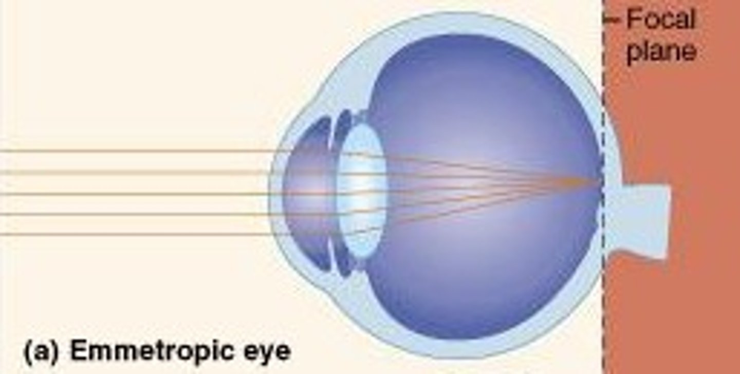
On an emmetropic eye (no refractive error), where does light from an object at infinity fall within the eye?
falls on the retina or image plane
When the object is near the eye, what does the lens do to focus the object on the retina? What happens to the power and focal length of the lens)
lens accommodates (increases in power, reduces focal length)
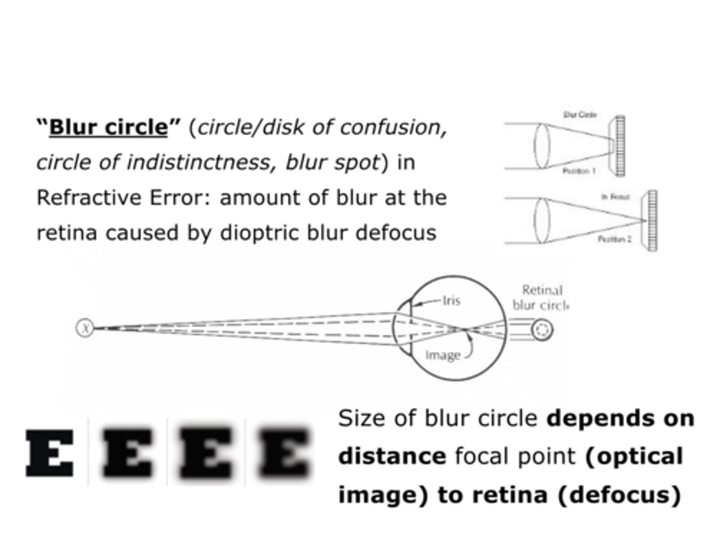
When is a blur circle generated?
when focused on a certain plane, objects in front of or behind that plane will be out of focus, generating a blur circle
What is an aperture stop? What is its effect on the field of view?
- a hole limiting the amount of light passing along an optical pathway placed near or at the lens
- has less effect on the field of view because light rays coming from oblique sources can still pass through the lens
What is a field stop? What is its effect on the field of view?
- a hole limiting somewhat the amount of light passing along an optical pathway placed at a distance from the lens
- has less effect on the amount of light passing through the lens, however it limits the field of view because light rays from oblique sources cannot pass through the hole in the stop to reach the lens
Does the iris act like an aperture or field stop?
aperture stop
What is depth of field?
the range of distance in object space within which an acceptable focus can be achieved
What is depth of focus?
- the corresponding range of distance in image space inside the eye within which focus is acceptable
- refers specifically to retinal image and perceived image clarity (absence of blur)
Does a smaller pupil lead to a smaller or larger depth of field and focus?
larger depth of field and focus
Does a larger pupil lead to a smaller or larger depth of field and focus?
smaller depth of field and focus
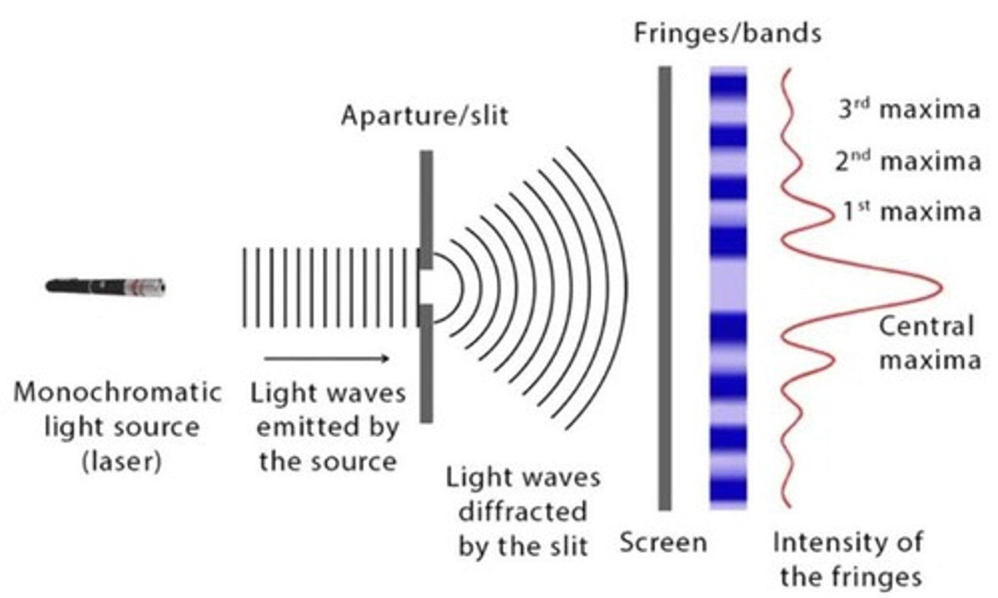
What is diffraction of light?
bending of light waves at the edges of an aperture (like pinhole or pupil)
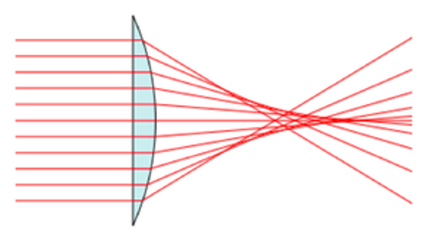
What are spherical aberrations?
changes in focal points for light rays entering off-axis in a lens
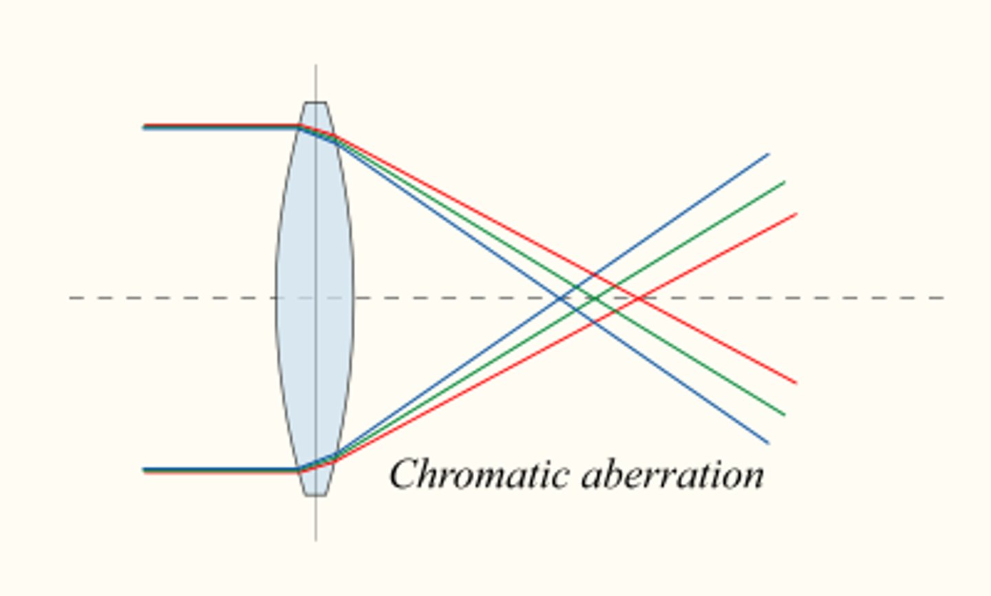
What are chromatic aberrations?
light of different wavelengths (colors) is refracted differently by a lens, thus a focal point varies with color
What is the entrance pupil?
image of the aperture stop formed by the optical elements in front of it (cornea)
Where does the entrance pupil appear to be? How large does it appear?
appears 1.13x larger, lies in a plane 0.5mm in front of the real pupil
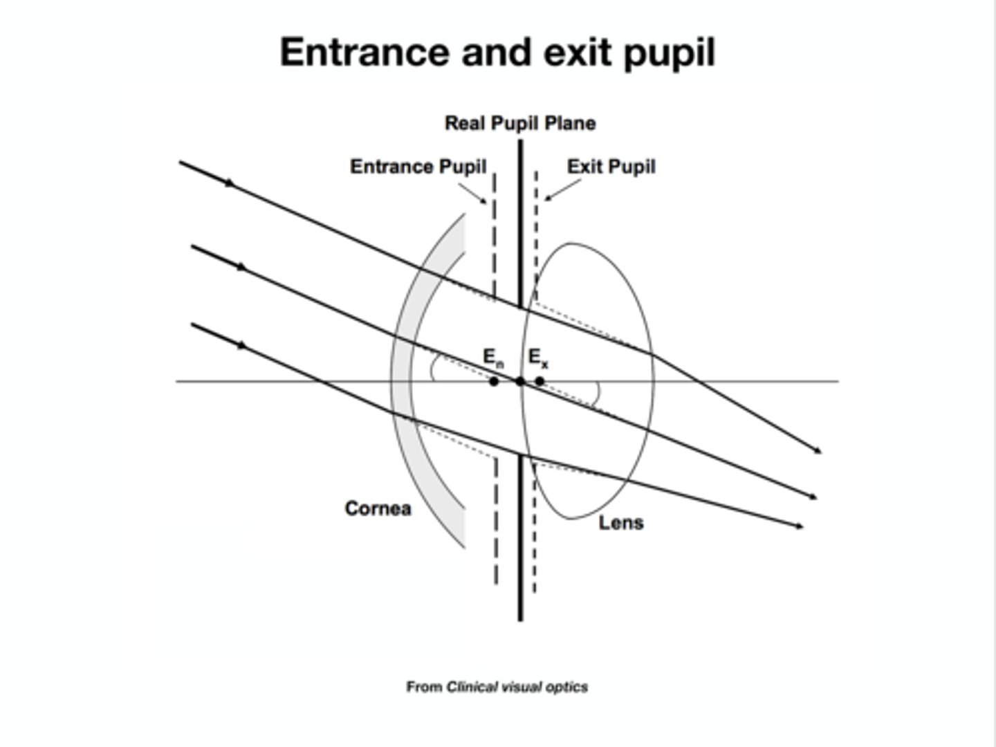
What is the exit pupil?
image of the aperture stop formed by the optical elements behind the the AS (lens)
Where does the exit pupil appear to be? How large does it appear?
appears 1.03x larger, lies 0.8mm behind the real pupil

Which pupil size measured in clinic? What is the range in young, healthy adults?
- entrance pupil size is measured in clinic
- ranges between 2-8mm in young, healthy adults depending on ambient illumination
What is the near response (triad)?
when you change your point of focus from a distant to a near object, three things occur: convergence, accommodation, pupillary constriction
As convergence angle increases in accommodation, what happens to the pupil diameter?
decreases (pupil constricts)
The pupil size/accommodation ratio (P/A) changes with age. In what age range does the pupillary near response become noticeably larger?
20-40 years of age
How do the pupils move in bright light (pupillary light reflex)?
pupils de-centered nasally
What is hippus?
hippus is rhythmic oscillations of the iris
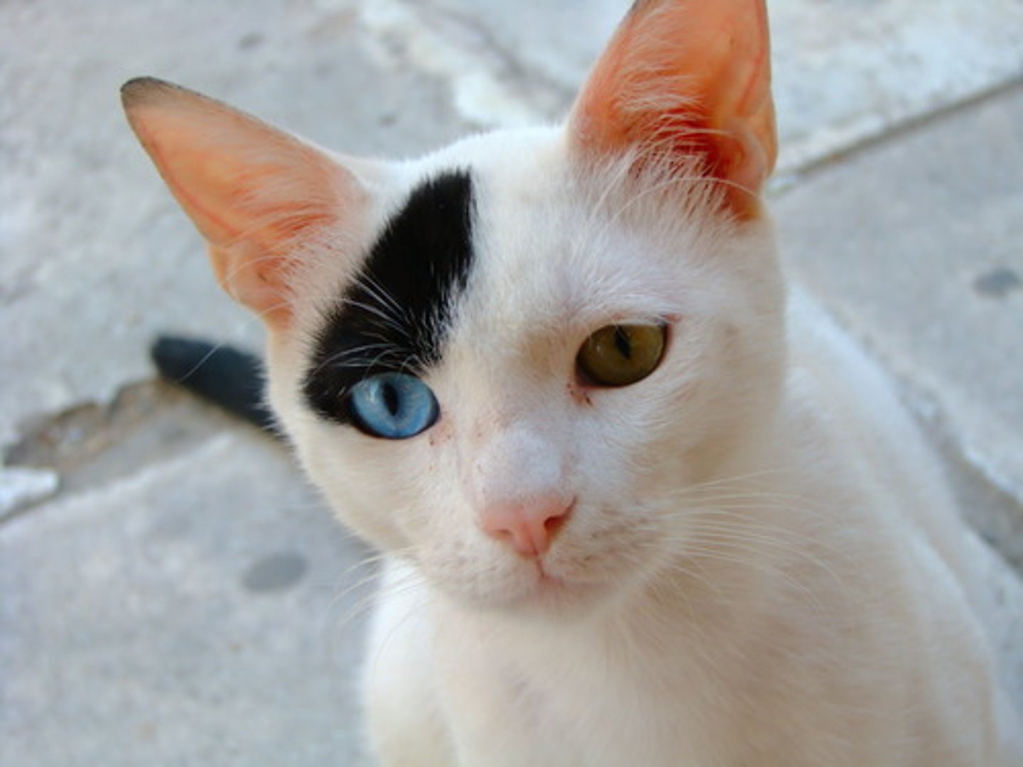
What is heterochromia? What is it caused by?
- heterochromia is a condition where a person has different colored eyes
- caused by the variation in the amount of melanin
- can be genetic, congenital, or acquired (trauma, eye disease, certain medications)
What is arcus senilis?
an arc or ring of white/gray coloration around the outer edge of the iris denotes deposition of fat or cholesterol within the cornea, usually age-related
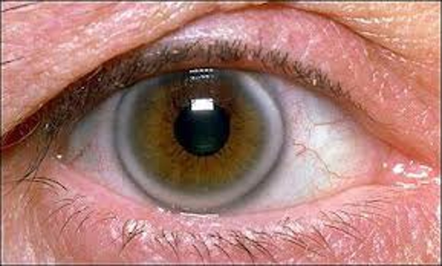
What condition may arcus senilis indicate?
may indicate systemic hyperlipidemia
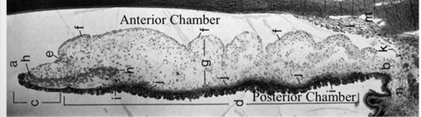
What are the five layers of the iris?
1) anterior border layer
2) stroma
3) muscular layer: sphincter, dilator
4) anterior iris epithelium
5) posterior iris epithelium
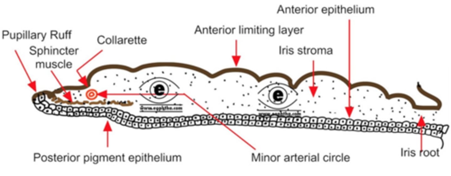
What is the collarette of the iris?
- anatomical border between pupillary and ciliary region of the iris, thickest region
- site of the minor arterial circle

What does the pupillary region of the iris contain?
thicker central region contains sphincter muscle

What does the ciliary region of the iris contain?
contains dilator muscle
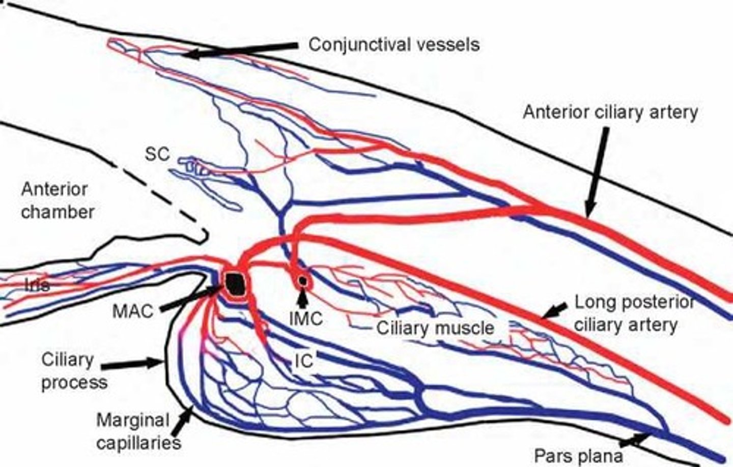
What is the minor arterial circle derived from?
major arterial circle (MAC)
What is MAC formed by?
by the anastomoses of the anterior ciliary arteries and long posterior ciliary arteries (LPCAs)
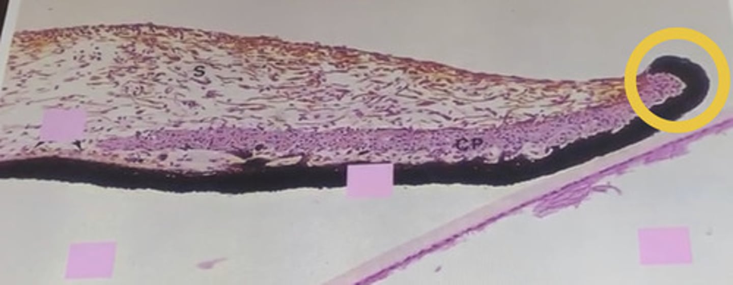
What types of cells does the anterior border layer of the iris contain? How are they organized?
melanocytes and fibroblasts; fibroblasts concentrate on the surface overlying melanocytes beneath
What is the function of the anterior border layer?
first pigmented cells to block light, helping the iris to serve as effective aperture stop, and to determine eye color
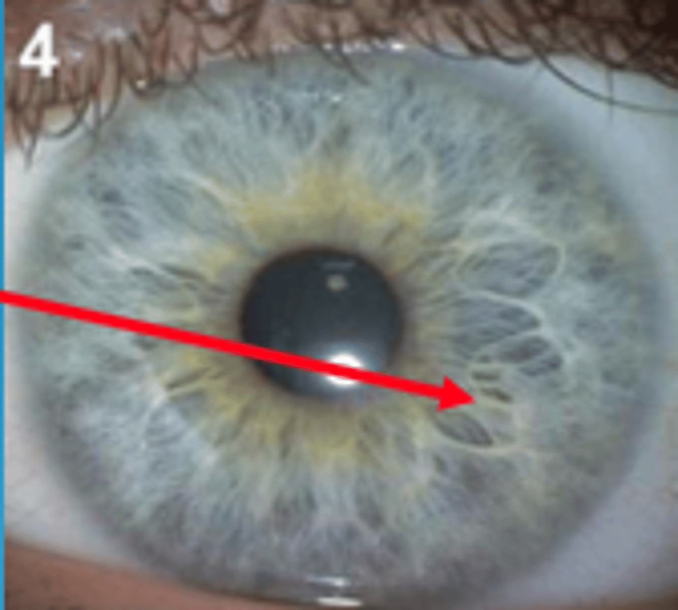
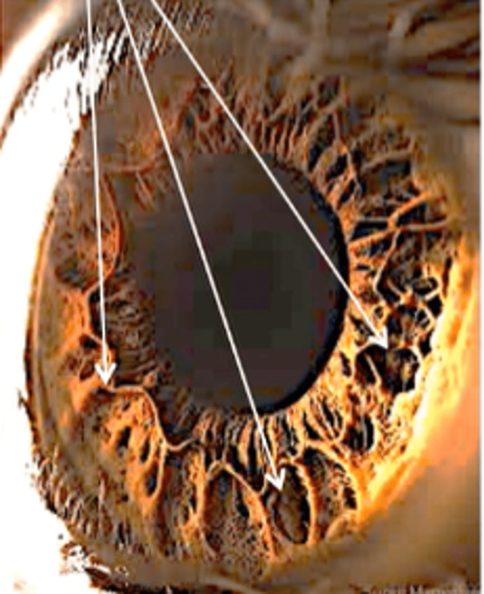
What are the holes in the anterior border layer of the iris?
Crypts of Fuchs
What do contraction folds of the iris block during dilation? How do they affect IOP?
contraction folds during dilation can block anterior chamber and increase IOP (beware of angle closure)
What is the purpose of the Crypts of Fuchs?
allow aqueous to enter stroma, serve as "iris fingerprint"
What is the iris stroma composed of?
spongy mixture of melanocytes, fibroblasts, collagen, and blood vessels
What are clump cells? Where do they appear and what do they do?
- clump cells appear in the vicinity of the sphincter muscle
- they clean up "spilled" pigment from melanocytes or pigmented epithelium
What does the blood supply to the iris (major/minor arterial circles) contain to make sure the vessel is tight and has no fluid (aqueous) leaks?
- tight junctions (lots of zonule occludens)
- overlapping endothelium
- secondary covering of pericytes and collagen
What does the blood supply to the RETINA contain to make sure the vessel is tight and has no fluid leaks?
- tight junctions (lots of zonule occludens)
- overlapping endothelium
- secondary covering of pericytes and collagen
Where is the minor arterial circle located on the iris?
about 2/3 of the way from the iris root to the pupil (also at the collarette)
What is the source of the intramuscular circle?
anterior ciliary artery
What is the source of the major arterial circle?
long posterior ciliary artery
What is the thickest portion of the iris?
sphincter muscle
Which region of the iris is the minor arterial circle located in?
pupillary region (with sphincter)
Is the ciliary region of the iris thick or thin?
very thin
Which muscle defines the ciliary region?
dilator
Are the nerves that control function of the sphincter and dilator muscles autonomic or somatic?
autonomic (non-voluntary)
What is the pupillary ruff?
inner edge of iris along pupil
In which ways/shapes do the muscle fibers of the sphincter and dilator run?
- sphincter: circular (around pupil)
- dilator: radial (perpendicular to sphincter fibers)
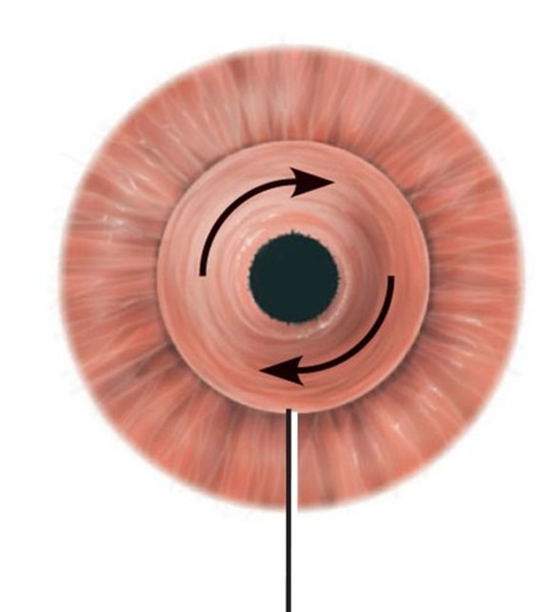
Does sympathetic or parasympathetic innervation stimulate the sphincter muscle (constrict pupil)? Which neurotransmitter is involved?
parasympathetic; acetylcholine
Does sympathetic or parasympathetic innervation stimulate the dilator muscle (dilate pupil)? Which neurotransmitter is involved?
sympathetic; norepinephrine
What is miosis?
constriction of the pupil
What is myadriasis?
dilation of the pupil
Which receptors does acetylcholine bind to stimulate the sphincter muscle?
muscarinic receptors (NOT beta-adrenergic)
Which receptors does norepinephrine bind to stimulate the dilator muscle?
alpha-adrenergic receptors
Which epithelial layer has heavy pigmentation?
posterior epithelial layer
Which epithelial layer forms the dilator muscle?
anterior epithelial layer -> it is the myoepithelium from which the dilator muscle arises
What does the most central tissue of the posterior epithelium layer form?
wraps around the pupil margin to form the pupillary ruff
What is a synerchiae?
condition where there is adherence of the iris to the cornea or lens (can be treated)