BLG 111 full midterm 1
 Call Kai
 Learn
Learn Practice Test
 Spaced Repetition
 Match
Match Flashcards
 Knowt Play
1/270

There's no tags or description
Looks like no tags are added yet.
Name | Mastery | Learn | Test | Matching | Spaced | Call with Kai | Chat |
|---|
No analytics yet
Send a link to your students to track their progress
271 Terms
cardiovascular system
consists of the heart, blood vessels, and blood
right side of heart
pumps deoxygenated blood to lungs
left side of heart
pumps oxygenated blood to body
pulmonary circuit
carries blood to lungs for gas exchange and returns it to heart
short, low-pressure circulation
systemic circuit
carries blood between heart and body tissues
fast, high-pressure circulation
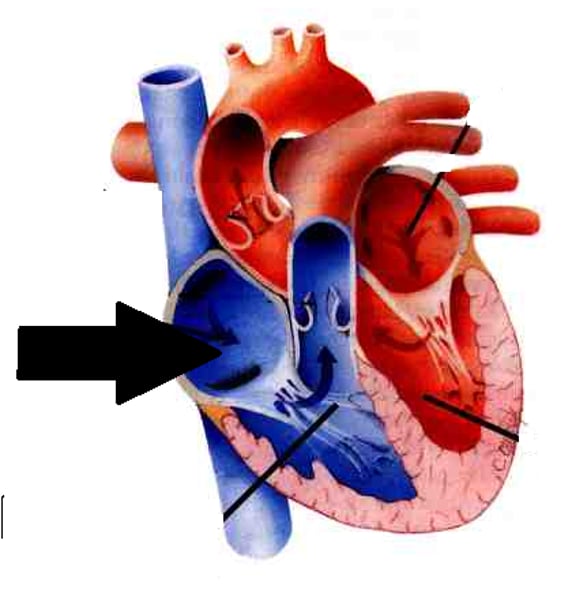
right atrium
Receives deoxygenated blood from body
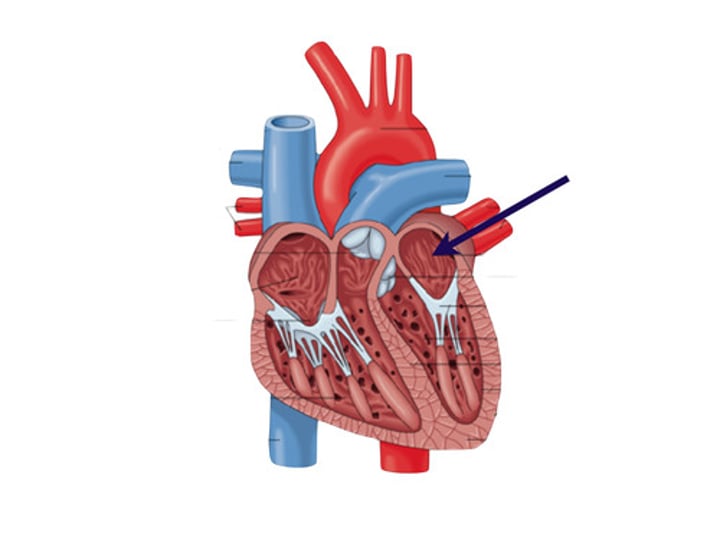
left atrium
Receives oxygenated blood from lungs
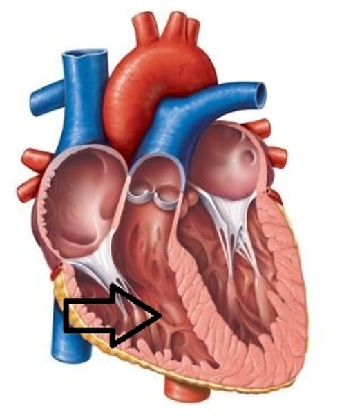
right ventricle
pumps deoxygenated blood to lungs
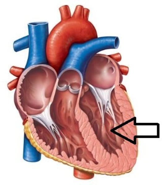
left ventricle
pumps oxygenated blood to body
Where is the heart located?
mediastinum, between lungs
Pericardium
Double-walled membrane surrounding heart
fibrous pericardium
fibrous CT
protects heart, anchors heart to surroundings, and prevents overfilling
parietal layer of serous pericardium
lines internal surface of fibrous pericardium
pericardial cavity
fluid-filled cavity that decreases friction between pericardial layers
percarditis
inflammation of pericardium causing pericardial friction rub
Cardiac tamponade
Excess fluid that leaks into pericardial space
can compress heart's pumping abilty
epicardium
visceral layer of serous pericardium
Myocardium
heart muscle
endocardium
Inner layer of heart
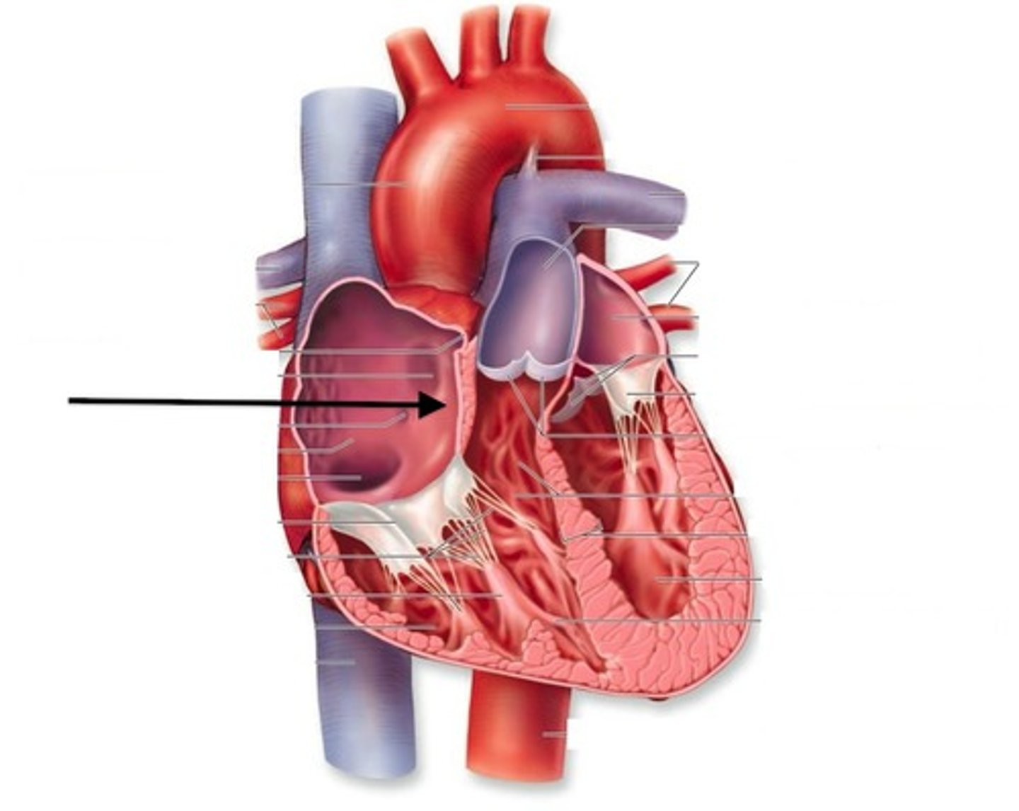
What separates the atria?
interatrial septum
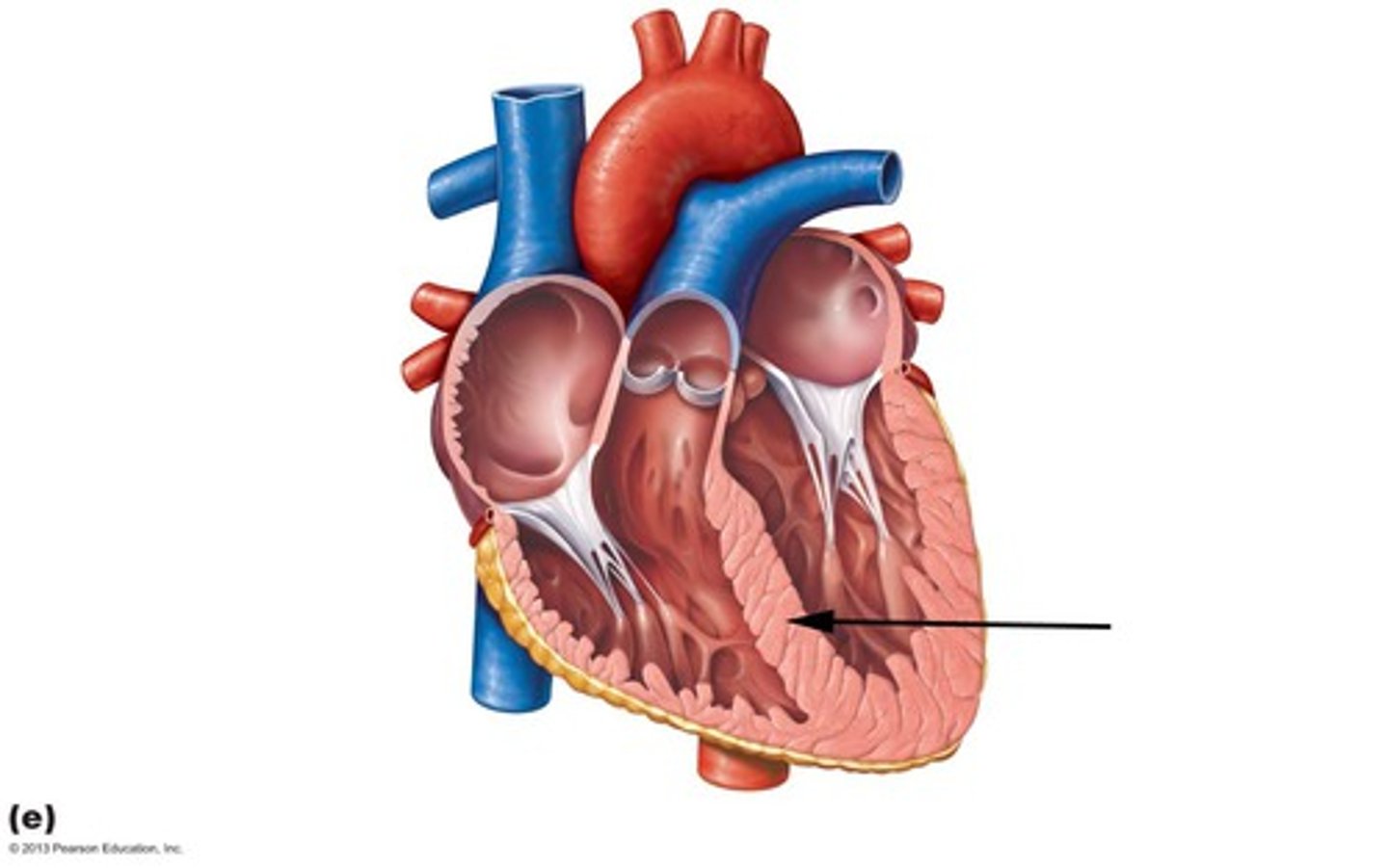
What separates the ventricles?
interventricular septum
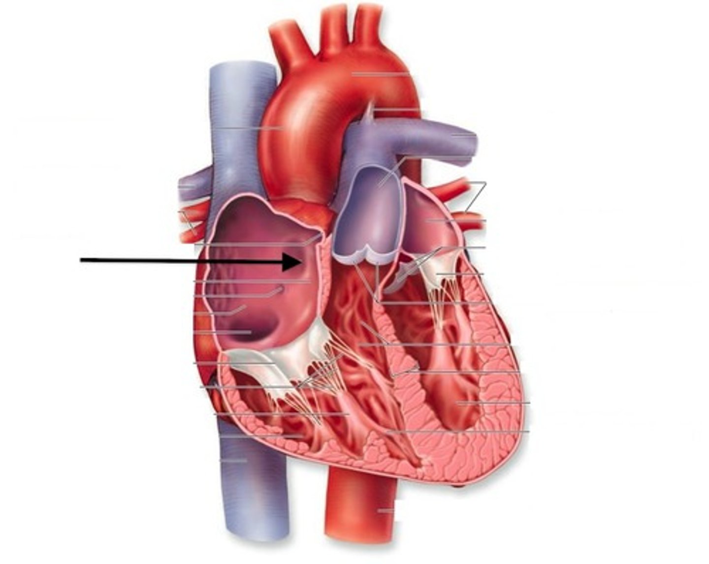
Fossa ovalis
remnant of foramen ovale of fetal heart
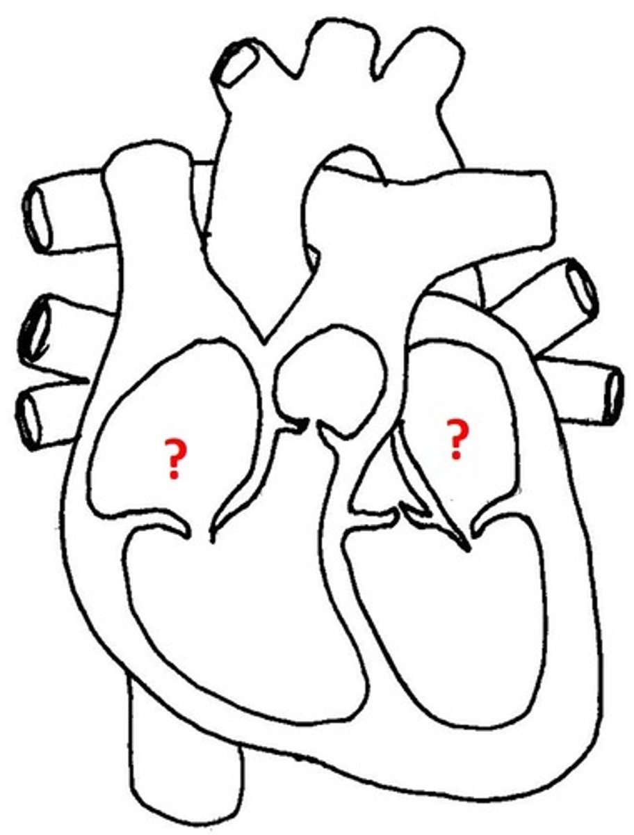
Atria
upper / receiving chambers
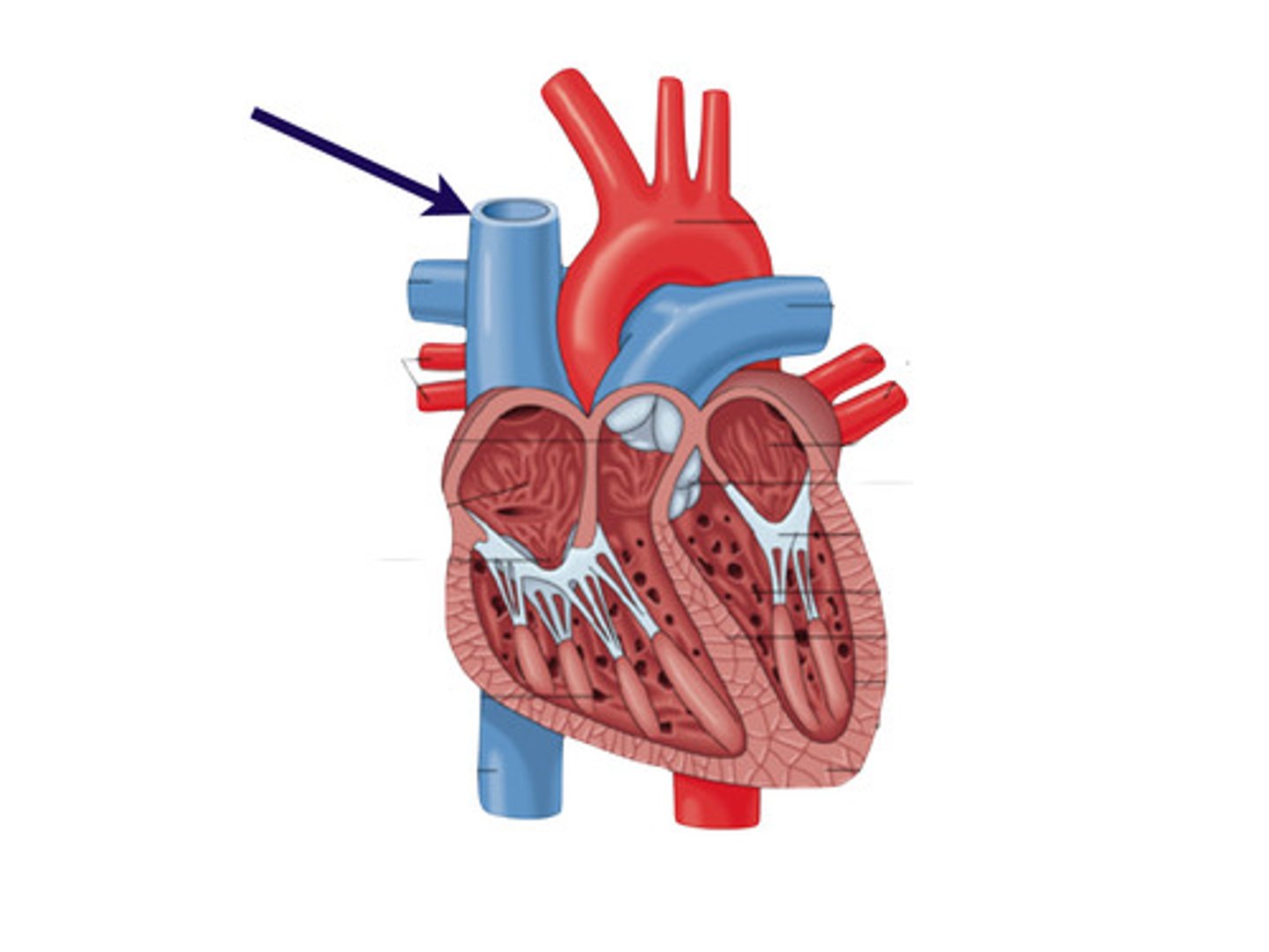
superior vena cava
returns blood from body regions above diaphragm
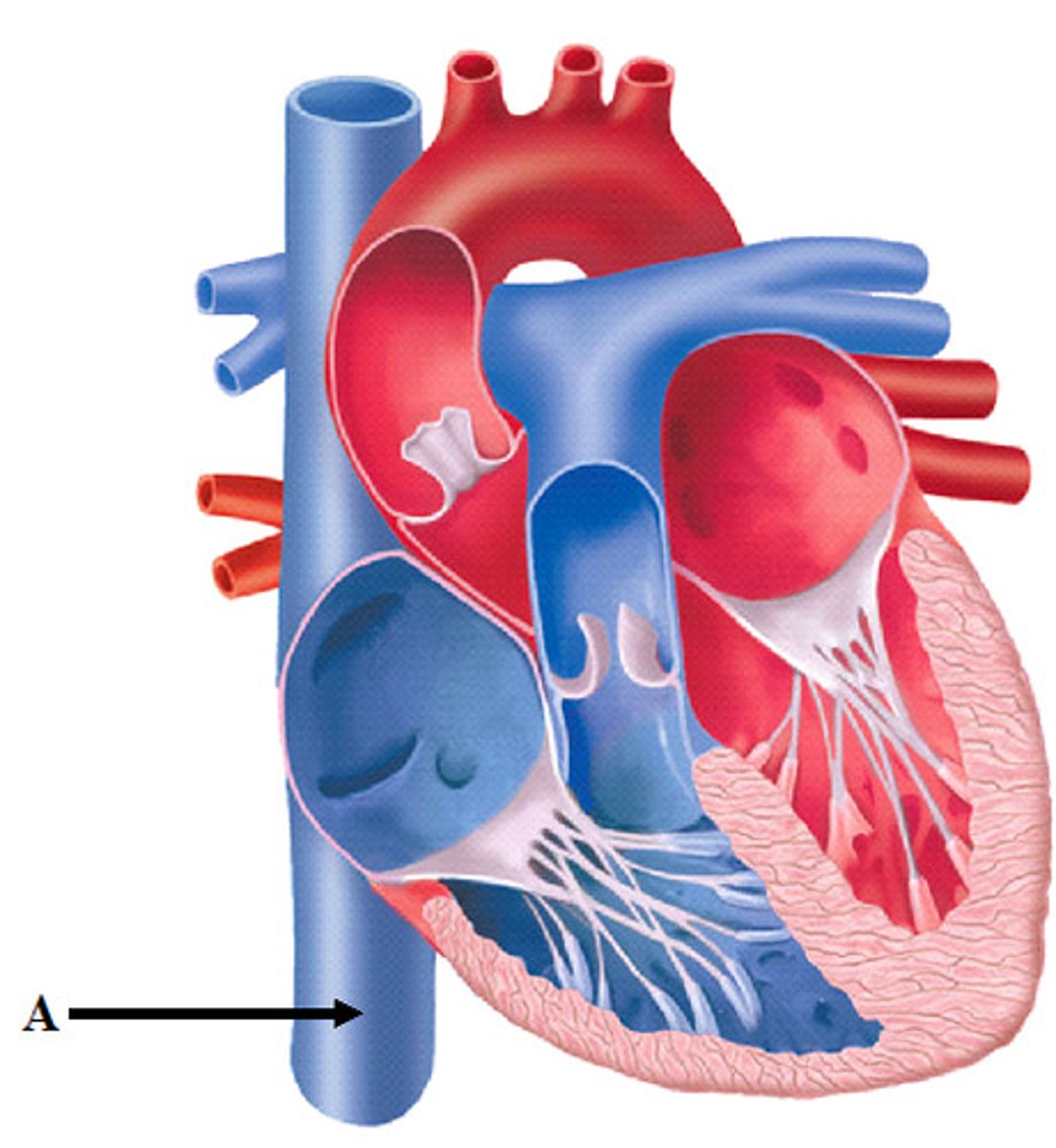
inferior vena cava
returns blood from body regions below diaphragm
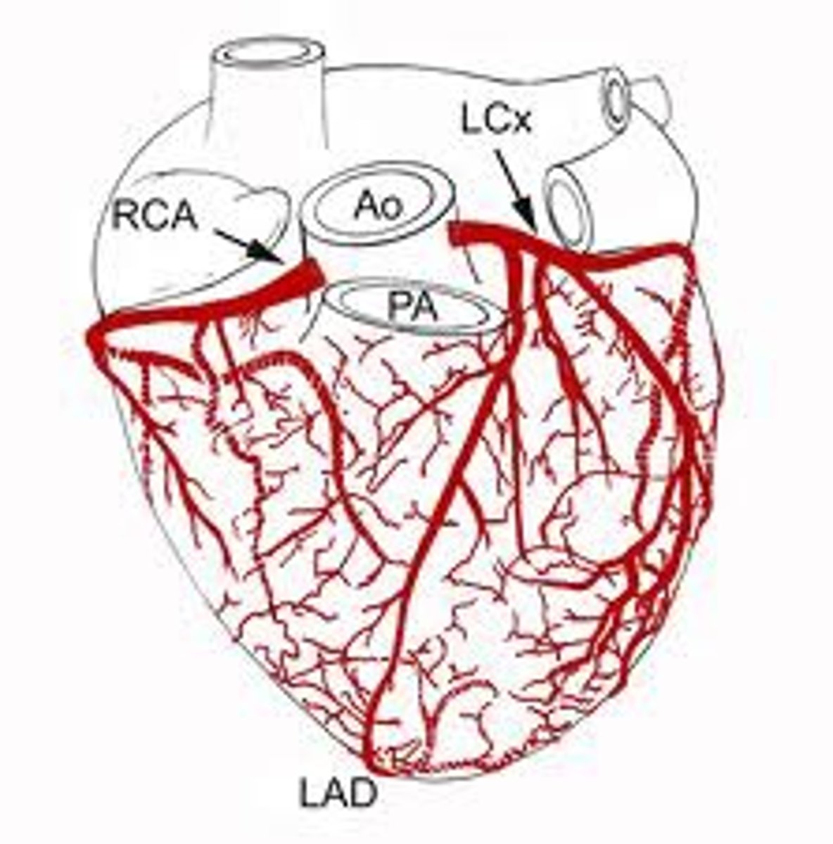
coronary arteries
two arteries that supply blood to heart muscle
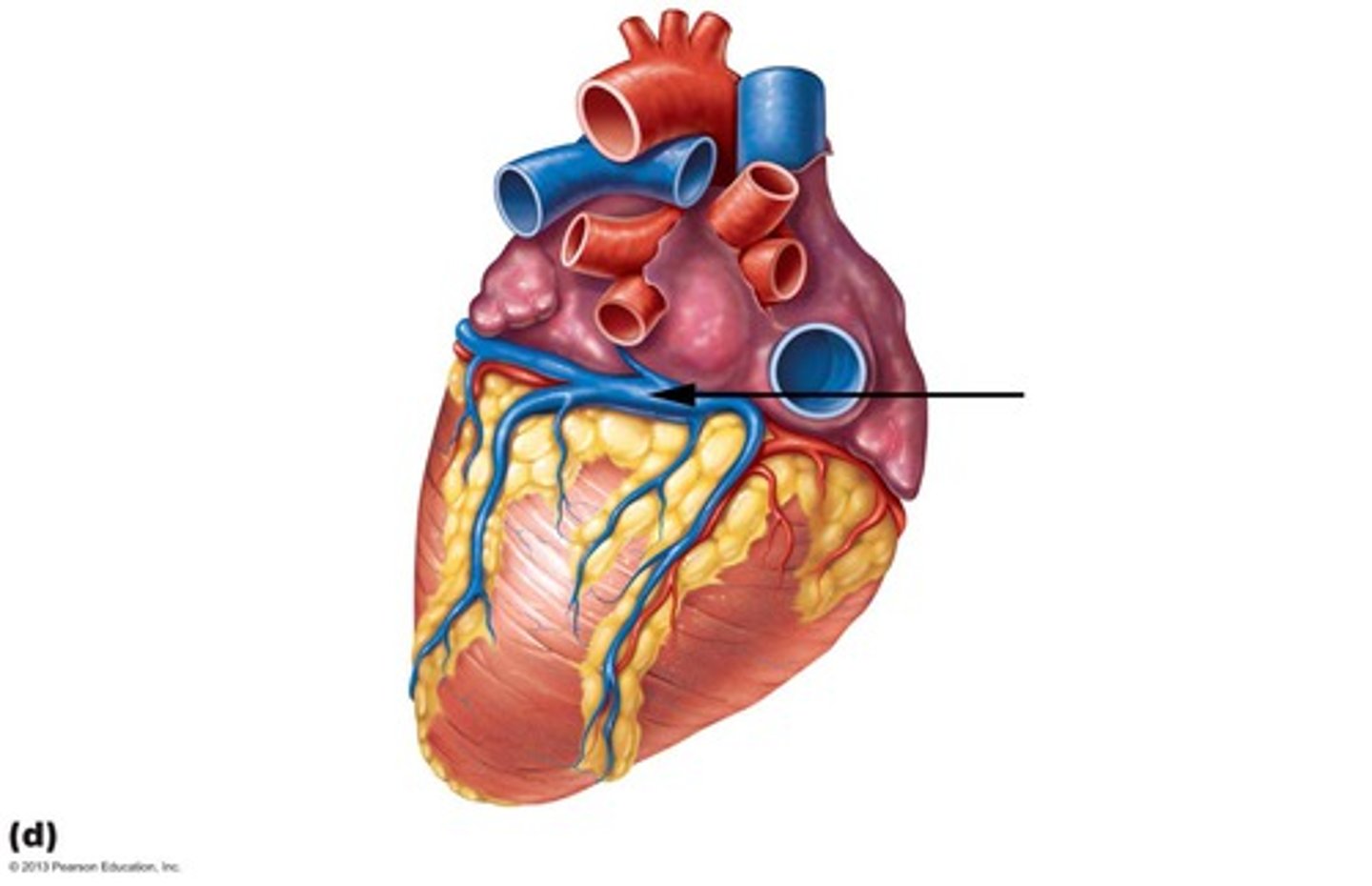
coronary sinus
returns blood from coronary veins
Ventricles
lower / discharging chambers
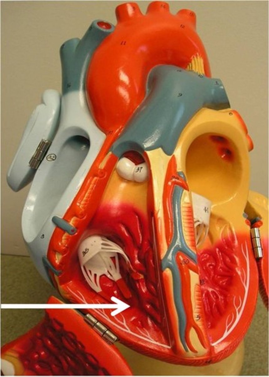
trabeculae carneae
muscular ridges on ventricular walls
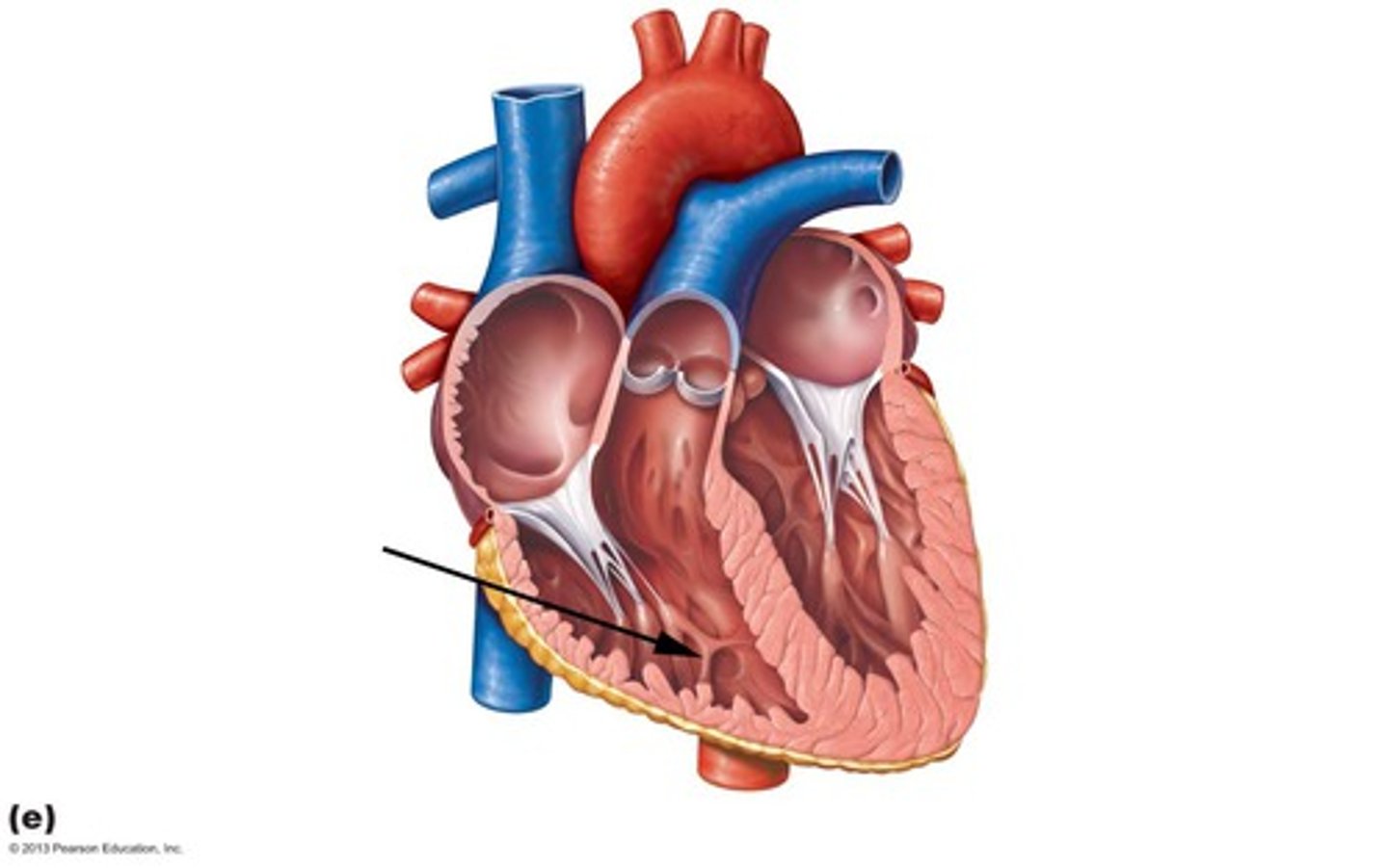
papillary muscles
anchor chordae tendineae
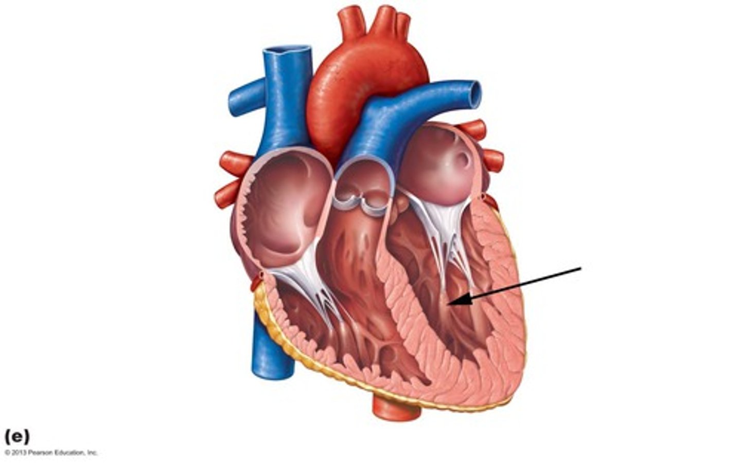
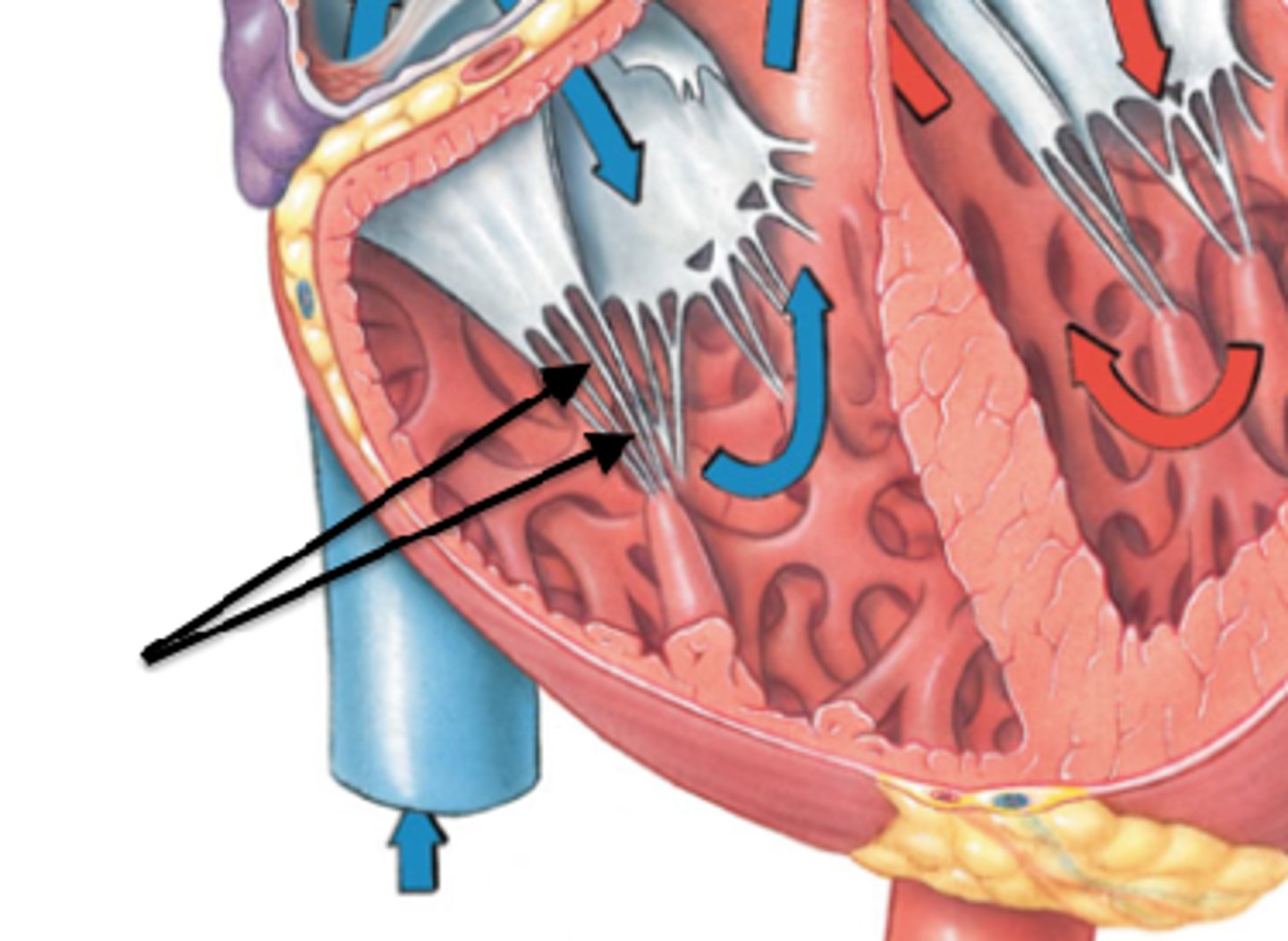
chordae tendineae
heart strings
prevent cusps everting back into atria
heart valves
ensure unidirectional blood flow
dependent on pressure changes
atrioventricular valves (AV)
prevent back flow into atria when ventricles contract
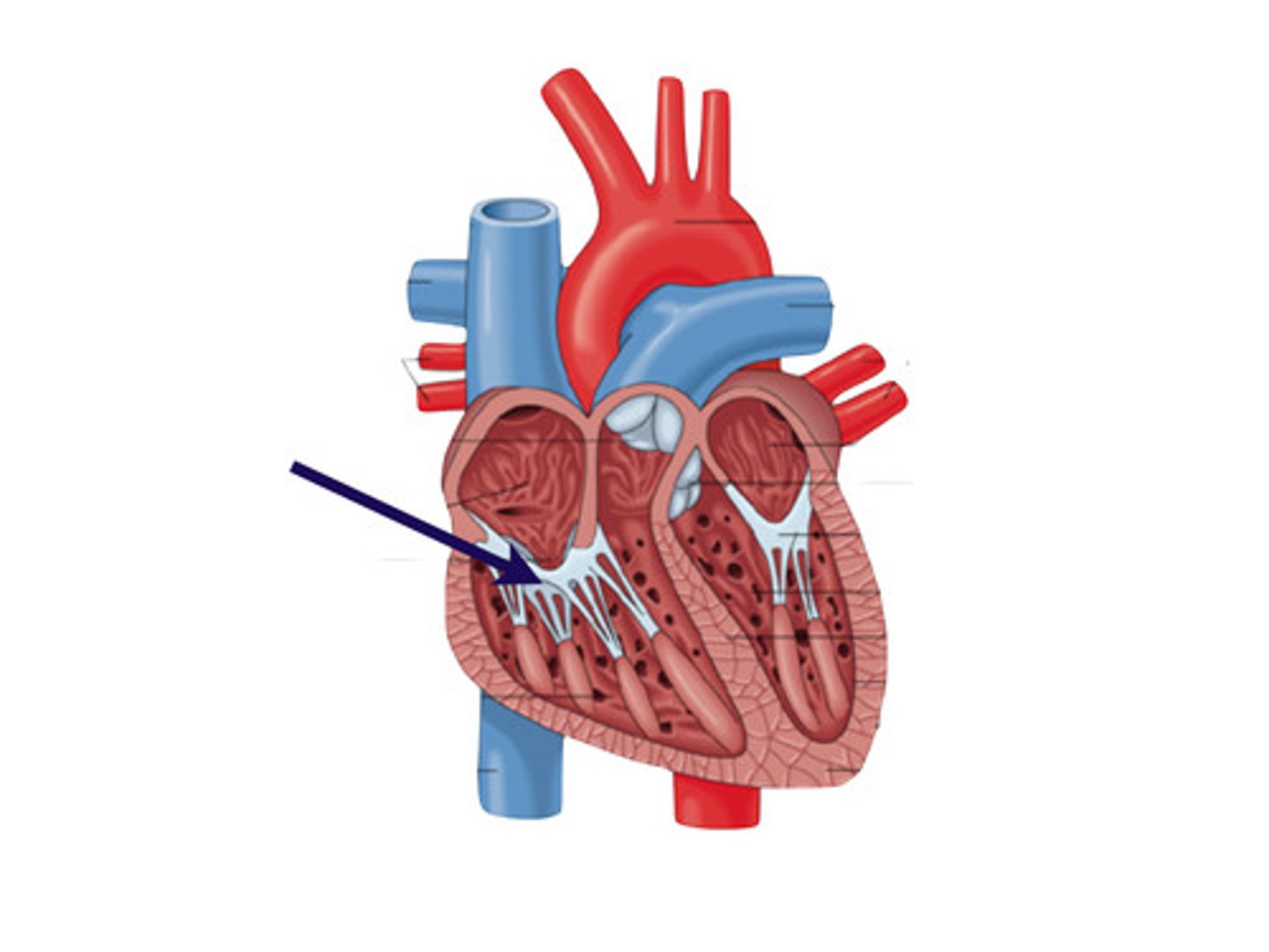
What is the valve with 3 cusps between the right atria and ventricle?
Tricupsid Valve
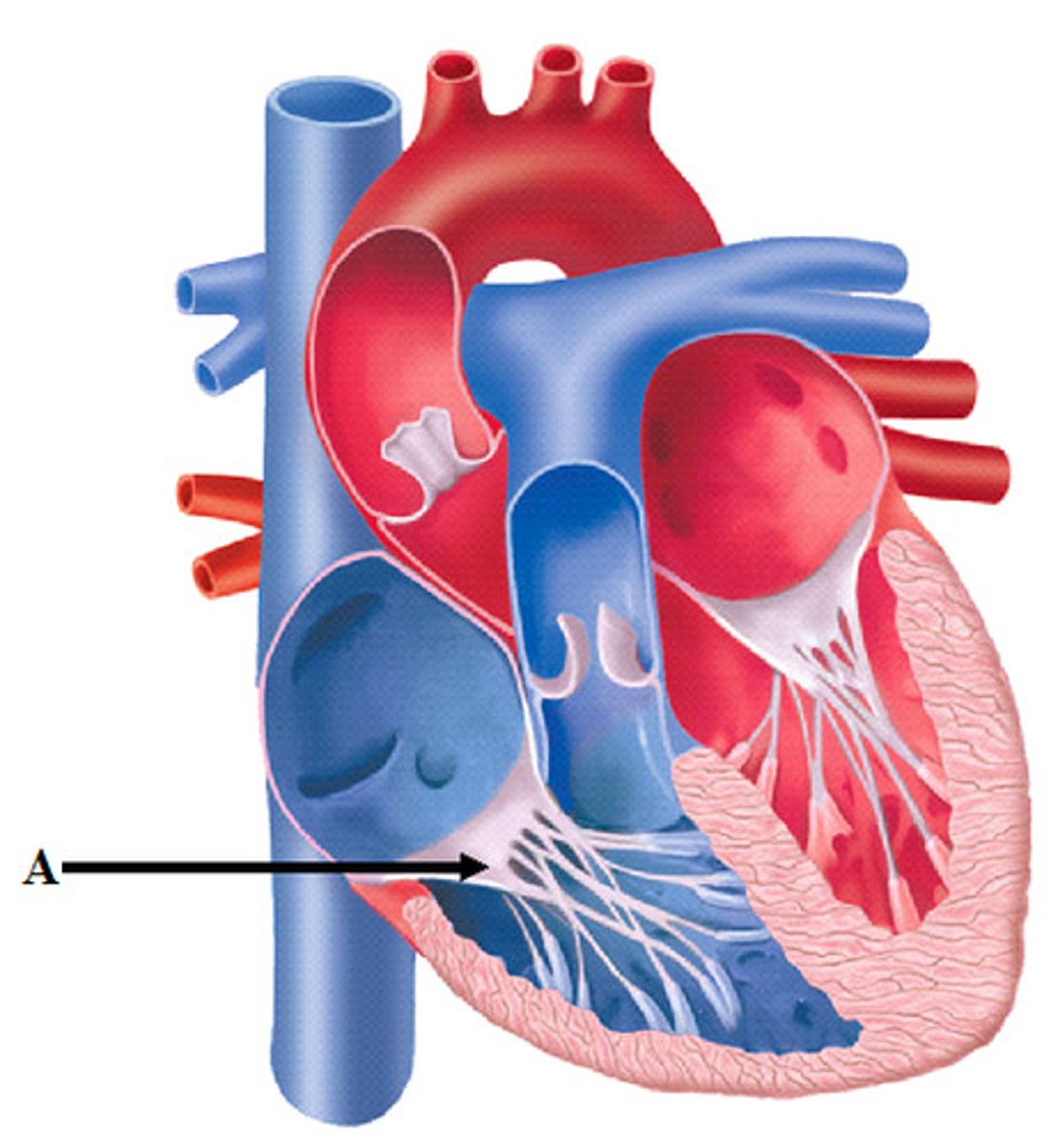
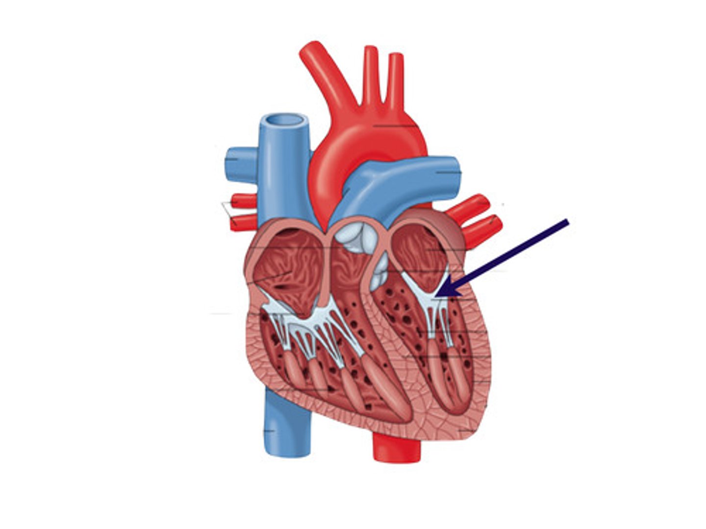
What is the valve with 2 cusps between left atria and ventricle?
Mitral valve
What happens when atrial pressure is greater than ventricular pressure?
1. increased pressure opens AV valves
2. ventricles fill
3. atria contracts, forcing more blood into ventricles
What happens when atrial pressure is less than ventricular pressure?
1. ventricles contract, forcing blood against cusps
2. AV valves close
3. papillary muscles and chordae tendineae tighten
semilunar valves (SL)
prevent back flow into ventricles when ventricles relax
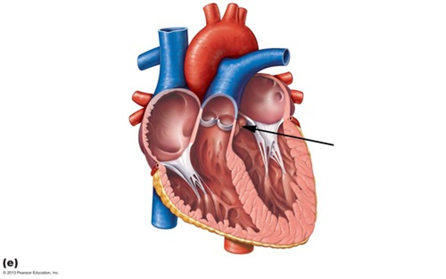
What is the SL valve on the right side of the heart?
pulmonary SL valve
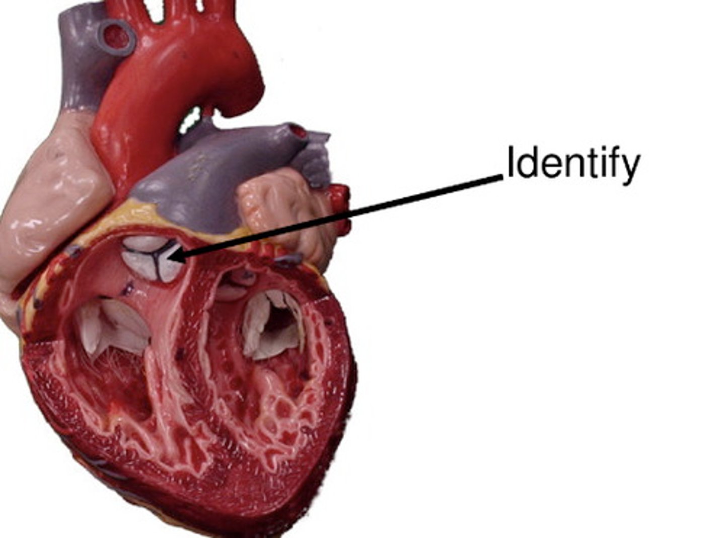
What is the SL valve on the left side of the heart?
aortic SL valve
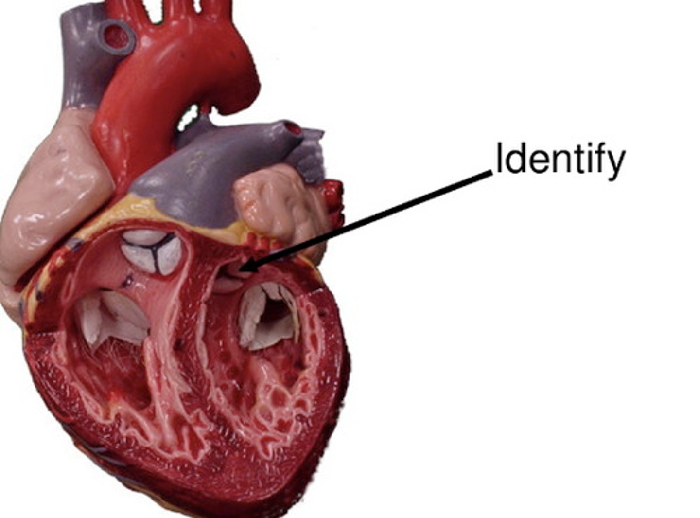
What happens when SL valves open?
1. ventricles contract, increasing pressure
2. pressure forces blood against SL valves, opening them
What happens when SL valves close?
1. ventricles relax, decreasing pressure
2. blood flows back, filling and closing cusps
incompetent valve
Blood backflows so heart repumps same blood over and over
- cause swishing sounds
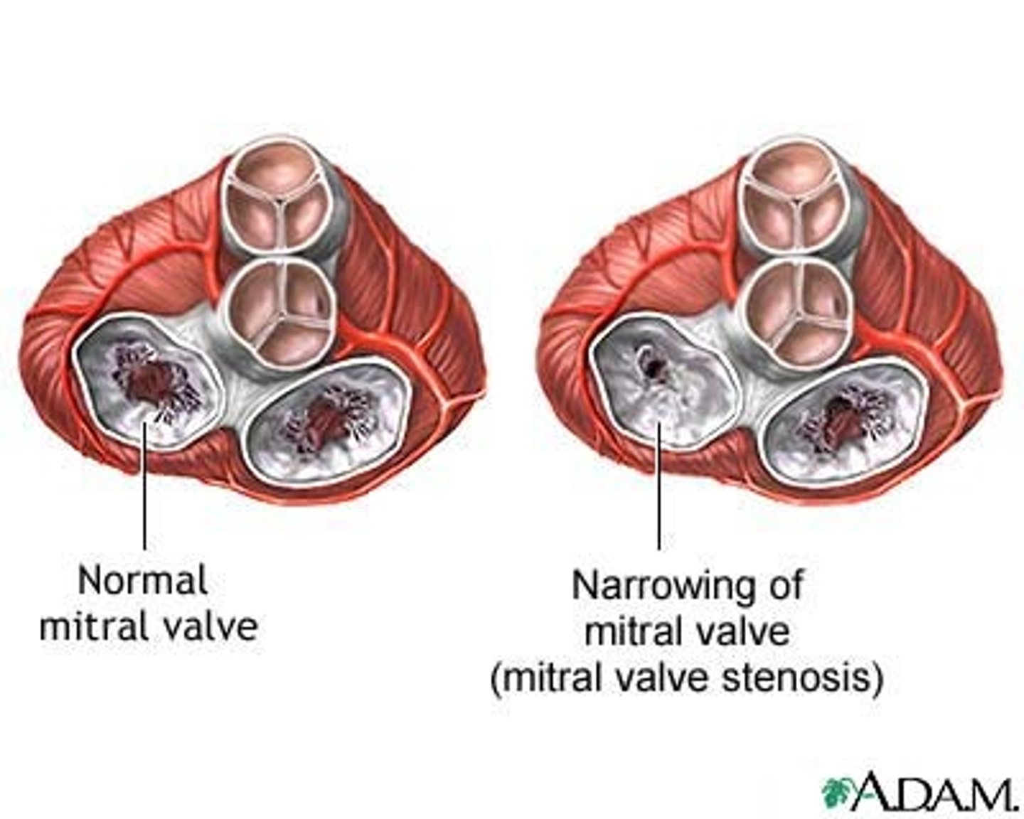
Valvular stenosis (narrowing)
stiff flaps constrict opening
heart exerts more force to pump blood
What is the shortest circulation in the body?
coronary circulation
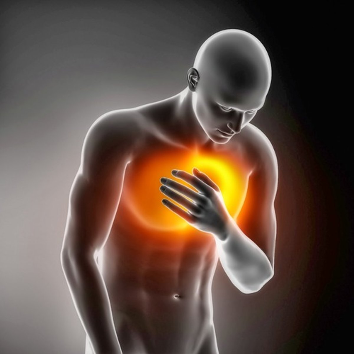
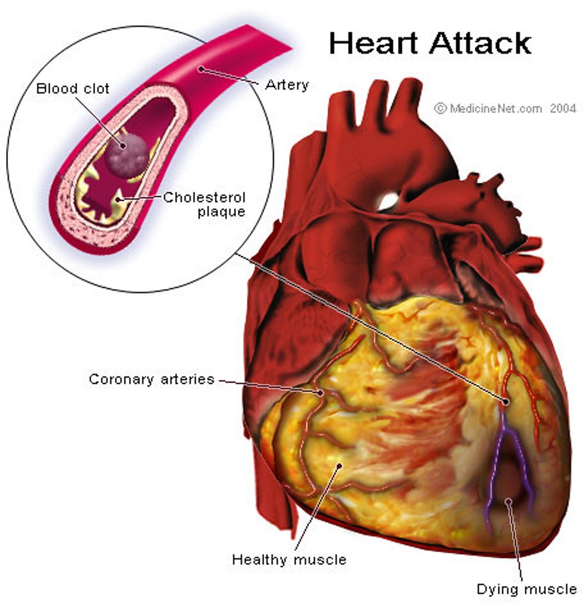
angina pectoris
chest pain that results when the heart does not get enough oxygen
myocardial infarction (MI)
heart attack caused by coronary blockage
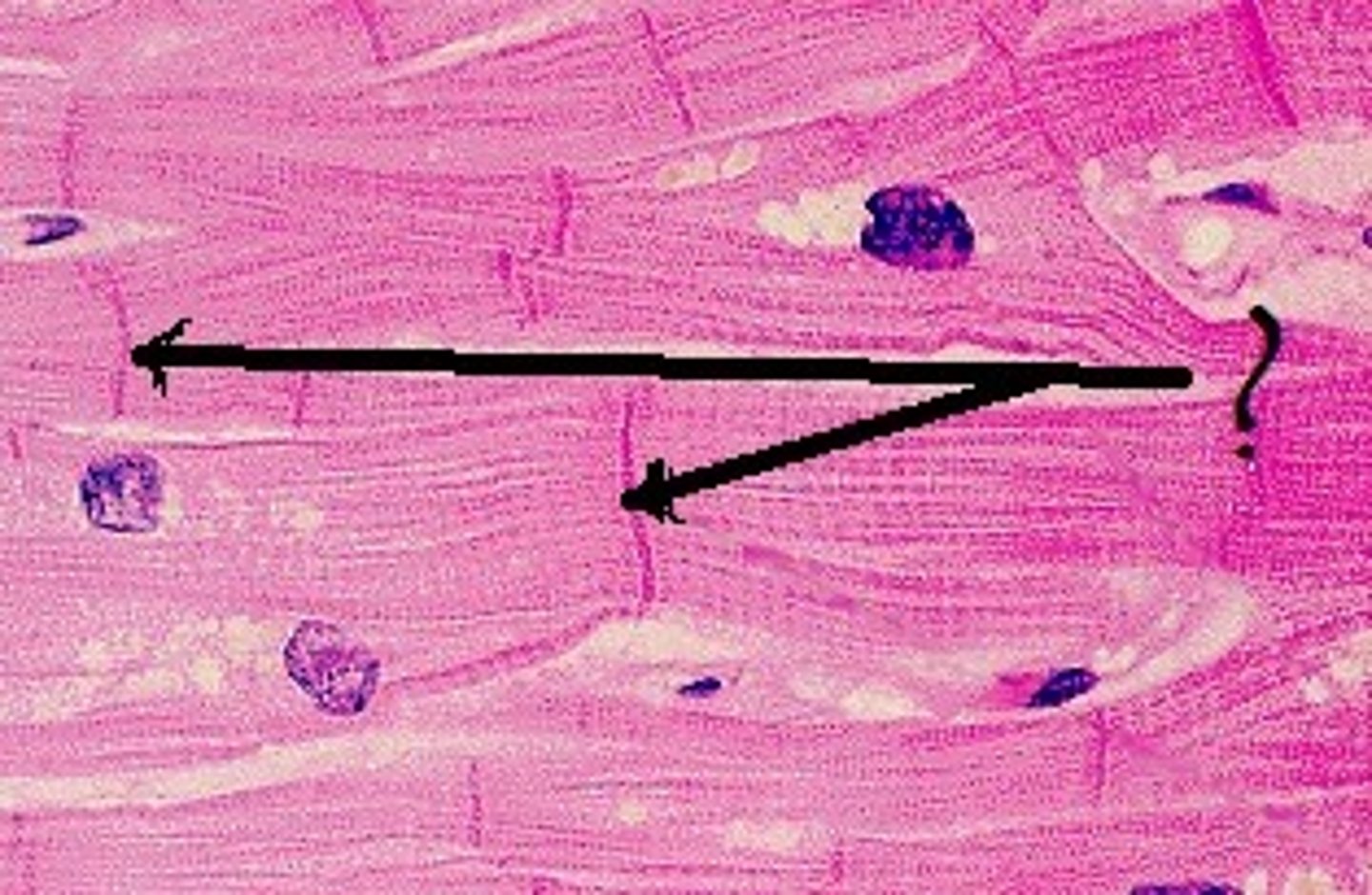
intercalated discs of cardiac cells
connecting junctions consisting of desmosomes and gap junctions
allows heart to be functional syncytium
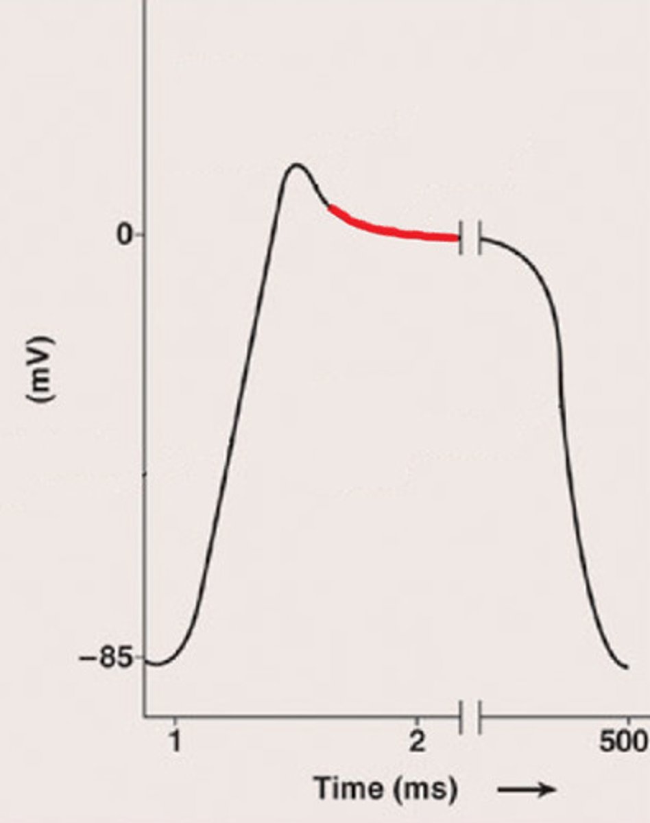
differences between cardiac and skeletal muscle
Some cardiac muscle cells are self-excitable
Heart contracts as a unit
Influx of Ca2+ from ECF triggers Ca2+ release from SR
Tetanic contractions cannot occur in cardiac muscles due to longer refractory period
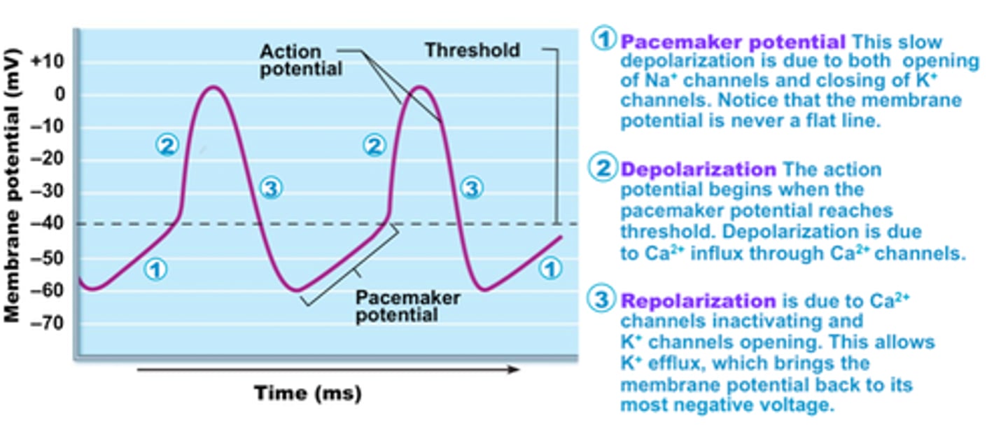
pacemaker cells
heart cells that spontaneously depolarize
intrinsic cardiac conduction system
network of noncontractile cells that initiate and distribute impulses to coordinate depolarization and contraction of heart
what is the sequence of excitation of the heart?
1. SA node
2. AV node
3. AV bundle
4. R and L bundles
5. subendocardial conducting network
1. Pacemaker Potential
slow depop due to opening of Na channels and closing of K channels (mp is never a flat line)
2. Depolarization
AP begins when pacemaker potential reaches threshold
depop due to ca2+ influx via ca2+ channels
3. Repolarization
Ca2+ channes inactivate, K channels open
ectopic focus
abnormal pacemaker that takes over pacing
Extrasystole (premature contraction)
ectopic focus in small region of heart generates impulse before SA node causing delay in next impulse
- heart has longer time to fill, thus next contraction is felt as a thud due to larger volume of blood being pumped out
- due to excessive caffeine or nicotine use
heart block
few or no impulses reach ventricles causing them to beat at their own rate
cardioacceleratory center
sends sympathetic signals to increase both rate and force
cardioinhibitory center
sends parasympethic signals to decrease rate
Why do cardiac muscle APs have a plateau?
due to Ca2+ influx through slow Ca2+ channels, keeping cell depolarized
--> prevents tetanus; gives muscle time to contract and relax
Electrocardiogram (ECG)
graphic recording of electrical activity
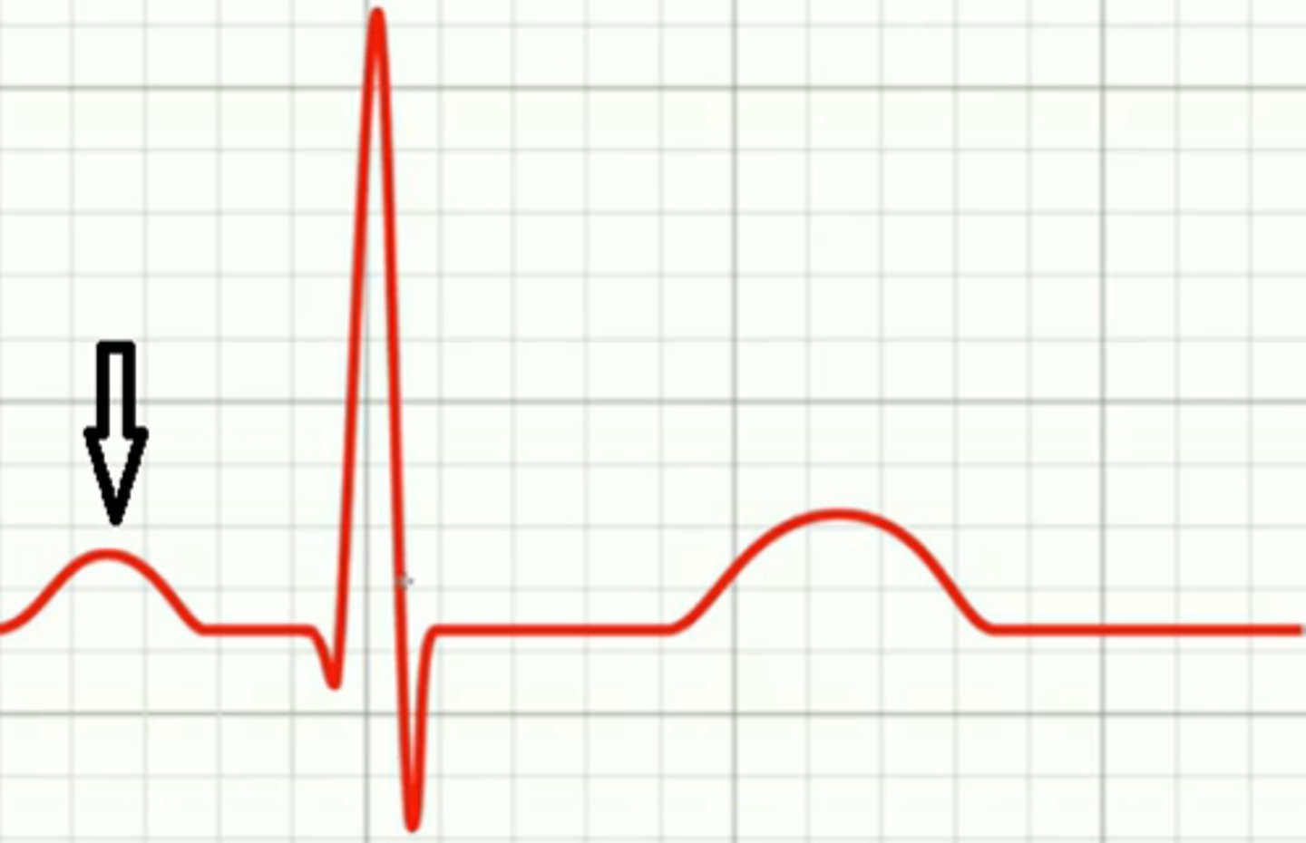
P wave
atrial depolarization caused by SA node
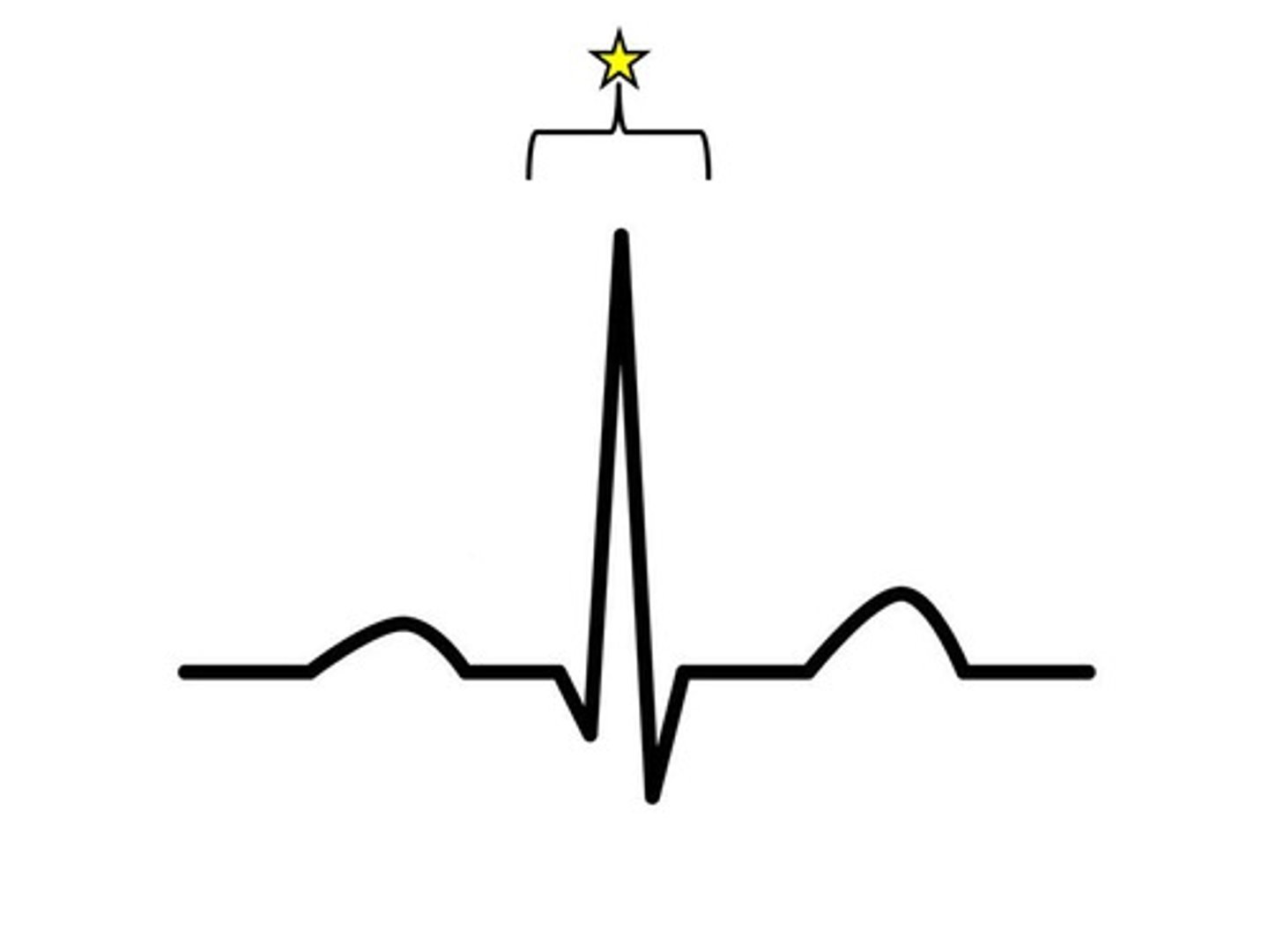
QRS complex
ventricular depolarization begins at apex, atrial repolarization occurs
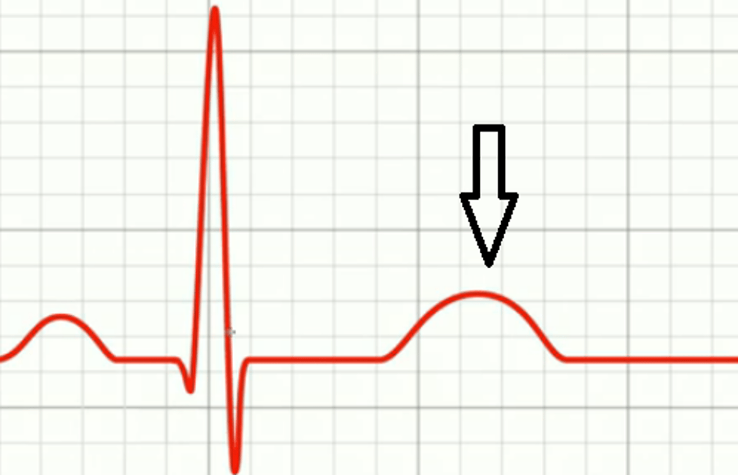
T wave
ventricular repolarization begins at apex
P-R interval
beginning of atrial excitation to beginning of ventricular excitation
S-T segment
entire ventricular myocardium depolarized
Q-T interval
from ventricular depolarization to ventricular repolarization
Ventricular fibrillation (V-fib)
abnormal heart rhythm which results in quivering of ventricles
Systole
Contraction of the heart
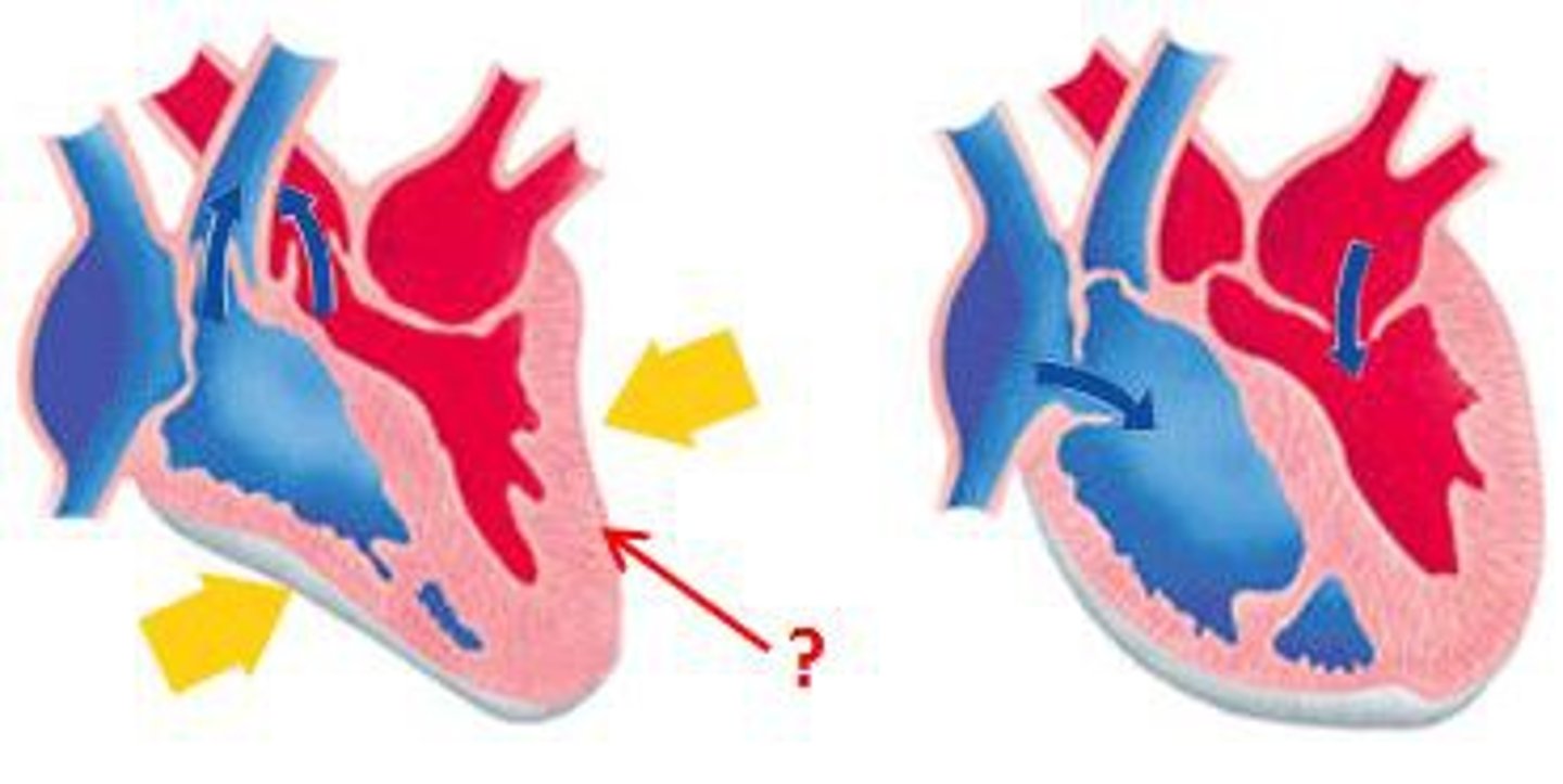
diastole
Relaxation of the heart
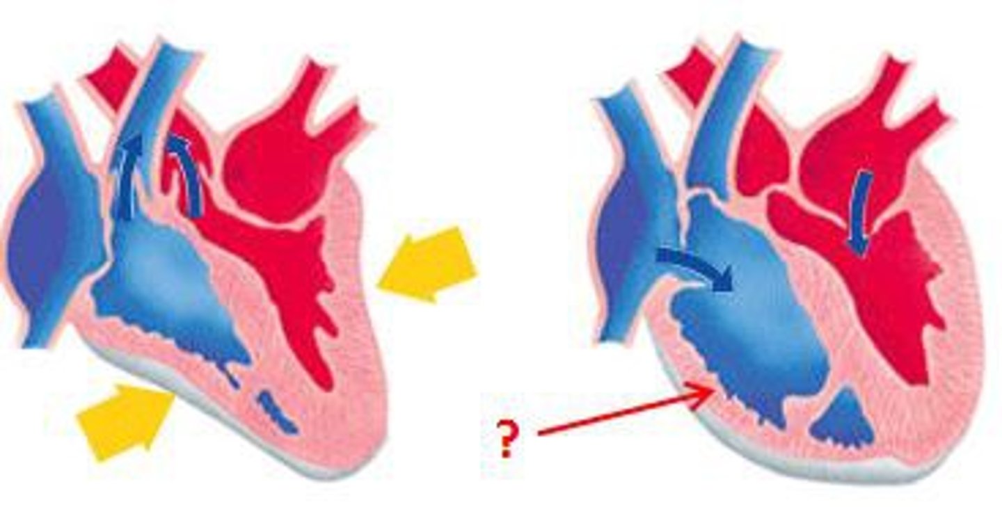
cardiac cycle
blood flow through heart during one complete heartbeat
Ventricular filling: mid-to-late diastole
- Pressure is low; 80% of blood flows from AV valves into ventricles
- atrial depop triggers atrial systole
- atria contracts, pushing remaining 20% of blood into ventricle
- depop spreads to ventricles (QRS wave)
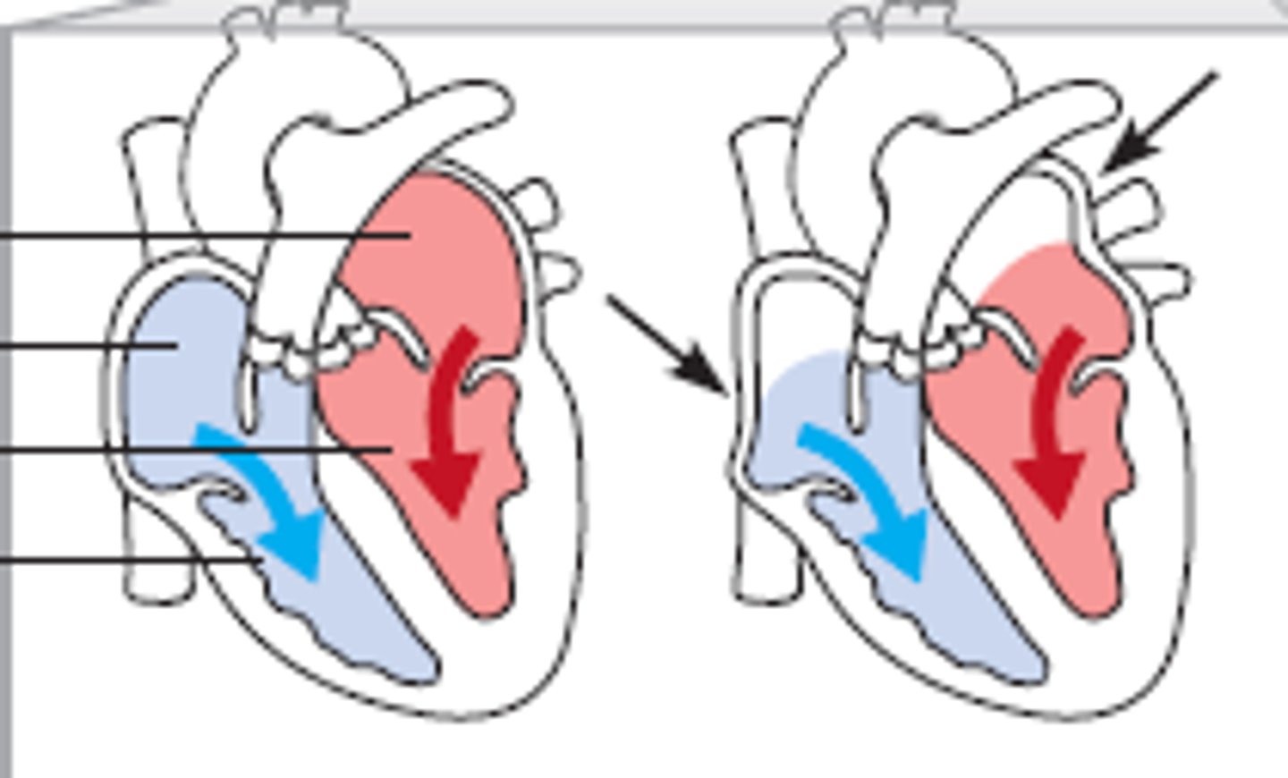
End Diastolic Volume (EDV)
volume of blood in each ventricle at end of ventricular diastole
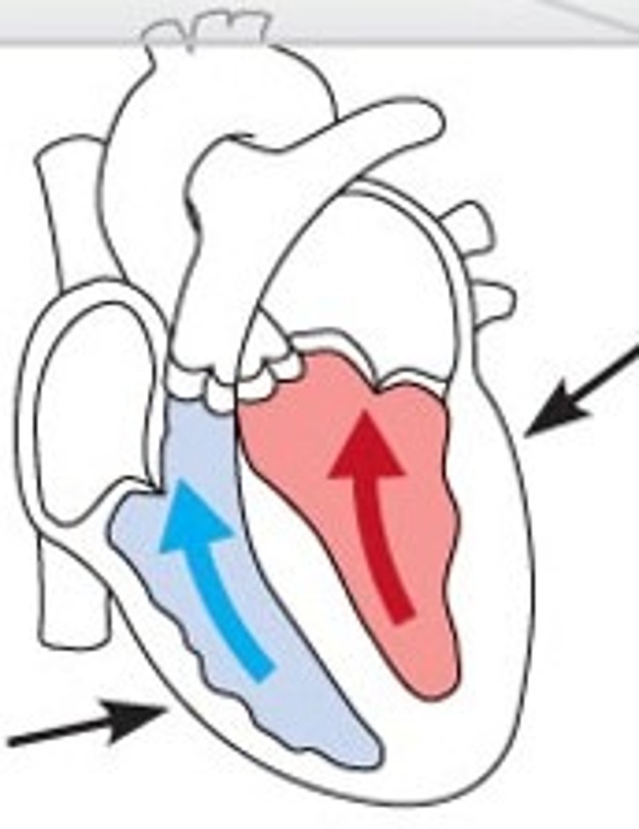
isovolumetric contraction
- atria relax; ventricles contract
- high pressure closes AV valves AND opens SL valves
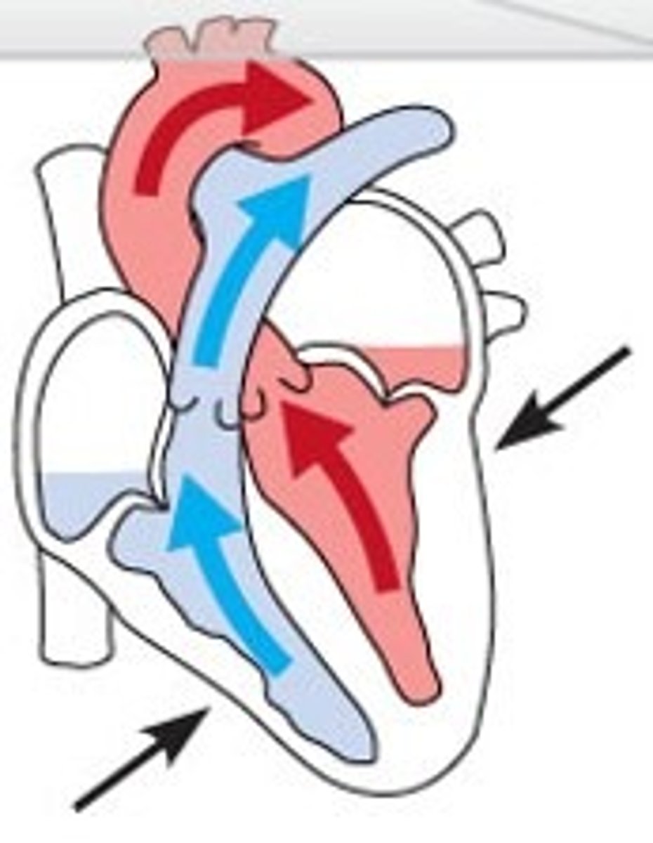
Ventricular ejection
- blood rushes into aorta and pulmonary trunk
- pressure of aorta: 120 mmHg
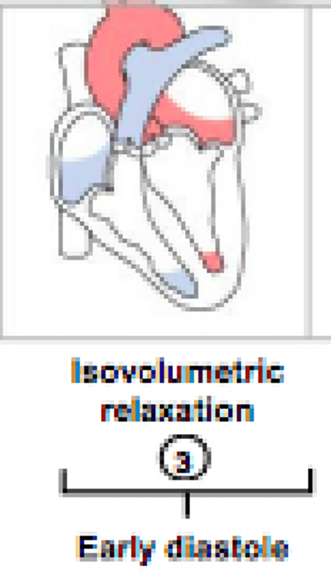
isovolumetric relaxation: early diastole
- ventricles repop (t wave); ventricles relax
- ventricular pressure decreases causing back flow of blood that closes SL valves
- atria fills; when atrial pressure > ventricular pressure, AV valves open = cycle begins again
end systolic volume (ESV)
volume of blood remaining in each ventricle after systole
What is the first heart sound?
closing of AV valves (lub) @ beginning of ventricular systole
What is the second heart sound?
closing of SL valves (dub) @ beginning of ventricular diastole
heart murmurs
abnormal heart sounds
stenotic valve
fails to open completely, restricting blood flow
- high-pitched sound
Cardiac Output (CO)
Amount of blood pumped in 1 minute (~5 L)
stroke volume
the volume of blood pumped out by a ventricle with each heartbeat
cardiac reserve
difference between resting and maximal CO
Preload
degree of stretch of heart muscle before contraction
(affects EDV = more blood is able to fill in ventricle @ beginning)
Frank-Starling Law of the Heart
greater the stretch, the greater the force of contraction
Venous Return (VR)
amount of blood returning to the heart
the higher the venous return =
higher EDV, SV, CO
Contractility of the heart
contractile strength at muscle length
increase contractility/force = increase amount of blood ejected (decreases ESV)
afterload
pressure ventricles must overcome to eject blood
hypertension
pressure in LV << systemic pressure = valves don't open properly = reduced amt of blood ejected
sympathetic nervous system regulation of heart rate
Ne causes:
- pacemaker to fire faster = increase HR
- increases Ca2+ movement = increase contractility
parasympathetic nervous system regulation of heart rate
Ach hyperpolarizes pacemakers by opening K+ channels = decreases HR
vagal tone
slowing of heart rate
What does epinephrine and thyroxine do to HR?
Epinephrine = increases HR and contractility
Thyroxine = increases HR; enhance Ne and E
What specific ions are needed to maintain normal heart function?
Ca and K
Hypocalcemia
depresses heart
Hypercalcemia
increases HR and contractility (can disrupt HR and cause arrythmias)
Hyperkalemia
alters electrical activity = can lead to heart block and cardiac arrest