Modification of Neural Circuits
1/24
There's no tags or description
Looks like no tags are added yet.
Name | Mastery | Learn | Test | Matching | Spaced |
|---|
No study sessions yet.
25 Terms
Hebb’s Postulate
Hypothesized that the coordinated activity of a presynaptic terminal and a postsynaptic neuron strengthens the synaptic connection between them
Components of Hebb’s Postulate
Originally formulated to explain the cellular process of learning and memory
Widely applied to situations involving long-term modifications of in synaptic strength (development)
Synaptic terminals strengthened by correlated activity will be retained or sprout new branches
Terminals weakened by uncorrelated activity will lose their connection with the post synaptic cell
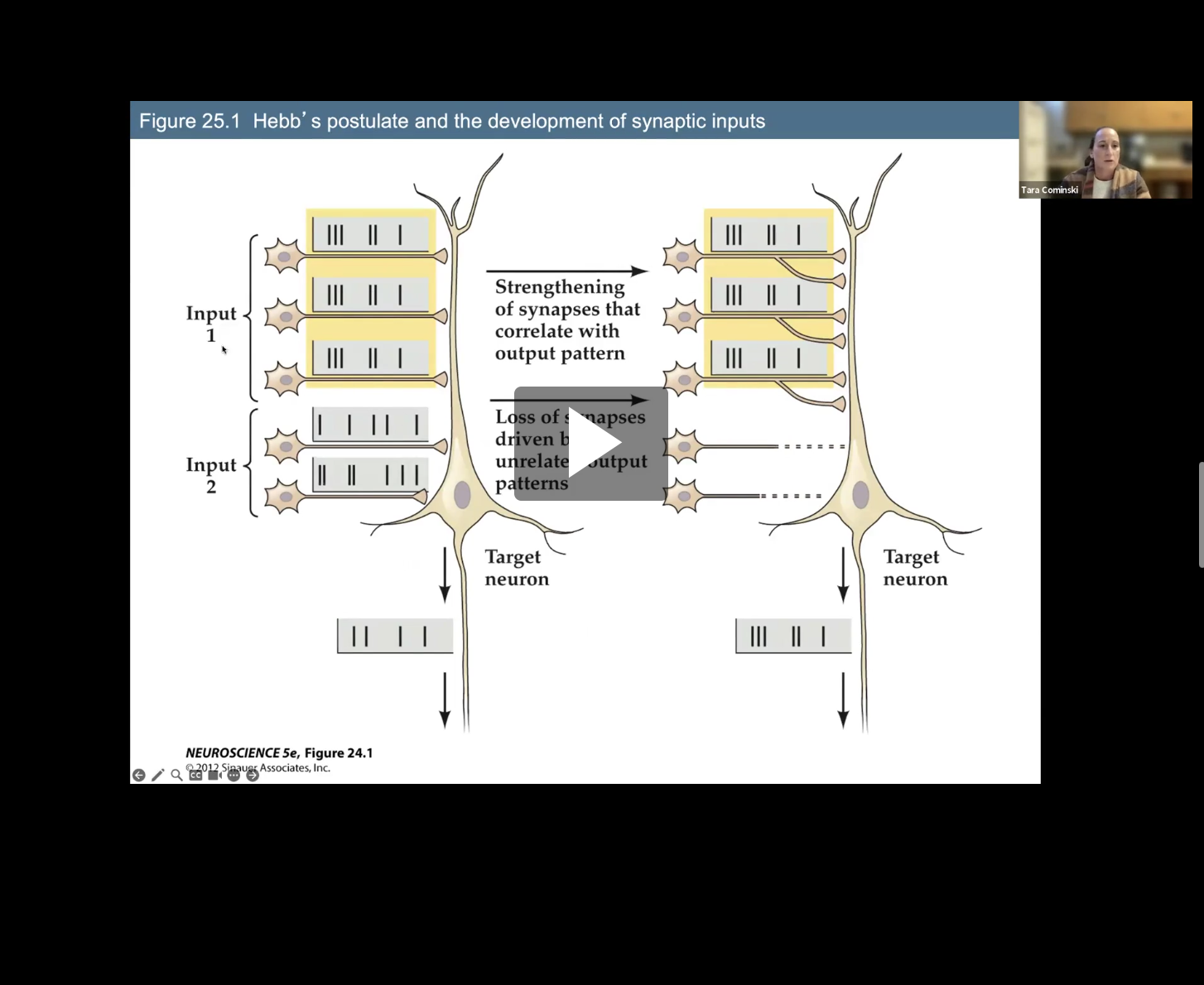
Figure 25.1- Hebb’s Postulate and the Development of synaptic inputs
Post synaptic neuron gets input, with pattern of action potentials, patterns that don’t correlate with the output pattern of the postsynaptic cell/target neuron get lost.
Those patterns that are correlated get strengthen and grow new terminals
Role of activity on neural circuits provide an initial understanding for which 2 components (Postnatal brain growth and development)
How new behaviors in newborns emerge and are shaped by experience
Why the brain continues to grow after birth
Parallel growth of the dendritic and axonal branches and the additional of synaptic connections accounts for most of postnatal brain growth.
Built in Behaviors- Imprinting- Box 25A
Innate abilities such as imprinting must be conducted during a critical period window ( 1 day for a baby chick), behavior will not develop properly and will be unable to correctly bond with the mother, affecting its ability to survive.
2 environmental factors that especially influence early life are
Critical periods
Parental Imprinting in hatchling birds
Critical Periods
time when experience and neural activity that reflects that experience have maximal effect on the acquisition or skilled execution of a particular behavior
Parental Imprinting in hatchling birds
hatchling recognizes its parent
expressed only if animals have specific experiences during a particular time (hours or days) in early postnatal development
Critical periods for sensorimotor skills and complex behaviors…
last much longer
Critical periods for sensorimotor skills and complex behaviors include examples:
Language aquisition in humans
Song birds: Song birds: male birds acquire the capacity to produce species-specific song by mimicking tutor bird
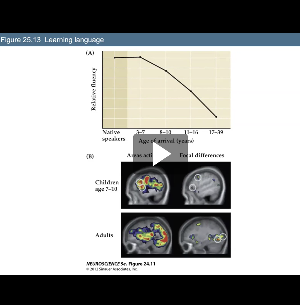
Explain Figure 23.13- Learning Language
Shows native speakers with high fluency as time proceeds with later ages, relative fluency decreases drastically
Changes in brain can be seen in Figure B (Different plasticity)
4 Basic Properties of Critical Period
Time during which a given behavior requires specific environmental influences in order to develop normally
Once a critical period ends, the core features of a behavior are largely unaffected by subsequent experience
If not exposed to appropriate stimuli during the critical period it is difficult or impossible to remedy
In most mammals, including humans, critical periods rely particularly on changes in organization and function of circuits in the cerebral cortex
Critical Periods in Visual System Development
• Experiments in animals with highly developed visual
abilities, cats or monkeys, provided key information that
defined how activity and experience shape connections
in the primary visual cortex
Effects of Visual Deprivation on Ocular Dominance
Illuminate one eye and record activity of individual neurons in the cortex
Visual cortical neurons divided into seven “ocular dominance” groups based on their degree of response to either the contralateral or ipsilateral eye.
Group 1 cells driven by stimulation of the contralateral eye (opposite side)
Group 7 cells driven by stimulation of the ipsilateral eye (same side)
Group 4 cells driven equally well by either eye
This normal distribution of ocular dominance can be altered by visual experience
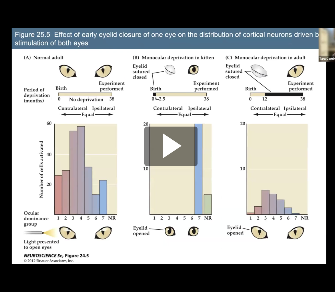
Fig 25.5- Effect of early eyelid closure of one eye on the distribution of cortical neurons driven by simulation of both eyes
Study in cats that show what the effects of depriving one eye during of visual simulation during development can do
(A) Ocular dominance distribution of single-unit recordings from a large number of neurons in the primary visual cortex of normal adult cats. Cells in group 1 were activated exclusively by the contralateral eye, cells in group 7 by the ipsilateral eye. There were no cells that were not responsive (NR) to light stimulation in the retina.
(B) One eye of a newborn kitten was closed from 1 week after birth until 2.5 months of age. After 2.5 months, the eye was opened and the kitten matured normally to 38 months. Note that the deprivation was relatively brief—the sutured eye had been open for 35.5 months of the cat’s life. Even so, light presented to the open but transiently deprived eye elicited no electrical responses in visual cortical neurons. The only visually responsive cells responded to the ipsilateral (non-deprived) eye.
(C) A much longer period of monocular deprivation in an adult cat showed little effect on ocular dominance, although overall cortical activity was diminished. Most of the responsive cells were driven by both eyes. In addition, some cells (groups 1 and 2) were uniquely or mostly responsive to the deprived eye
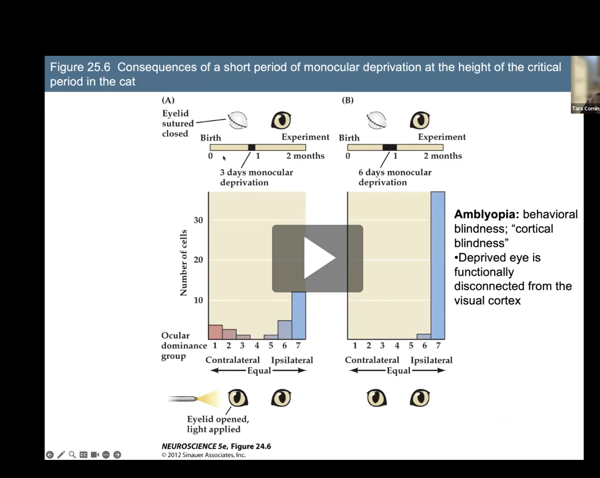
Figure 25.6- Consequences of a short period of monocular deprivation at the height of the critical period in the cat
Same experiment as before but deprivation period is shorter and done within same timeframe
1st image ( 3 day monocular deprivation in kitten)
3 days isn’t enough, still so activation
2nd image (6 days in kitten)
after 6 days, blindness in one eye (amblyopia), 6 days after critical period, so only one eye
Amblyopia
Behavioral blindness; “cortical blindness”
Deprived eye is functionally disconnected from the visual cortex
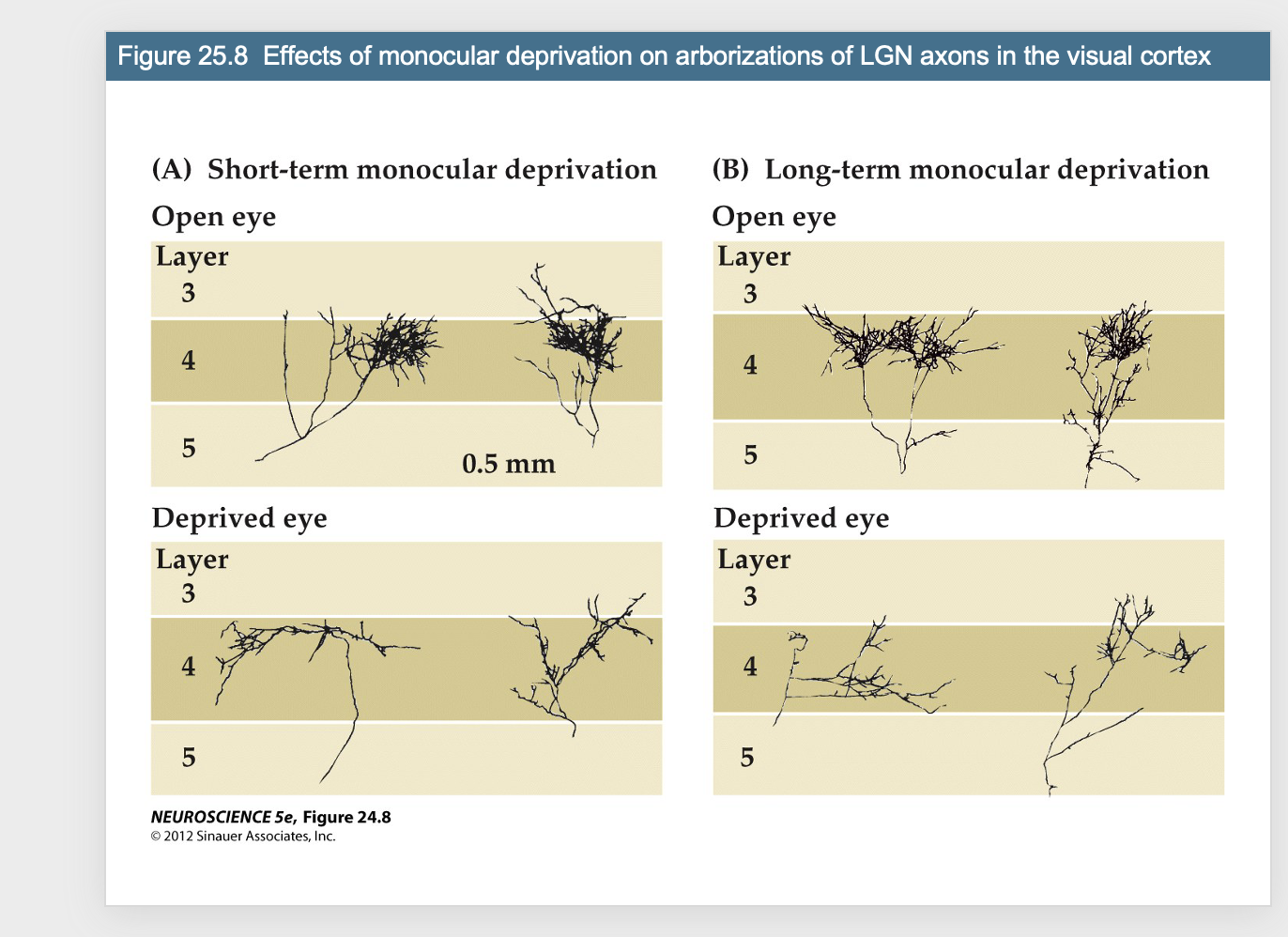
Figure 25.8- Effects of monocular deprivation on arborizations of LGN axons in the visual cortex
Shows neurons of open eye vs deprived eye, can see the dramatic decrease in neuronal processes in deprived eye.
Short term vs long term, you see a similar decrease in processes because of no receiving the simulation in inputs like they normally would in critical period
Thus processes retract back
How does experience change neural circuits during critical periods? How do patterns of activity modify connections?
Must rely on signals generated by synaptic activity associated with sensory experience, perceptual and cognitive processing, or motor performance
NMDA receptors – excitatory neurotransmission
Regulation of the number and placement of local inhibitory synapses and the expression of GABA receptors on post synaptic sites are sensitive to changes in levels of electrical activity during early postnatal life
Neurotransmitters and neurotrophins that influence critical periods are thought to modulate intracellular calcium levels
Studies in rhesus monkeys in the late 1980’s demonstrated that:
The number synapses throughout the cortex increased during prenatal and a limited period of postnatal life
Declined during adolescence
Reached a steady state in adulthood
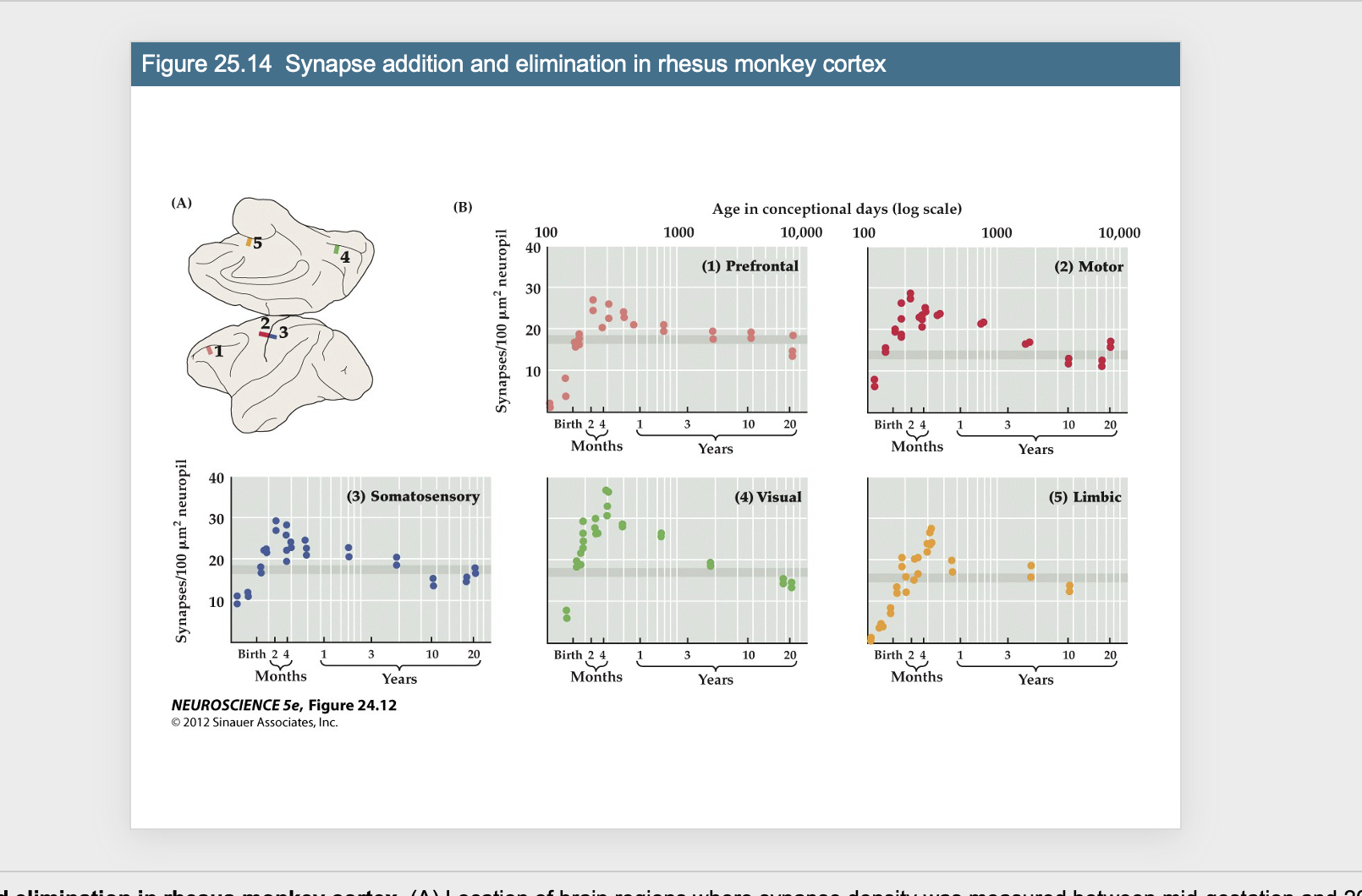
Synapses- Monkey Experiment: Figure 25.14- Synapse addittion and elimination in rhesus monkey cortex
Looking at different parts of the cortex
Can see the same pattern of: from birth, the number of synapses increases, then a little drop and leveling out when into adult hood,
(A) Location of brain regions where synapse density was measured between mid-gestation and 20 years of age.
(B) Rapid addition followed by gradual decline of synapse density in the cerebral cortex. Age has been converted to a logarithmic scale of “conceptional days” in order to fit the entire life span onto one graph. Synapse addition apparently continues through early life, gradually declines throughout most of adolescence, and reaches a steady state (shaded horizontal bar) after puberty (between 2 and 3 years of age in the rhesus monkey
Gray Matter volumes in humans:
Researchers at NIMH (1990 -2010) – performed a longitudinal MRI study in 13 children ranging from ages 4-20 to measure the growth of gray matter
Gray matter grows throughout the cortex during early life; declines in adolescence
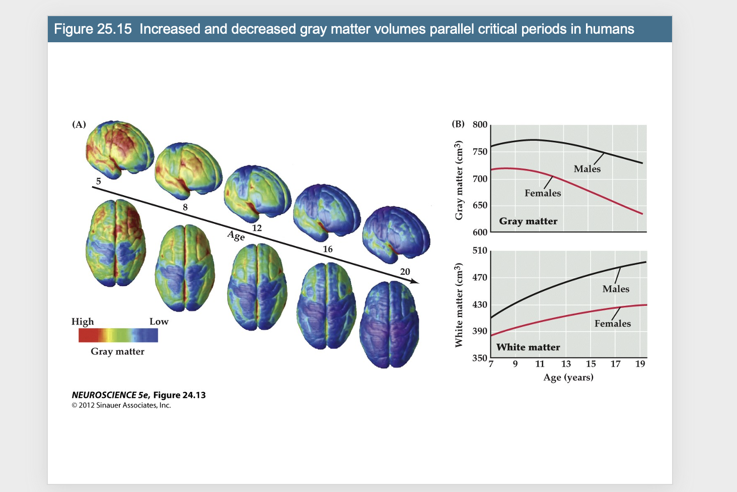
Measured 13 kids throughout study
the amount of brain matter is different, but can see the pattern of amount of gray matter (cell bodies) increase, and then decrease and level out
White matter (axons) is increasing, correlates with the pattern of gray matter but flipped
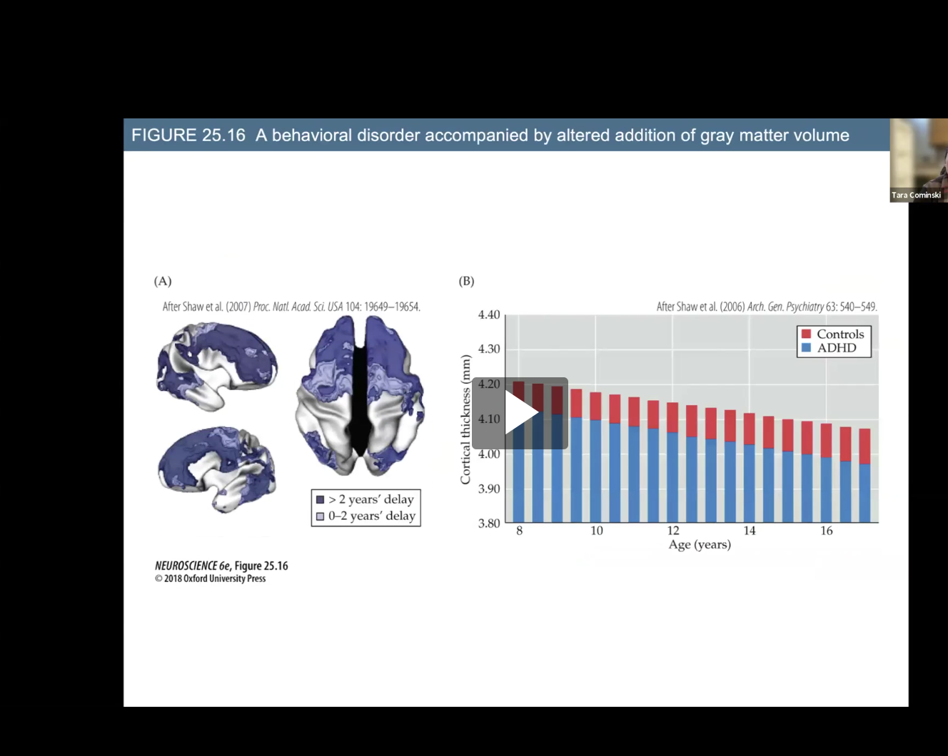
Figure 25.16- A behavioral disorder accompanied
Study in patients with ADHD
Map of cortical regions in which gray matter volume increases more slowly in children with ADHD vs typical developing children
Slight delay in gray matter volume increase in ADHD children
Cortical thickness is slightly decreased in ADHD patients.