MLS 409 Exam 2
 Call Kai
 Learn
Learn Practice Test
 Spaced Repetition
 Match
Match Flashcards
 Knowt Play
1/94

There's no tags or description
Looks like no tags are added yet.
Name | Mastery | Learn | Test | Matching | Spaced | Call with Kai | Chat |
|---|
No analytics yet
Send a link to your students to track their progress
95 Terms
What are the general requirements for setting up a urine culture?
Media: BAP & MAC
Incubation: 35-37C, ambient air, 18-24 hours
Which type of urine specimen do you use calibrated loops for?
Midstream clean catch, straight & indwelling catheter, nephrostomy/cytoscopy
How do you collect a specimen from an indwelling catheter source?
Collect from sampling port, not the bag
How do you culture a suprapubic aspirate urine specimen?
Plate specimen directly
What is the purpose of doing colony counts on urine specimens?
To distinguish a UTI from contamination
> 100,000 CFU/mL indicates a UTI
Isolation of colonies
What does it mean when there are 1-2 organisms vs multiple organisms on a urine plate?
Less organisms = infection, more = contamination
What are common urinary pathogens?
E. coli (most common!)
Klebsiella pneumoniae
Proteus mirabilis
Enterococcus faecalis
Staphylococcus saprophyticus (UTIs in young women)
Pseudomonas aeruginosa
Describe the following characteristics common to the Neisseriaceae:
Gram stain morphology
Oxidase production
GNDC
Oxidase positive (aerobic)
Describe the specific specimen collection, transport, and processing (including nutritional and atmosphere) requirements of Neisseria gonorrhoeae and Neisseria meningitidis cultures.
Culture immediately (temperature sensitive, transport at room temp)
Capnophilic (2-8% CO2)
N. gonorrhoeae
Media (enriched): CHOC and modified Thayer-Martin agar
Susceptible to drying (directly inoculate)
Inoculate with dacron or rayon swabs (others are inhibitory)
If delayed, use enriched selective media with transport system
N. meningitidis
Media: SBA, CHOC, modified Thayer-Martin
What is Thayer-Martin media? (Including its purpose, proper use, significant ingredients, inhibitory/selective properties)
CHOC agar with added antibiotics
Vancomycin, colistin, nystatin, trimethoprim
Selective for pathogenic Neisseria sp.
N. gonorrhoeae: small, gray to tan, translucent, raised
N. meningitidis: small, tan, sometimes mucoid, convex
N. lactamica (non-pathogenic, but still grows): small, tan, convex
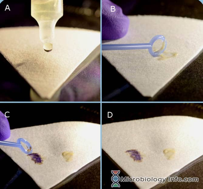
Explain the oxidase test
Purpose: detect cytochrome c oxidase
Positive: purple color change
Negative: no color change
Explain the nitrate reduction test
Purpose: detect reduction of nitrate to nitrite
Positive: red after adding reagent (nitrite), or colorless after zinc (N2 gas)
Negative: red only after adding zinc
Explain the butyrate esterase test
Purpose: detects butyrate esterase enzyme
Positive: blue color change
Negative: no color change
Explain the beta-lactamase test
Purpose: Detects the penicillinase/beta-lactamase enzyme
Positive: color change
Negative: no color change
Predict the carbohydrate utilization test results & growth on Thayer-Martin for the Neisseria sp and Moraxella catarrhalis
| Glucose | Maltose | Lactose | Sucrose | TM |
N. gonnorhoeae | Positive | Negative | Negative | Negative | Growth |
N. meningitidis | Positive | Positive | Negative | Negative | Growth |
N. lactamia | Positive | Positive | Positive | Negative | Growth |
M. catarrhalis | Negative | Negative | Negative | Negative | No Growth |
What tests can help differentiate between Moraxella and Neisseria sp?
Butyrate esterase
Neisseria: negative
Moraxella: positive
Nitrate
Neisseria: negative
Moraxella positive
Growth on Thayer-Martin
Neisseria: Growth
Moraxella: No Growth
What are the expected test results from a BactiCard for Neisseria sp and Moraxella catarrhalis?
Note: these tests are performed from enriched selective media only
BactiCard Performed on isolates from enriched selective media only | ||||
| N. gonorrhoeae | N. meningitidis | N. lactamica | M. catarrhalis |
IB Pos: teal | Negative | Negative | Negative | Positive |
PRO Pos: purp | Positive | Variable | N/A | N/A |
GLUT Pos: purp | Negative | Positive | N/A | N/A |
BGAL Pos: teal | Negative | Negative | Positive | N/A |
Draw the flowchart for GN(D)C
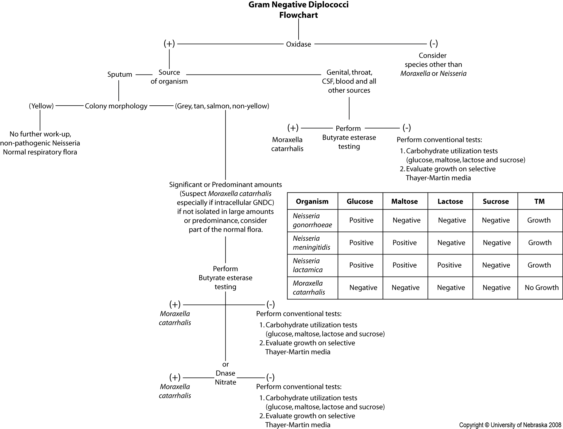
What are some examples of transport media used for Neisseria sp?
Jembec
Transgrow
Gono-Pack
Describe Neisseria gonorrhoeae, including its clinical significance, diseases caused, and treatment/prevention.
Clinical Significance
Present in genital, rectal, throat, eye, synovial fluid specimens
Infects any site containing columnar or transitional epithelial cells
Primarily spreads through sexual contact
Always pathogenic
Diseases
Causes gonorrhea (reported to the health department)
Males: acute urethritis, prostatitis, epididymitis, intracellular GNDC
Females: can be asymptomatic or cause severe discharge
Can lead to cervicitis, leading to pelvic inflammatory disease (PID), sterility, and ectopic pregnancies
Can also cause: Disseminated gonococcal infection
Treatment/Prevention
Treated with ceftriaxone or cefixime
Diseases prevented by screening mothers and providing eye treatments for infants within 2 hours of delivery
Describe Neisseria meningitidis, including its clinical significance, diseases caused, and treatment/prevention.
Clinical Significance
Source of infection: oral secretions and respiratory droplets, can be found in CSF
People can be asymptomatic carriers
Can enter the bloodstream = septicemia and/or meningitis
AST not performed
Serotypes A, B, C, W-135, and Y cause most cases
Diseases
Endemic Meningitis (Reported to Health Dept)
Risk factors: college-age young adults
Symptoms: fever, lethargy, irritability, headache, photophobia, stiff neck, seizures
Septicemia = purpura with petechial rash
Treatment and Prevention
Penicillin for meningitis (AST not performed)
3rd gen cephalosporins for meningococcemia
Prevent with vaccines and prophylaxis treatment to close contacts
Describe Neisseria lactamica including its clinical significance, diseases caused, and treatment/prevention.
Clinical Significance:
Normal flora of nasopharynx, rarely implicated in disease
Sterile sites (may req full identification)
Describe Moraxella catarrhalis including its clinical significance, diseases caused, and treatment/prevention.
Clinical Significance:
Normal flora of upper respiratory, alimentary, and genitourinary tracts but opportunistic pathogens in children and elderly
Most beta-lactamase positive
Diseases
Otitis media and maxillary sinusitis (3rd common cause)
Acute bronchitis & Pneumonia (in immunocompromised)
What is the colonial morphology of Moraxella catarrhalis?
Morphology: hockey-puck, smooth, opaque, gray to white that develops into a pink/salmon color.
Describe the following characteristics common to the Enterobacterales:
Gram stain morphology
Oxidase reaction
Glucose fermentation
Nitrate reduction
Growth on MacConkey agar
GNR / GNCB
Oxidase negative (except Plesiomonas)
Glucose fermenters
Nitrate reduction positive
Growth on MAC + HE/XLD agar
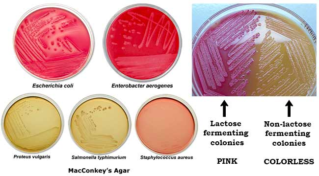
Describe MAC agar, including its purpose, proper use, and significant inhibitory/selective properties
Select for GNR (bile silts and crystal violet inhibit GP)
Differentiates lactose fermenters/non-fermenters
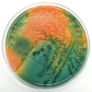
Describe Hektoen enteric agar (HE), including its purpose, proper use, and significant inhibitory/selective properties
Selective for GNR (higher concentration of bile salts than MAC)
Still inhibits many normal flora Enterics
Differentiates: lactose and sucrose fermenters; ferric salt producers (H2S - hydrogen sulfide)
Interpretation
Lactose &/ sucrose fermenter = yellow/orange colonies
Lactose &/sucrose non-fermenter = colorless, blue/green
H2S production = black precipitate
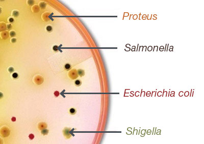
Describe Salmonella-Shigella agar (SS) including its purpose, proper use, and significant inhibitory/selective properties
Selective for GNRs (very inhibitory, but other Enterics can grow)
Differentiates lactose fermenters and H2S producers
Interpretation
LF: pink colonies
NF: colorless colonies
H2S: black precipitate
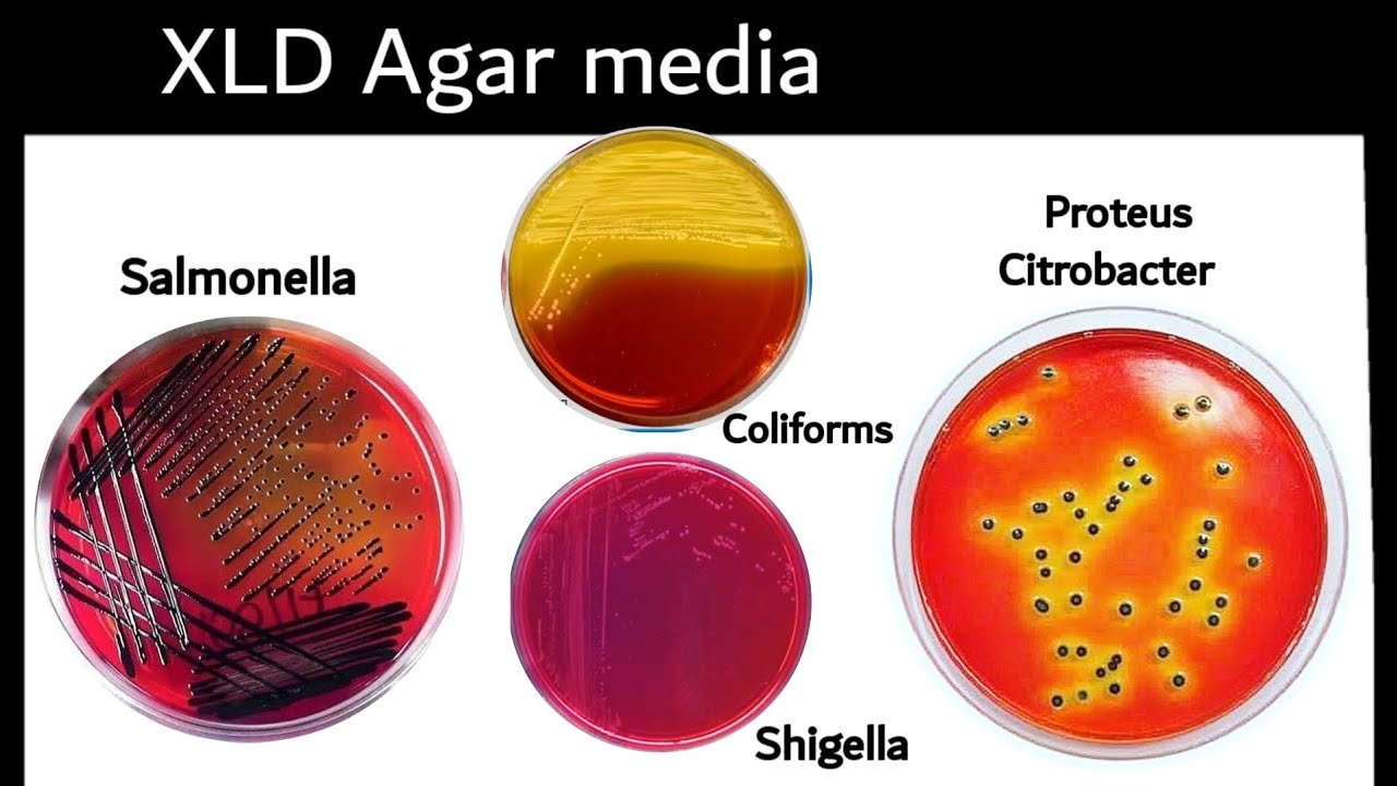
Describe Xylose-lysine-deoxycholate agar (XLD) including its purpose, proper use, and significant inhibitory/selective properties
Selective for GNRs
Sodium desoxycholate inhibits GPC and some GNR
Lower concentration of bile salts = better Shigella recovery
Differential for lactose, sucrose, xylose fermenters; lysine decarboxylation, H2S producers
Interpretation
Fermenters: yellow colonies
+ Xylose fermenters that do not decarboxylate lysine
Non-fermenters: red
Xylose fermenters that decarboxylate lysine, turning pH back to red
H2S: black precipitate
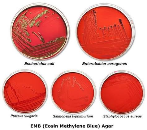
Describe Eosin-methylene blue agar (EMB) including its purpose, proper use, and significant inhibitory/selective properties
Selective for GNRs
Differential for lactose/sucrose fermenters
Interpretation
Fermenters: purple, strong = metallic
Non-fermenters: clear colonies
E. coli: metallic green sheen
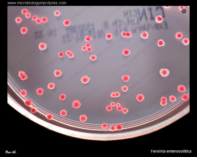
Describe Cefsulodin-irgasan-novobiocin agar (CIN) or Yersinia selective agar including its purpose, proper use, and significant inhibitory/selective properties
Selective for GNRs; used to recovery primarily Yersinia enterocolitica
Ingredients inhibit most organisms found in the stool
Differentiates mannitol fermenters
Interpretation
Fermenter: pink
Yersinia: bulls eye colonies with pink center and clera periphery
Nonfermenter: clear
What is the purpose of using Gram-negative broth?
Selectively enriched to promote Salmonella and Shigella sp
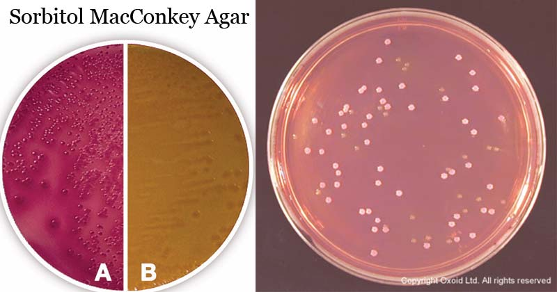
Describe Sorbitol MacConkey Agar (SMAC) including its purpose, proper use, and significant inhibitory/selective properties
A specialized MAC agar
Selects for GNRs
Differentiates sorbitol fermentation (MAC: lactose fermentation)
Interpretation
Fermenter: pink
Nonfermenter: clear
What is the most common etiologic agent of subacute bacterial endocarditis?
Viridans streptococci
Discuss the appropriateness of performing direct Gram stains on blood cultures
Guides empirical therapy
Differentiate pathogens from contaminants
Facilitates AST testing
What organisms are indicative of septicemia/bacteremia?
S. aureus
E. coli
Streptococcus sp
Enterococcus sp
Candida sp
What organisms are indicative of skin contamination?
CoNS
Bacillus sp
Micrococcus sp
Explain the indole test
Detects tryptophanase, which hydrolyzes tryptophan to indole
Positive: blue/purple
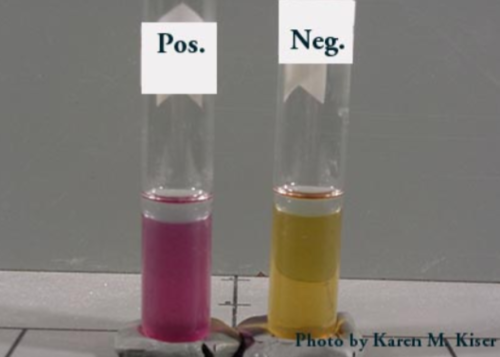
Explain the lysine and ornithine decarboxylase (LDC, ODC) test
Detects lysine decarboxylase, turning lysine into cadaverine
Detects ornithine decarboxylase, turning ornithine into putrescine and CO2
Bacteria first ferments glucose, lowering pH = yellow
Acidic environment triggers decarboxylation, raising pH = purple
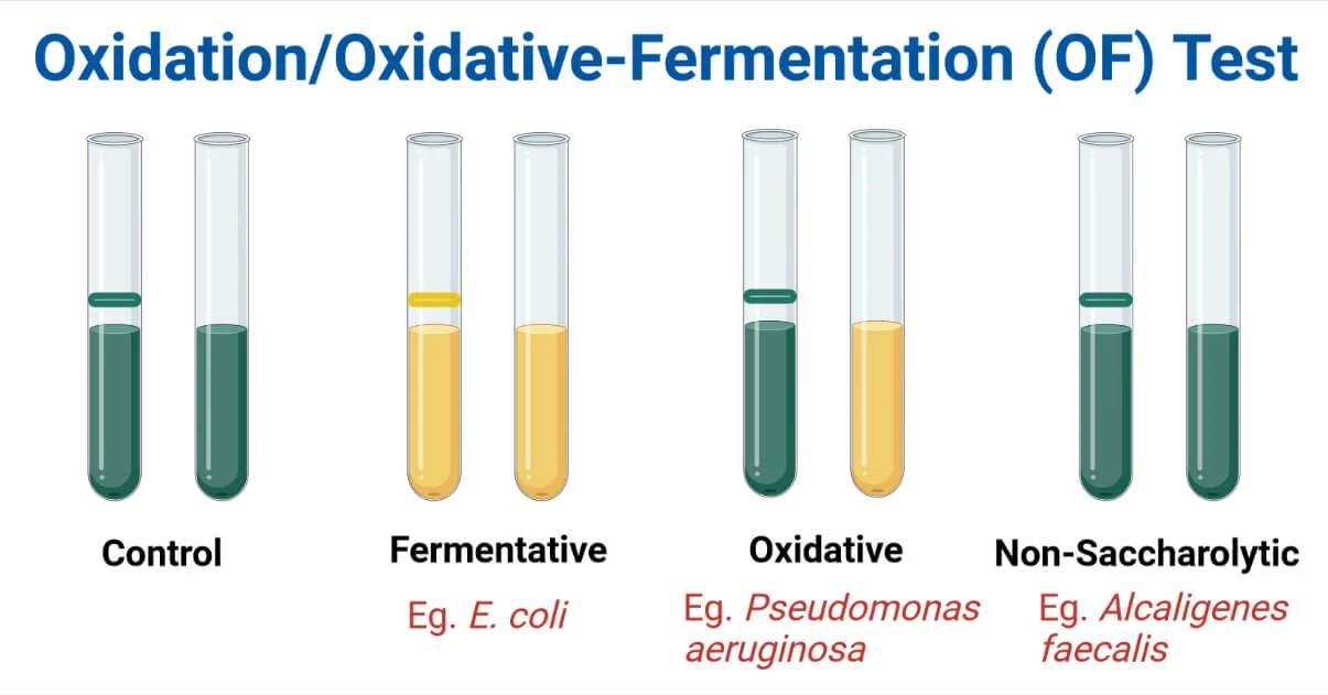
Explain the glucose oxidation-fermentation reaction
Two test tubes = one aerobic and anaerobic
Fermentative: both tubes turn yellow (can break down glucose with/without oxygen)
Oxidative: only the aerobic tube turns yellow (needs oxygen)
Inert/Non-saccharolytic: bacteria cannot use glucose at all
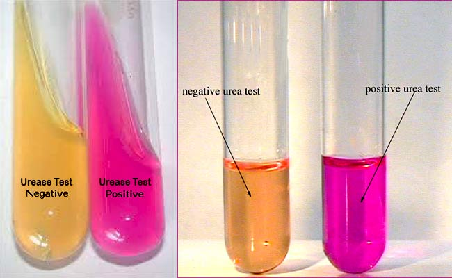
Explain the urease production test
Detects urease, which breaks down urea into ammonia and CO2
Positive: pink agar
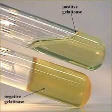
Explain the gelatin liquefaction test
Detects gelatinase, which breaks down gelatin into smaller amino acids, causing the media to lose its ability to solidify at cold temperatures
Positive: medium remains liquid after chilling
Negative: medium solidifies upon chilling
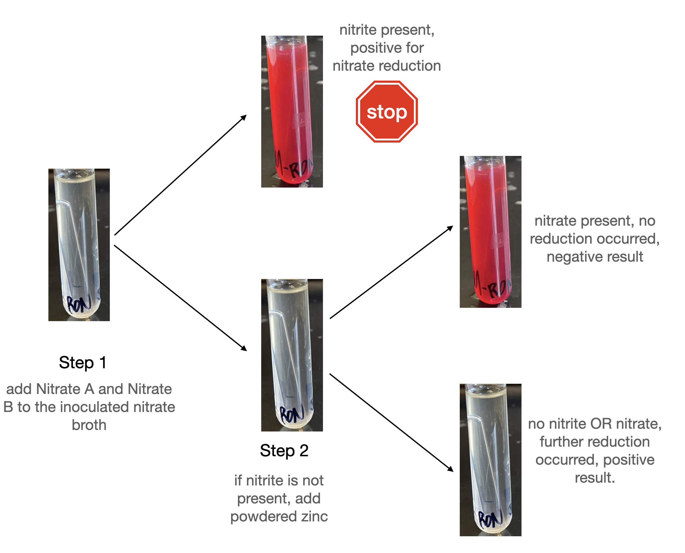
Explain the nitrate reduction test
Detects if bacteria can reduce nitrate to nitrite
Positive: medium turns red after adding reagent; or medium remains colorless after adding reagent and zinc (N2 gas)
Negative: medium remains colorless after adding reagent; or medium turns red after adding zinc
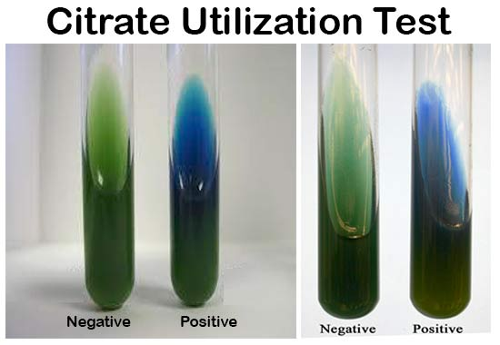
Explain the citrate utilization test
Detects if bacteria can use citrate as a carbon and energy source
Positive: blue medium
Negative: medium remains green
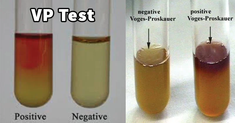
Explain the Voges-Proskauer (VP) test
Determines if 2,3-butanediol is a produce of glucose fermentation
Positive: red/purple color near top of tube
Negative: no color change
Explain the deaminase reaction test (tryptophan, lysine, phenylalanine)
Determines if bacteria can remove an amine group (NH2) from an amino acid
Tryptophan: indole test
Phenylalanine: positive result is a green color on a slant
Lysine: positive result is a red color on a slant
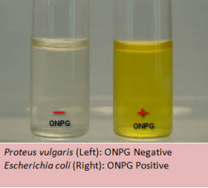
Expalin the orthonitropheynl-beta-D-galactopyranoside (ONPG) test
Detects beta-galactosidase, which is needed to break down lactose
Positive: liquid medium turns yellow
What are the clinically relevant lactose-fermenting members of the order Enterobacterales?
Escherichia coli
Citrobacter freundii
Klebsiella pneumoniae
Klebsiella oxytoca
Enterobacter cloacae
Klebsiella (Enterobacter) aerogenes
What are the clinically relevant species that are non-lactose fermenting members of the order Enterobacterales and also LDA/TDA positive and ONPG negative?
Proteus vulgaris
Proteus mirabilis
Morganella morganii
What are the clinically relevant species that are non-lactose fermenting members of the order Enterobacterales and also LDA/TDA negative?
Shigella sonnei
Shigella species
Salmonella species
Salmonella typhi
Serratia species
Citrobacter freundii
Citrobacter koseri
Yersinia entercolitica
What are the general characteristics of Enterobacterales?
GNRs
Glucose fermenters
Oxidase negative
Nitrate reduction positive
Identify the key test results of LDC, LDA/TDA, H2S, ODC, Motility, Indole, and ONPG to differentiate:
Salmonella sp
Shigella sp
Shigella sonnei
Lactose Nonfermenters; LDA/TDA negative
Organism | LDC | LDA/TDA | H2S | ODC | Motility | Indole | ONPG |
Salmonella sp | Pos | Neg | Pos | Pos | Pos | Neg | Neg |
Shigella sp | Neg | Neg | Neg | Neg | Neg | -/+ | Neg |
Shigella sonnei | Neg | Neg | Neg | Pos | Neg | Neg | Pos |
Identify the key test results of ODC, LDC, Indole, Citrate, VP, Motility, and H2S to differentiate:
Escherichia coli
Klebsiella pneumoniae
Klebsiella oxytoca
Klebsiella (Enterobacter) aerogenes
Enterobacter cloacae
Lactose Fermenters, ONPG positive
Organism | ODC | LDC | Indole | Citrate | VP | Motility | H2S |
E. coli | Pos/Neg | Pos/Neg | Pos | Neg | Neg | Pos/Neg | Neg |
K. pneumoniae | Neg | Pos | Neg | Pos | Pos | Neg | Neg |
K. oxytoca | Neg | Pos | Pos | Pos | Pos | Neg | Neg |
K. (E.) aerogenes | Pos | Pos | Neg | Pos | Pos | Pos | Neg |
E. cloacae | Pos | Neg | Neg | Pos | Pos | Pos | Neg |
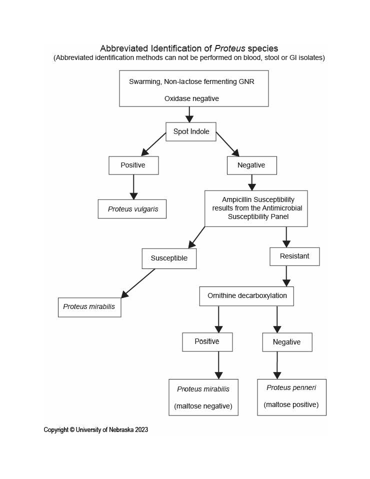
Identify the key test results of Urease, H2S, ODC, Indole, LDA/TDA to differentiate:
Proteus mirabilis
Proteus vulgaris
Organism | Urease | H2S | ODC | Indole | LDA/TDA |
Proteus mirabilis | ++ | + | + | - | + |
Proteus vulgaris | ++ | + | - | + | + |
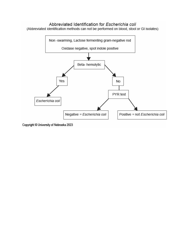
Draw out the abbreviated ID scheme for E. coli
Draw out the abbreviated ID scheme for Proteus sp
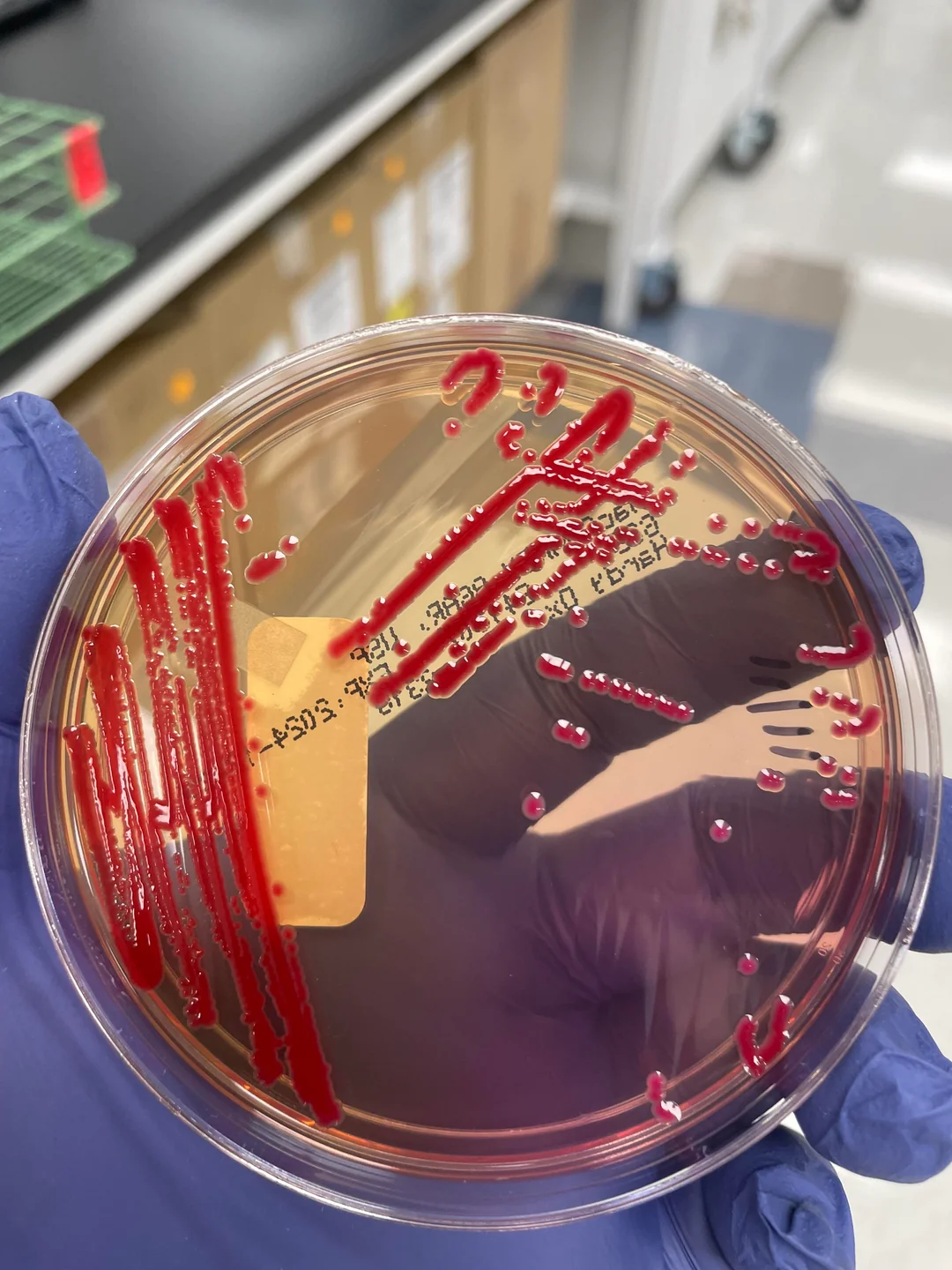
What does Serratia marcescens look like on SBA and MAC?
SBA: creamy, gray white
MAC: translucent/pale
Produces a red pigment (prodigiosin) at room temperature
What does Yersinia enterocolitica look like on CIN/Yersinia agar?
Smooth colonies with a bulls-eye appearance
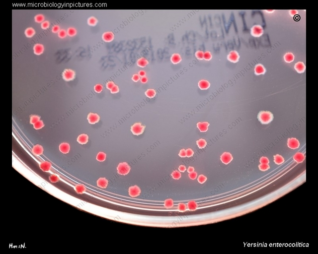
What is the only lactose fermenting GNR that also produces H2S?
Citrobacter freundii
Define the following terms:
a. Capsular antigen (K)
b. Somatic antigen (O)
c. Flagellar antigen (H)
d. Vi antigen
Capsular antigen (K): surrounds bacterial cell outside the O antigen, protection from phagocytosis and complement
Somatic antigen (O): heat-stable polysaccharide portion of the lipopolysaccharide (LPS) in the outer membrane of GN bacteria
Flagellar antigen (H): heat-labile protein (flagellin) that makes up flagelal
Vi antigen: surface capsular polysaccharide antigen that overlies O antigen, inhibiting phagocytosis
T/F: AST is routinely performed on suspected pathogenic Enterobacterales isolates except Salmonella (except typhoidal isolates) and Shigella
True
Biochemical ID of Shigella sp should be confirmed with what?
Serogrouping
Which Enterics are commonly responsible for GI infections?
Salmonella sp
Shigella sp
E. coli
Yersinia enterocolitica
Which Enterics are commonly responsible for opportunistic/nosocomial infections?
K. pneumoniae = destructive pneumonia
Enterobacter cloacae
Serratia sp
Citrobacter sp
Proteus sp = implicated frequently in UTIs
Morganella morganni
Which Enterics are commonly responsible for UTIs?
E. coli (#1 etiologic agent of UTI)
Proteus sp
Which Enterics are commonly responsible for typhoid fever?
Salmonella typhi
The most pathogenic of the Salmonella sp
Rose spots appear
Constipation followed by bloody diarrhea
Which Enterics are commonly responsible for neonatal meningitis?
E. coli
Which Enterics are commonly responsible for hemolytic uremic syndrome (HUS)?
E. coli
Which Enterics are commonly responsible for respiratory infections?
K. pneumoniae
Which Enterics are commonly responsible for wound infections?
Preotus
E. coli
K. pneumoniae
Which Enterics are always pathogens?
Shigella sp
Salmonella sp
Yersinia
E. coli O157:H7
Describe the specific specimen processing requirements for Haemophilus species, including nutritional requirements, atmosphere requirements, and length of incubation.
Haemophilus sp are fastidious organisms that are capnophilic, facultative anaerobes that require V (NAD) and X (Hemin) growth factors
Avoid drying and extreme temperatures, culture immediately
CHOC
CO2
Moist environment
Incubate for 24-48 hours
Describe the characteristic Gram stain and colonial morphology of Haemophilus species.
GNCB
Tannish
Translucent
Mousy odor
H. ducreyi: small, grey, yellow/tan, hockey-puck
What tests/characteristics are used to identify Haemophilus sp?
X and V requirements
Disk test, watch for growth (contains either X, V, or XV)
Growth around either X/V/XY = requires that factor
Growth around all disks: does not require any factor
Porphyrin test (ALA test)
Determines if organism can synthesize X factor from ALA
Positive: red/orange fluorescence after reagent/UV light, does not require X factor, organism produces porphyrin
Hemolysis on horse blood agar
Detects organism’s ability to produce hemolysins that lyse horse RBCs
Helps differentiate between hemo/nonhemolytic species
Satellitism
Demonstrates dependence on V factor (NAD)
S. aureus inoculated, lyses RBCs, releasing X favor and secretes V factor, which allows Haemophilus to grow
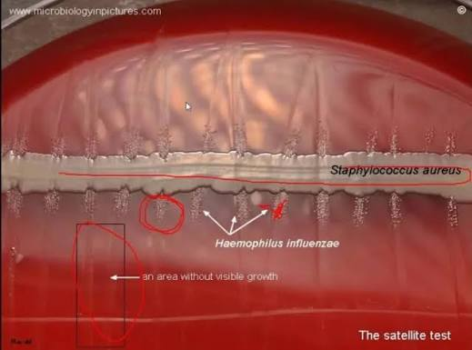
Describe the difference in test results between H. influenzae and H. parainfluenzae (X/Y test; Porphyrin; Horse blood; Satellitism)
Influenzae | Parainfluenzae | |
X/Y Test (Growth) | XV | V & XV |
Porphyrin | Negative | Positive |
Horse Blood | No | No |
Satellitism | Positive | Positive |
Describe the function and clinical significance of the capsule as a virulence factor for Haemophilus influenzae.
H. influenzae’s polysaccharide capsule is its most significant virulence factor (6 antigen types a-f, based on capsular polysaccharides). Helps prevent phagocytosis.
Encapsulated strains, especially Hib cause severe invasive infections, especially in unvaccinated children younger than 5
Vaccines are available
Un-encapsulated species can still cause disease
Localized infections, risk factors for elderly and immunocompromised
What kind of diseases do H. influenzae cause?
Respiratory infections
Meningitis
Otitis media
Conjunctivitis (ssp. aegypticus)
What disease does H. ducreyi cause?
Chancroid/Soft Chancre
Should you perform AST on Haemophilus sp?
Yes (ish)
Traditionally, ampicillin was the drug of choice, but resistance has been seen
Beta-lactamase should be performed
Other AST can be done
What are the GNR glucose non-fermenters?
Pseudomonas aeruginosa
Stenotrophomonas maltophilia
Acinetobacter baumannii
Burkholderia cepacia
Fill in this table for Glucose NF GNRs
| Oxidase | 42C | ADH | ODC | LDC | Gelatin | Polymyxin B |
P. aeruginosa | |||||||
S. maltophilia | |||||||
B. cepacia |
| Oxidase | 42C | ADH | ODC | LDC | Gelatin | Polymyxin B |
P. aeruginosa | + | Growth | + | - | - | +/- | Susceptible |
S. maltophilia | - | No | - | - | + | + | Susceptible |
B. cepacia | +/- | +/- | - | +/- | + | +/- | Resistant |
What are the expected results of a glucose oxidation-fermentation test for glucose non-fermenting GNRs?
Aerobic tube turns yellow
Anaerobic tube remains green
Describe the following characteristics for P. aeruginosa
Colony morphology
Gram morphology
Glucose utilization
Oxidase
Pigment
Growth at 42C
Colony: rough, spreading, may have green sheen and be beta-hemolytic
GS: thin GNR
Glucose oxidizer
Oxidase positive
Growth at 42C
Describe key characteristics/tests of S. maltophilia
Strong maltose oxidizer
Opportunistic
Oxidase negative
Glucose “O” or “I”
BAP: pale yellow to lavender pigment
ADH: negative
ODC: negative
LDC: positive
DNase: positive
Polymyxin B: sensitive
Describe key characteristics/tests of P. aeruginosa
Glucose oxidizer
Strict aerobe
Pigments: pyoverdin/cyanin/rubin/mealnin
Beta-hemolytic
Metallic sheen/mucoid
Non LF
Grape smell
Oxidase: positive
Indole: negative
Always perform AST
Draw the ID scheme for GNF GNRs
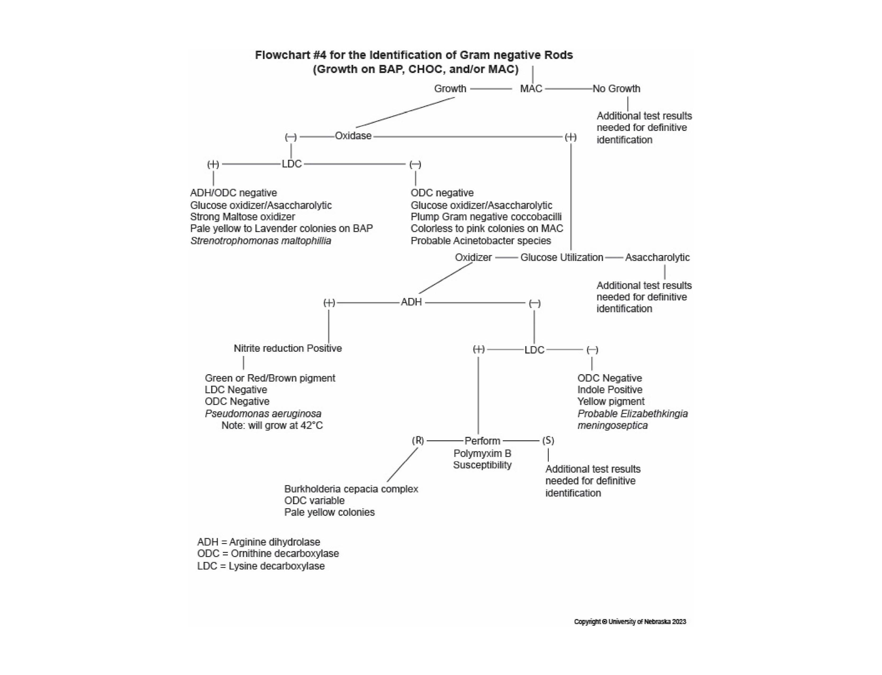
How do GNF GNRs cause disease?
Predominant opportunistic - affecting immunocompromised or hospitalized patients
Survive well in moist environments, form biofilms, have antimicrobial resistance
Other VFs: pili, endo/exotoxins, proteolytic enzymes, extracellular capsule
What kind of diseases can P. aeruginosa cause?
Opportunistic infections
Wounds
Respiratory
Otitis externa
Corneal ulcers
UTI
Cystic fibrosis
Burns
Osteomyelitis
What kind of diseases can S. maltophilia cause?
Respiratory
Wound
Opportunistic
When do you reject a respiratory specimen for culture using a direct specimen Gram stain?
When there are more than 25 epithelial cells/10X field
Describe the procedures for setting up a culture on a sputum specimen, including the types of media used and incubation requirements:
Direct specimen gram stain done first before culture set up
Media: BAP, CHOC, MAC
Sample areas that are purulent or bloody
Streak for isolation
BAP & CHOC - CO2; MAC - ambient
Temp: 35C
Time: 18-24 overnight, minimum 48
Describe the procedures for setting up a culture on a throat specimen, including the types of media used and incubation requirements:
Direct gram stain first
Throat swabs done to find Group A Strep only
Media: strep selective blood agar
Use swab to make primary streak, use loop to streak for isolation
Make cuts in primary streak or incubate anaerobically to allow detection of streptolysin O
Temp: 35C
Incubate in anaerobic conditions
Time: overnight 18-24 hours, minimum 48
Evaluate the normal flora of the following respiratory sites for culture interpretation:
a. Upper respiratory tract
b. Lower respiratory tract
Upper
Viridans strep
CoagN Staph
Nonpathogenic Neisseria
Diphtheroids
Anaerobes
Yeast
Lower
Normally sterile
Evaluate pathogens commonly found in the following respiratory sites for culture interpretation:
a. Upper respiratory tract
b. Lower respiratory tract
Clinical group | Organisms |
|---|---|
Community-acquired pneumonia | Streptococcus pneumoniae, Haemophilus influenzae, Moraxella catarrhalis |
Hospital-acquired/opportunistic | Klebsiella pneumoniae, E. coli, Enterobacter spp., Serratia marcescens, Staphylococcus aureus |
Hospital-acquired/opportunistic nonfermenters | Pseudomonas aeruginosa, Stenotrophomonas maltophilia, Acinetobacter spp. |
Other respiratory pathogen | Streptococcus pyogenes |
Cystic fibrosis-associated | Burkholderia cepacia complex |
Haemophilus species appear on Gram stain as:
GNCB