Learn: Anatomy lecture mideterm 2
 Call Kai
 Learn
Learn Practice Test
 Spaced Repetition
 Match
Match Flashcards
 Knowt Play
1/73

There's no tags or description
Looks like no tags are added yet.
Name | Mastery | Learn | Test | Matching | Spaced | Call with Kai | Chat |
|---|
No analytics yet
Send a link to your students to track their progress
74 Terms
functional classifications of joints
synarthrosis, amphiarthrosis, diarthrosis
synarthroses
immovable; common in the axial skeleton
amphiarthroses
slightly movable; common in axial skeleton
diarthroses
freely movable; common in appendicular skeleton (all synovial joints)
structural classifications of joints
fibrous, cartilaginous, synovial
structure of fibrous joints
connected by fibrous connective tissue, no joint cavity, immovable or slightly movable
types of fibrous joints
sutures, synchondroses, gomphoses

sutures
bones are tightly bound by a minimal amount of dense fibrous connective tissue, only between skull bones, allows bone growth so the brain can expand during childhood
syntoses
closed sutures
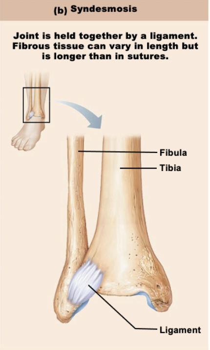
syndesmoses
bones connected exclusively by ligaments, amount of fibers depends on length of fibers
examples of syndesmoses
tibiofibular joint (immovable synarthrosis), interosseous membrane (between radius and ulna, freely movable diarthrosis)

gomphoses
peg in socket joint, only example is teeth in socket connected by periodontal ligament
structure of cartilaginous joints
bones are united by cartilage, lack a joint cavity
types of cartilaginous joints
synchondroses, symphyses
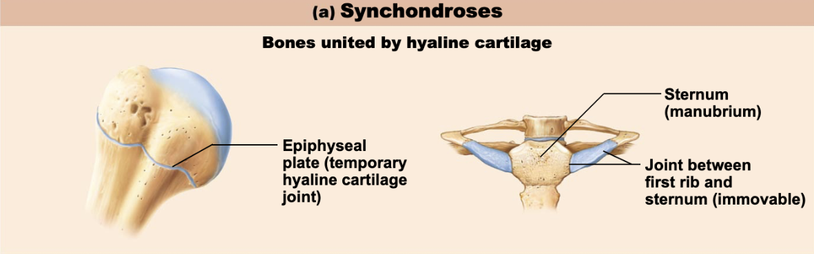
synchondroses
hyaline cartilage unites bones; epiphyseal plates and first sternocostal joint (between 1st rib and manubrium)
symphyses
fibrocartilage unites bones, resists tension and compression; hyaline cartilage present as articular cartilage; slightly movable joints that provide strength with flexibility
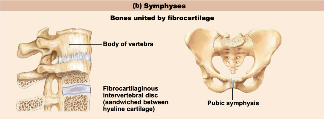
examples of symphyses
intervertebral discs, pubic symphysis
synovial joints
most movable; all are diarthroses (freely movable); each contains a fluid filled joint cavity (unique to synovial joints)

structure of all synovial joints - articular cartilage
ends of epiphyses are covered with hyaline cartilage; absorbs compression
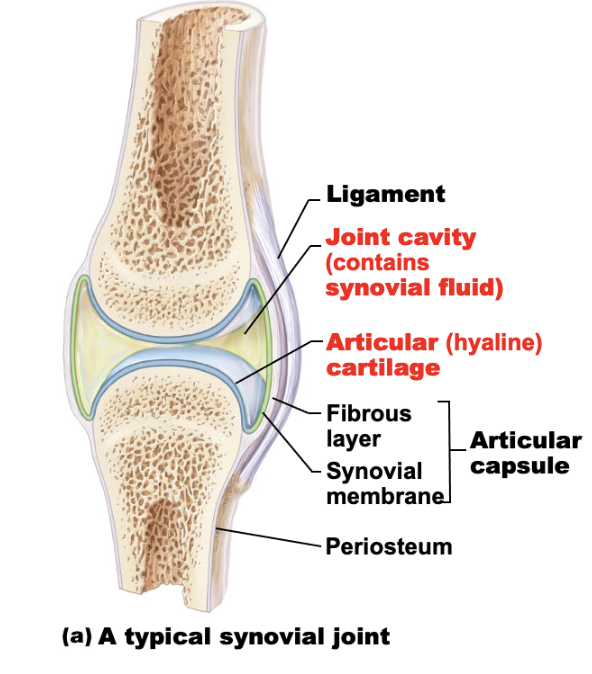
structure of all synovial joints - joint (articular) cavity
unique to synovial joints; cavity is a potential space that holds a small amount of synovial fluid

structure of all synovial joints - articular capsule
enclosed in a 2 layered capsule; fibrous layer (dense irregular connective tissue which strengthens joint), synovial membrane (loose connective tissue)
synovial membrane
lines joint capsule and covers internal joint surfaces; functions to make synovial fluid

structure of all synovial joints - reinforcing ligaments
often are thickened parts of the fibrous layer; sometimes are extrascapular ligaments located outside the capsule (fibular and tibial collateral ligaments of the knee; sometimes are intrascapular ligaments (located internal to the capsule)
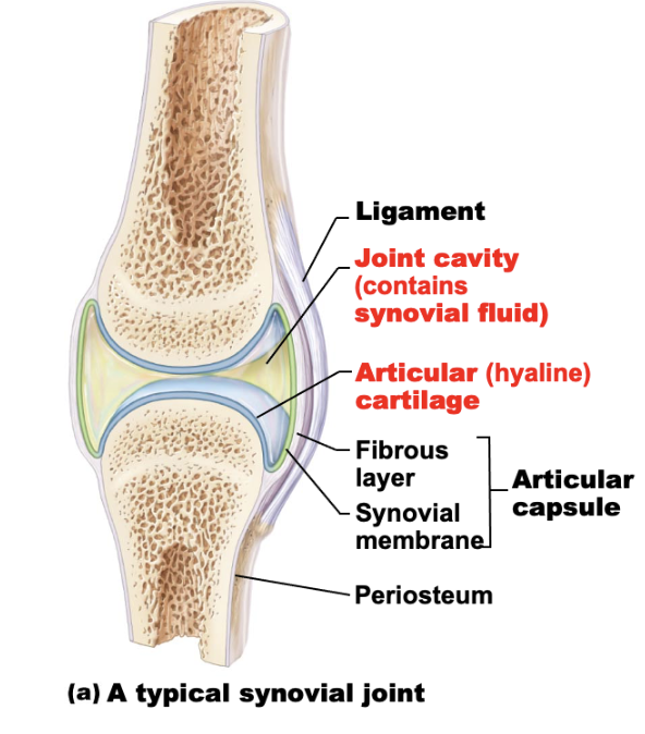
structure of all synovial joints - synovial fluid
a viscous fluid similar to raw egg white; filtrate of blood (leaks from capillaries in synovial membrane); contains glycoprotein molecules secreted by fibroblasts
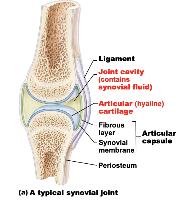
structure of all synovial joints - richly supplied with sensory nerves
detect pain; most monitor how much the capsule is being structured

structure of all synovial joints - rich blood supply
most supply the synovial membrane; extensive capillary beds produce basis of synovial fluid; branches of several major nerves and blood vessels
weeping lubrication
pressure on joints squeezes synovial fluid into and out of the articular cartilage (avascular); lubricates the joint; prevents overheating and subsequent destroyal of joint tissue
articular discs
in some synovial joints (meniscus); occurs in temporomandibular, sternoclavicular, and knee joint; occurs in joints whose articulating bones have somewhat different shape; distributes load more evenly and minimizes wear and damage; may also allow two different movements at the same joint (jaw)
bursae
a flattened fibrous sac lined by a synovial membrane; closed bags of lubricant; reduce friction between body sheaths
tendon sheaths
an elongated bursa that wraps around a tendon; reduce friction between body sheaths
synovial joint movements
gliding: one bone slides across the surface of another
angular movements: movements that change the angle between bones
rotation: movement around a bone’s long axis

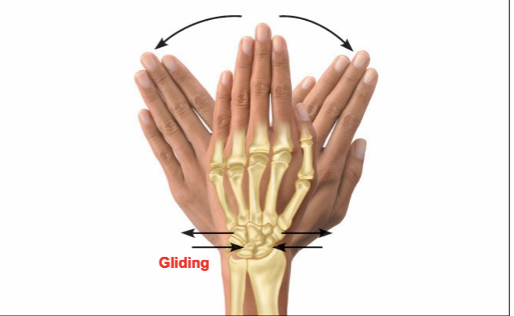
gliding
flat surfaces of 2 bones slip across each other; occurs between carpals, tarsals, and articular processes of vertebrae
angular movements
increase or decrease angle between bones; usually in the sagittal plane
flexion - decreasing angle
extension - increasing angle
abduction - moving a body part away from the midline in the frontal plane
adduction - moving a body part towards the midline in the frontal plane
circumduction - moving as a cone (circle) in space

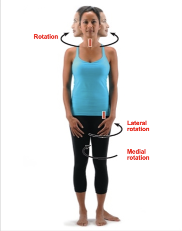
rotation
involves turning movement of a bone across its longitudinal axis; only movement allowed between Atlantoaxial joint; occurs at hip and shoulder joint
medial rotation - rotating towards the median plane
lateral rotation - rotating away from median plane

special movements
elevation - lifting body part superiorly
depression - moving elevated part inferiorly
protraction - non angular movement anteriorly
retraction - non angular movement superiorly


special movements
supination - forearm rotates laterally, palm faces anteriorly
pronation - forearm rotates medially, palm faces posteriorly; brings radius across the ulna
opposition - thumb moves across the palm to touch the tips of other fingers


special movements
inversion - special movement at the foot; turns sole medially
eversion - special movement at the foot; turns sole laterally
dorsiflexion - up movement of the foot; lifting the foot so its superior surface approaches the shin
plantar flexion - depressing the foot, elevating the heel

types of synovial joints
can be described functionally; nonaxial, uniaxial, biaxial, multiaxial
nonaxial
adjoining bones dont move around a specific axis

nonaxial joints - plane joint
articular surfaces are flat planes; short gliding movements are allowed; doesnt involve rotation around any axis
ex: intertarsal and intercarpal joints
uniaxial
movement occurs around a single axis; flexion/extension or rotation

uniaxial joints - hinge joint
cylindrical end of 1 bone fits into a trough on another bone; angular movement is allowed on 1 plane
ex: elbow joint, ankle joint, and joints between phalanges

uniaxial joints - pivot joint
sleeve and axle or facet and process articular surfaces; rotating bone turns only around its long axis
ex: proximal radioulnar joint, Atlantoaxial joint
biaxial
movement occurs around 2 axes; thus the joint enables motion along both the frontal and sagittal planes; flexion/extension and abduction/adduction (rotation is generally restricted or absent)

biaxial joints - condylar (or ellipsoid) joints
oval articular surfaces; side to side (abduction/adduction), back and forth (flexion/extension)
ex: metacarpopharangeal (knuckle) joints, wrist joint

biaxial joints - saddle joints
each articular surface has concave and convex surfaces; side to side (abduction/adduction), back and forth (flexion/extension)
ex: 1st carpometacarpal (allows opposition of the thumb)
multiaxial
movement can occur around all 3 axes and along all 3 body olanes; flexion/extension, abduction/adduction, and full rotation

multiaxial joints - ball and socket joint
spherical head of 1 bone fits into round socket of another
ex: shoulder joints, hip joints

temporomandibular joint features
modified hinge joint; condylar process of mandible articulates with mandibular fossa of temporal bone; multiaxial but not ball and socket


temporomandibular joint movements
2 surfaces of the articular disc allow hinge-like movement and gliding of superior surface anteriorly
sagittal axis (lateral excursion): side to side
vertical axis (hinge): elevation/depression
horizontal axis (gliding): protrusion/retraction
temporomandibular joint information
most easily dislocated joint because of the shallow socket
almost always dislocates anteriorly
condylar process glides anteriorly, ending up in the infratemporal fossa of the skull
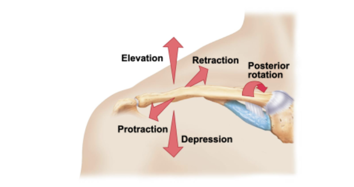
sternoclavicular joint
saddle joint; performs multiple complex movements; 4 ligaments surround joint
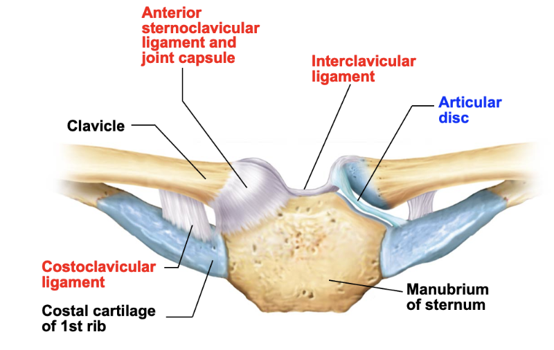
sternoclavicular joint ligaments
anterior and posterior sternoclavicular ligaments
interclavicular ligament
costoclavicular ligament

glenohumeral joint features
ball and socket joint; head of humerus and shallow glenoid cavity of scapula (glenoid cavity is slightly deepened by a rim of fibrocartilage called the glenoid labrum
glenoid labrum extends from margin of cavity to anatomical neck of humerus


glenohumeral joint ligaments
only strong thickening of capsule is superior coracohumeral ligament (helps support weight of upper limb)
anterior part of capsule thickens slightly into 3 weak glenohumeral ligaments
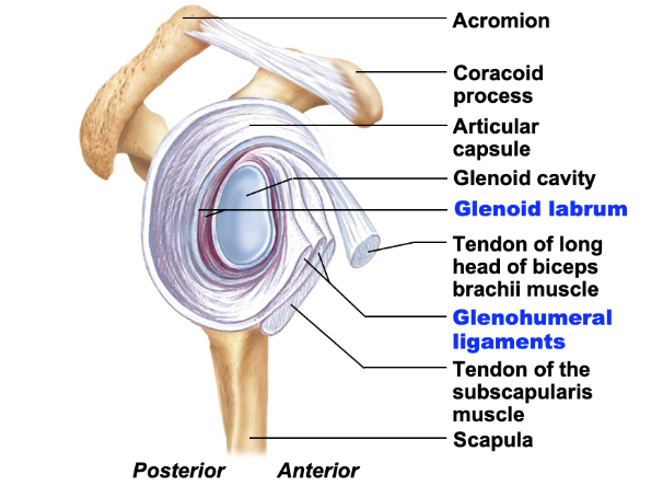

glenohumeral joint and rotator cuff tendons
muscle tendons contribute to joint stability
rotator cuff is made of 4 muscles and associated tendons
subscapularis
supraspinatus
infraspinatus
teres minor
rotator cuff injuries are common shoulder injuries

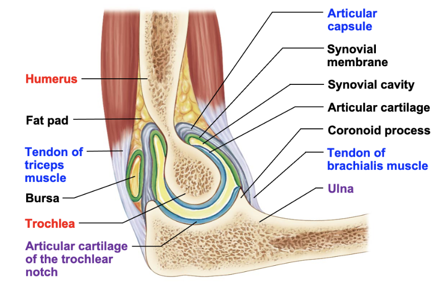
humeroulnar joint features
hinge joint (allows only flexion/extension); articulation of humerus with trochlear notch of ulna forms hinge
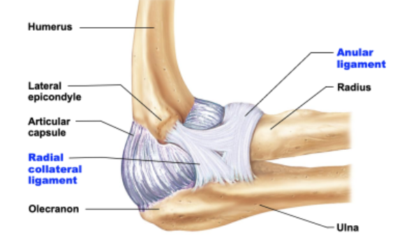
humeroulnar joint ligaments
articular capsule attaches to humerus and ulna and to anular ligament of radius (ring around radius’ head)
radial collateral and ulnar collateral ligaments prevent lateral and medial movements
tendons of biceps and triceps brachii provide stability


radiocarpal joint features
condylar joint; composed of radiocarpal and intercarpal joints
radiocarpal joint: joint between radius and proximal carpals (scaphoid + lunate); allows for flexion/extension, abduction/adduction, circumduction
intercarpal joint: joint between proximal and distal rows of carpals; allows for gliding movements
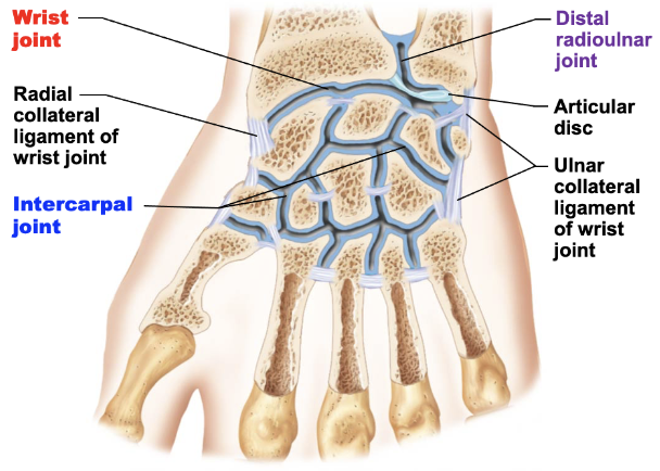

radiocarpal joint ligaments
stablized by numerous ligaments
palmar radiocarpal ligament anteriorly
dorsal radiocarpal ligament posteriorly
radial collateral ligament laterally
ulnar collateral ligament medially

hip joint features
ball and socket; movement occurs in all axes (limited by ligaments and acetabulum); head of femur articulates with acetabulum

hip joint ligaments
stability comes chiefly from acetabulum and capsular ligament
acetabulum labrum fibrocartilage prevents slippage from socket
muscle tendons contribute somewhat to stability

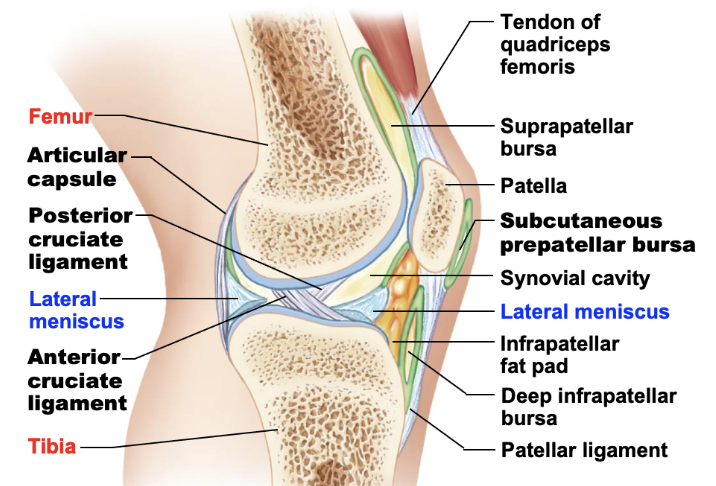
knee joint features
largest and most complex; acts primarily as a hinge joint; wheel shaped condyles of femur roll along almost flat condyles of tibia; has some capacity for rotation when leg is flexed

knee joint structure
structurally considered compound and bicondyloid
2 fibrocartilage menisci occur within joint cavity
femoropatellar joint: shares joint cavity; allows patella to glide across distal femur

knee joint capsule
covers tibial and femoral condyles (posterior and lateral aspects of knee but not anterior
reinforced by several capsular and extracapsular ligaments
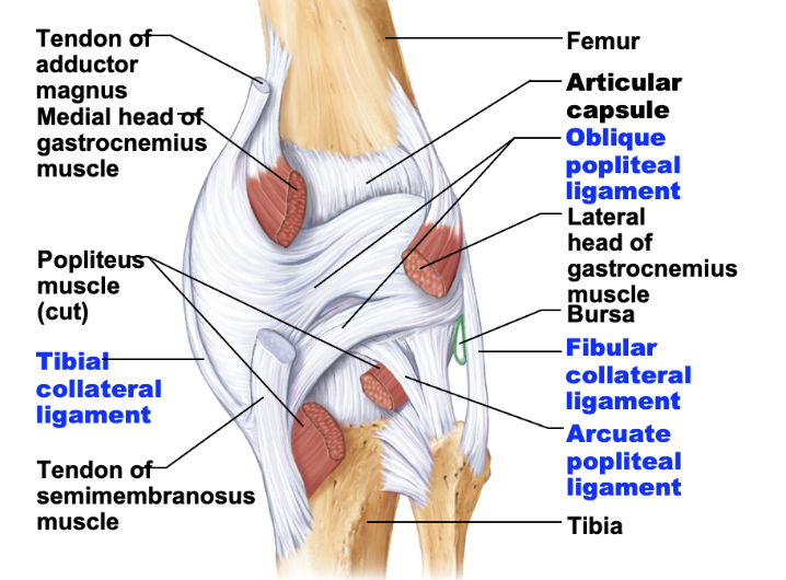
knee joint extracapsular and capsular ligaments
become taut when knee is extended
fibular and tibial collateral ligaments
oblique popliteal ligament
arcuate popliteal ligament

knee joint anterior ligaments
patellar ligament
medial and lateral patellar retinacula
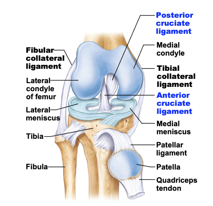
knee joint intracapsular ligaments
cruciate ligaments: cross each other like an “X” ; runs from proximal tibia to distal femur; prevent undesirable movements at the knee
anterior cruciate ligament: prevents anterior sliding of the tibia
posterior cruciate ligament: prevents anterior sliding of the femur or posterior displacement of the tibia


ankle joint features
hinge joint between united inferior ends of tibia and fibula + talus of the foot
allows dorsiflexion and plantar flexion only
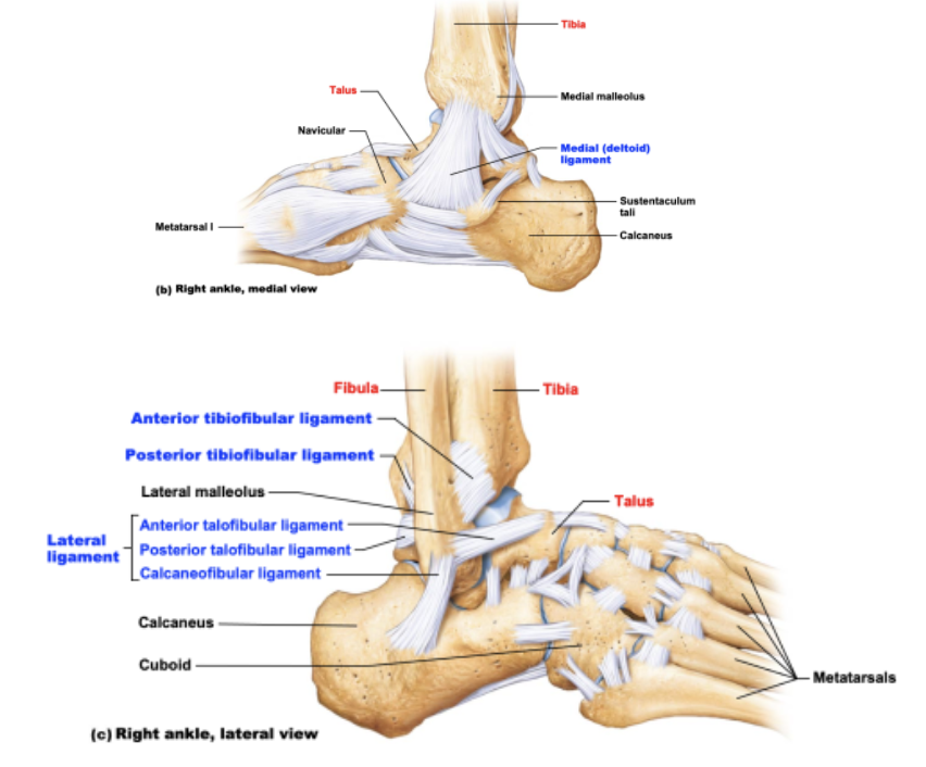
ankle joint ligaments
medially and laterally stabilized by ligaments
medial (deltoid) ligament
lateral ligament
inferior ends of tibia and fibula are joined by ligaments
anterior and posterior tibiofibular ligaments
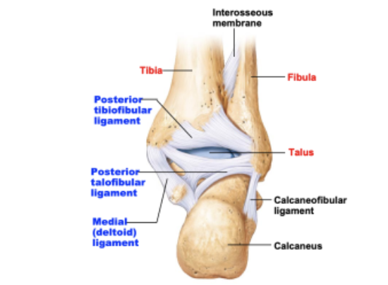
arthritis
describes more than 100 kinds of joint damaging dieases
osteoarthritis
most common type of wear and tear arthritis
rhuematoid arthritis
chronic inflammatory arthritis
gouty arthritis (gout)
uric acid buildup causes pain in joints