12. Somatic Sensory Nervous System
1/150
There's no tags or description
Looks like no tags are added yet.
Name | Mastery | Learn | Test | Matching | Spaced |
|---|
No study sessions yet.
151 Terms
What are sensory receptors?
Specialized receptors that detect changes in environment.
They receive stimuli and respond accordingly.
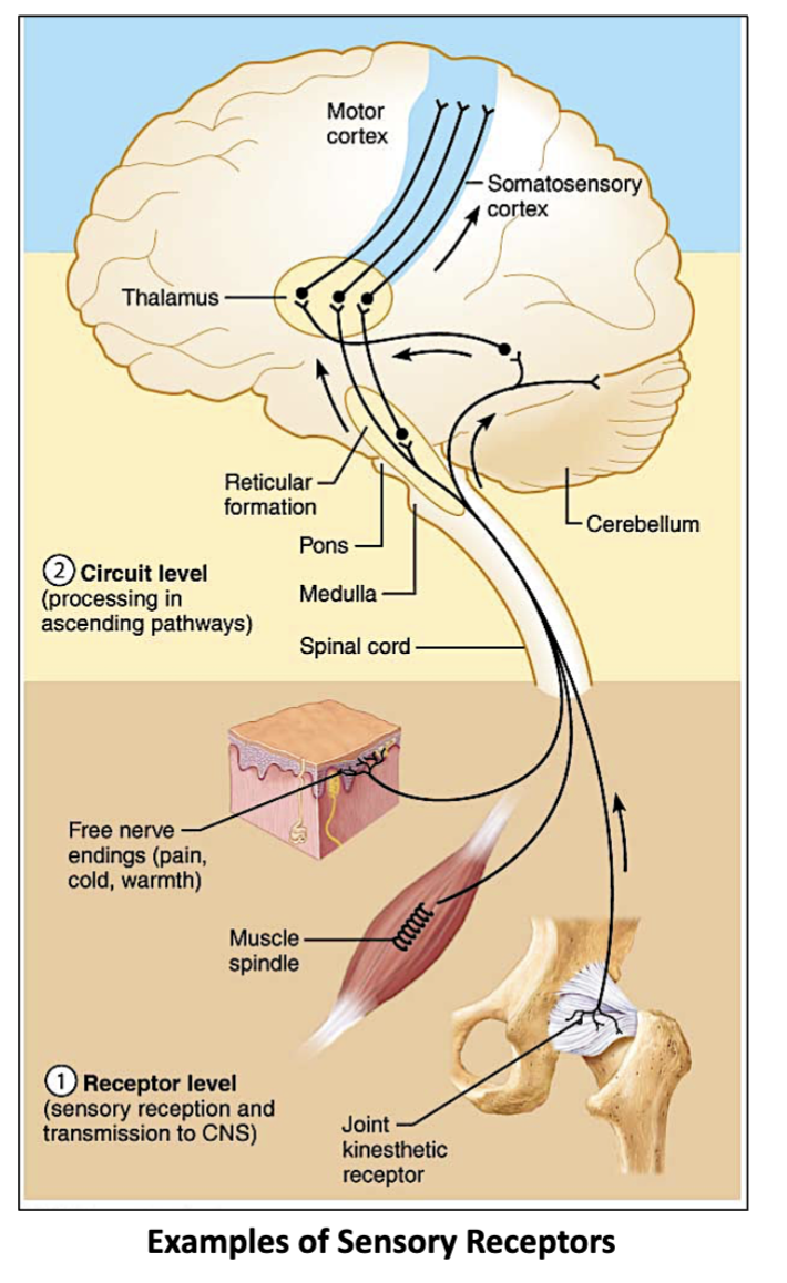
2 Examples of Sensory Receptors & what do they contain?
Cutaneous & Visceral Sensory Receptors
Cutaneous (skin) - Contains the…
Thermoreceptors which respond to temperature change
Mechanoreceptors respond to touch, pressure, and vibrations
Visceral (organs)- Contains the …
Chemoreceptors: respond to chemical changes in blood (like ph & blood)
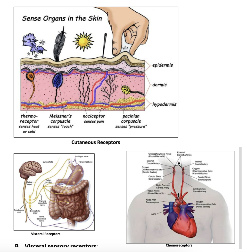
The Thermoreceptors & Mechanoreceptors are a part of which Sensory Receptor?
the Cutaneous Sensory Receptors, responsible for detecting temperature, touch, pressure, and vibrations.
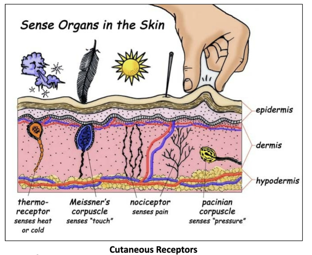
What is the Thermoreceptors job?
To detect temperature changes in the environment.
(part of the Cutaneous Sensory Receptors)
What is the Mechanoreceptors job?
To respond to touch, pressure, and vibrations.
(part of the Cutaneous Sensory Receptors)
What is the tongue’s surface covered by?
small elevations called tongue papillae.
Some papillae contain taste buds?
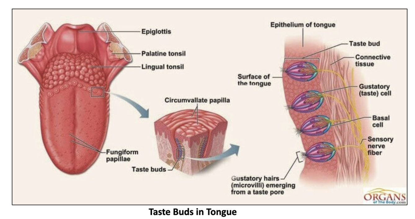
What does the tongue papillae contain?
taste buds that detect flavors.

What do taste buds contain?
specialized gustatory recpetors that respond to taste stimuli.

Where are the Gustatory (taste) receptor cells?
in taste buds.
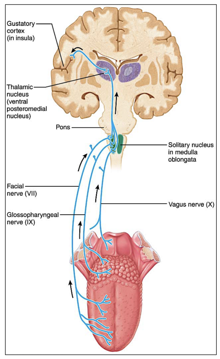
What do the Gustatory(taste) receptor cells activate?
Sensory neurons that are transmitted by facial (VII) 7 and
glossopharyngeal (IX) 9 nerves to brain.

Through which cranial nerves do Gustatory (taste) receptors send signals to the brain?
Facial (VII) and Glossopharyngeal (IX) nerves.
(7 & 9)

What is the Olfactory system in charge of?
the sense of smell, detecting and processing odors through olfactory receptors in the nasal cavity.
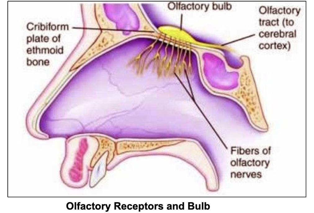
Where are the olfactory (smell) receptor neurons?
within the olfactory epithelium of the nasal cavity.

Definition of Olfactory Epithelium
the Olfactory Epithelium is the tissue inside the nasal cavity that detects smells
Where do the Axons of the Olfactory receptor neurons project through?
they project through the cribriform plate of the ethmoid bone and synapses with the neurons of the olfactory bulb

Where is the Cribriform plate?
located in the ethmoid bone
→ it separates the nasal cavity from the cranial cavity.

Axons of the ______ receptor neurons project through the ______ plate of the ______ bone and synapse with the neurons of the _______ bulb
Olfactory, cribriform, ethmoid, olfactory
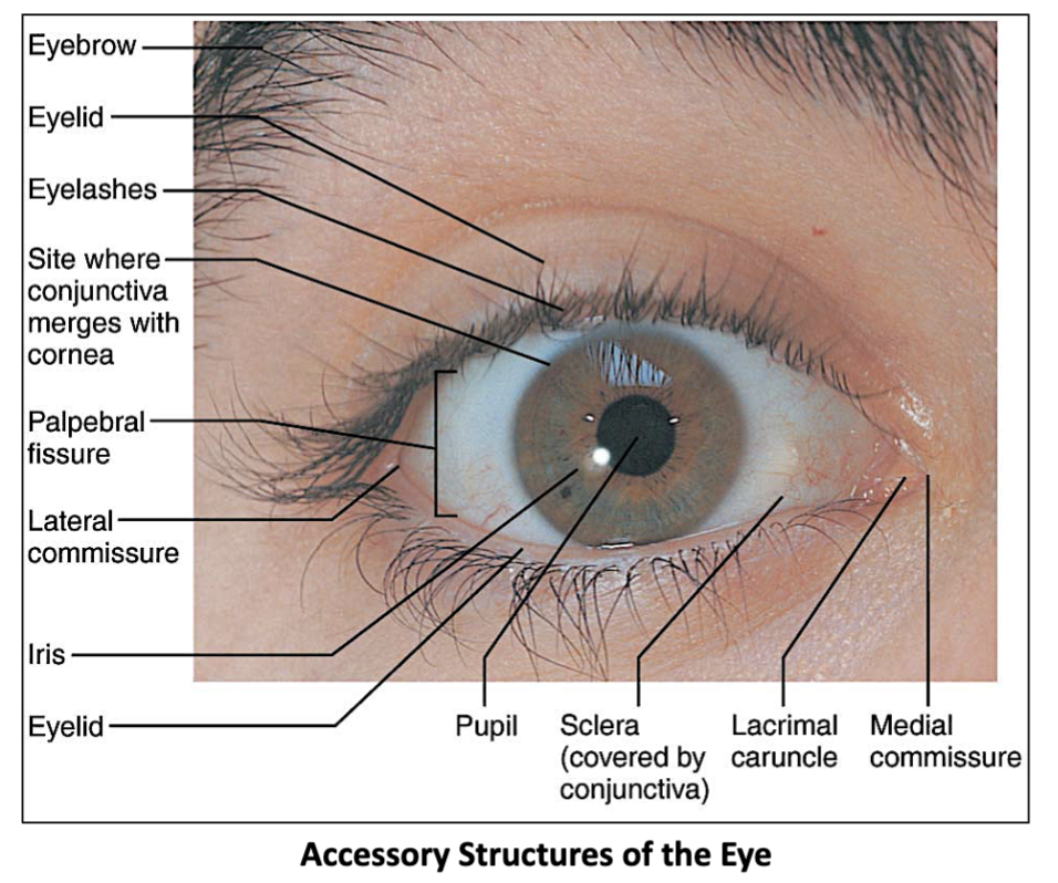
5 Accessory structure of the eye?
Eyebrows
Palpebrae (Eyelids)
Conjunctiva
Lacrimal Apparatus
Extrinsic Eye Muscles
3 Major parts of the Lacrimal Apparatus
Lacrimal gland (produces & secretes tears) → Excretory Lacrimal ducts → Nasolacrimal duct

Job of the lacrimal gland?
produces & secretes tears

Job of the Excretory Lacrimal ducts?
transport tears from the lacrimal gland to the nasal cavity
(also what covers the eye in tears)

6 step process of the lacrimal apparatus to produce tears
Lacrimal gland produces tears
Tears enter the conjunctival sac via the excretory ducts of the lacrimal gland
Tears flow down & across the eyeball
Tears enter the lacrimal canaliculi at openings called lacrimal puncta.
Tears drain into the Lacrimal sac
From the lacrimal sac, tears empty via the nasolacrimal
duct into the inferior meatus of the nasal cavity.

What is the lacrimal canaliculi?
Small channels that collect tears from the eyelids through openings called lacrimal puncta and channel them into the lacrimal sac.
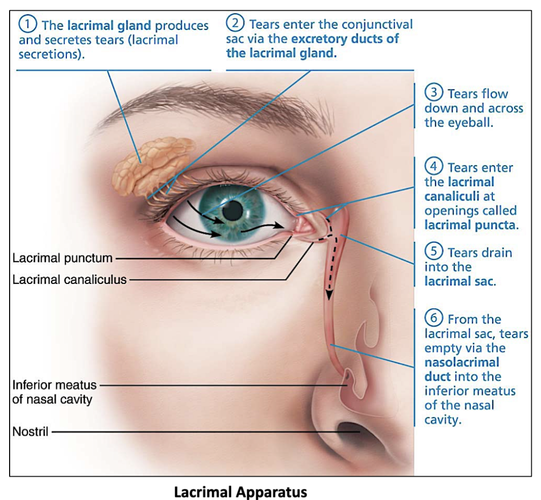
What is the lacrimal puncta?
Small openings on the eyelids that allow tears to go into the lacrimal canaliculi.

6 Extrinsic Eye Muscles
4 rectus - superior, inferior, medial & lateral
(moves eye in straight lines)
2 oblique - superior and inferior
(rotates eye)
- Eye muscles are innervated by Cranial nerves III, IV, and VI.
These muscles control eye movement.
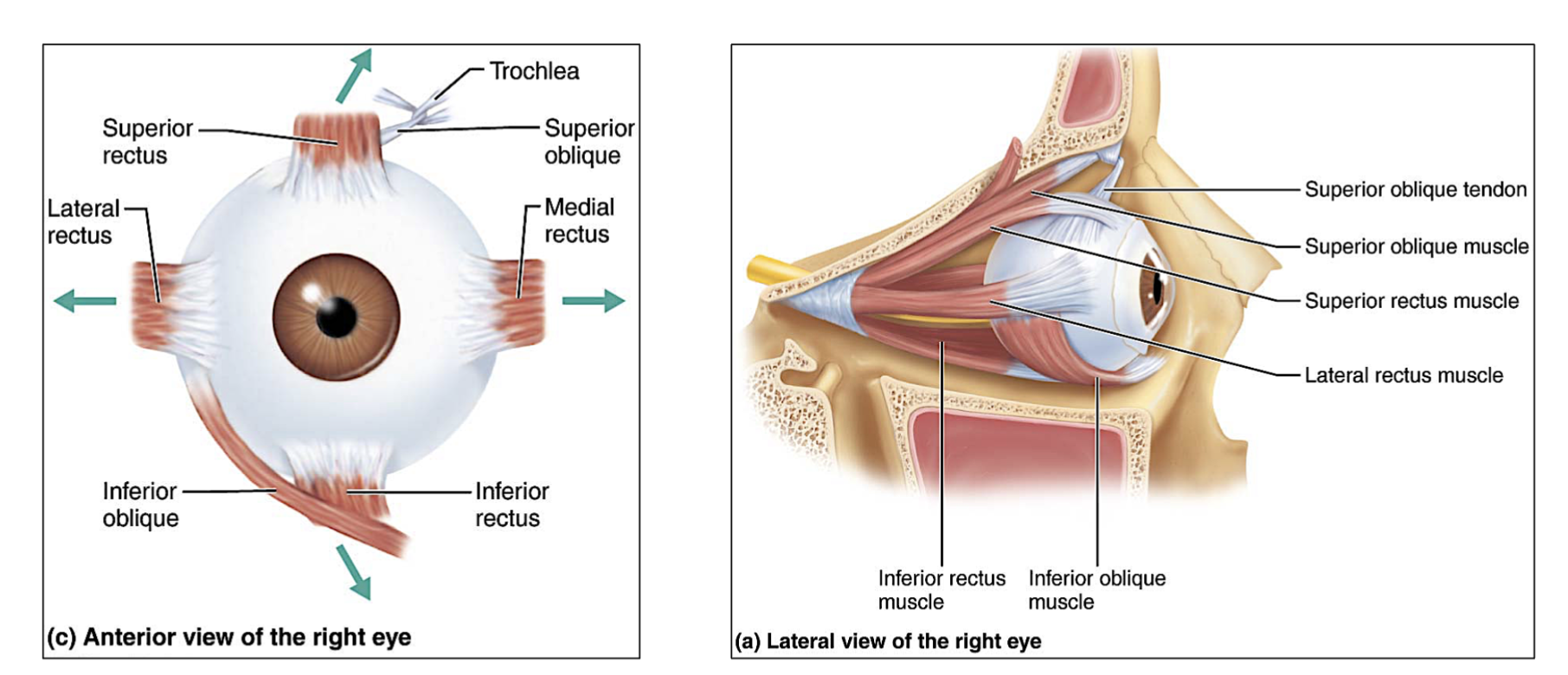
Which eye muscles moves eye in straight lines?
The 4 rectus muscles.
superior, inferior, medial & lateral

Which eye muscles rotates the eye?
The superior and inferior oblique muscles. (2 muscles)

Which cranial nerves control the eyes?
3 Cranial nerves→ III, IV, and VI. (3,4 & 6)
lll Oculomotor , IV Trochlear , lV Abducens
Which eye muscle is responsible for → helping you move the eye at angles and help it roll or stay steady when the head moves/rotates
the oblique muscles.(rotates the eye)
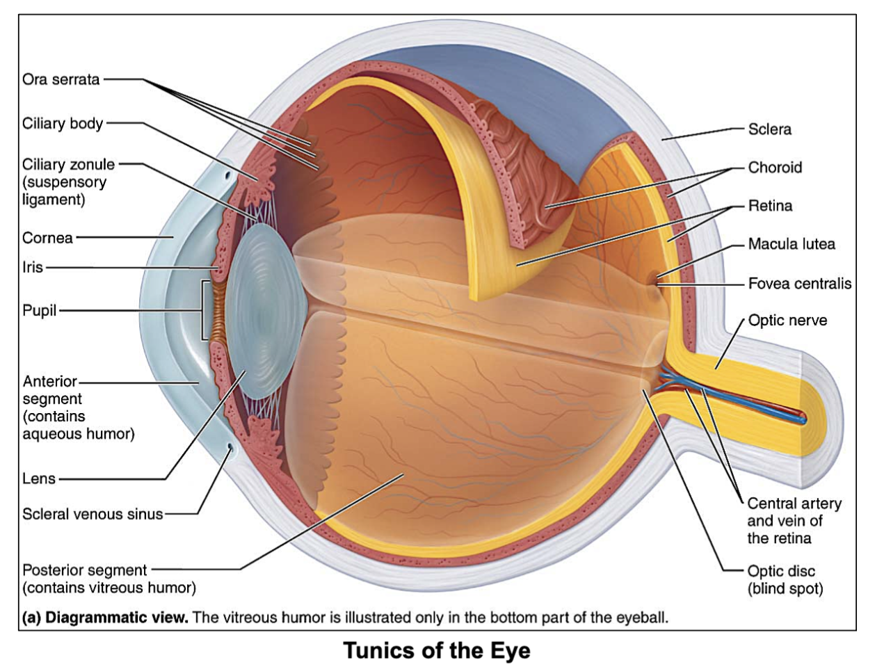
3 Tunics of the eye? (outermost to inner)
Fibrous Tunic (outermost)
Sclera - attachment of muscle
Cornea - transparent, lets light in
Vascular Tunic (Uvea/middle)
Choroid - under sclera
Ciliary body - secretes aqueous humor and holds lens in place
Iris : Color, changes the pupil size
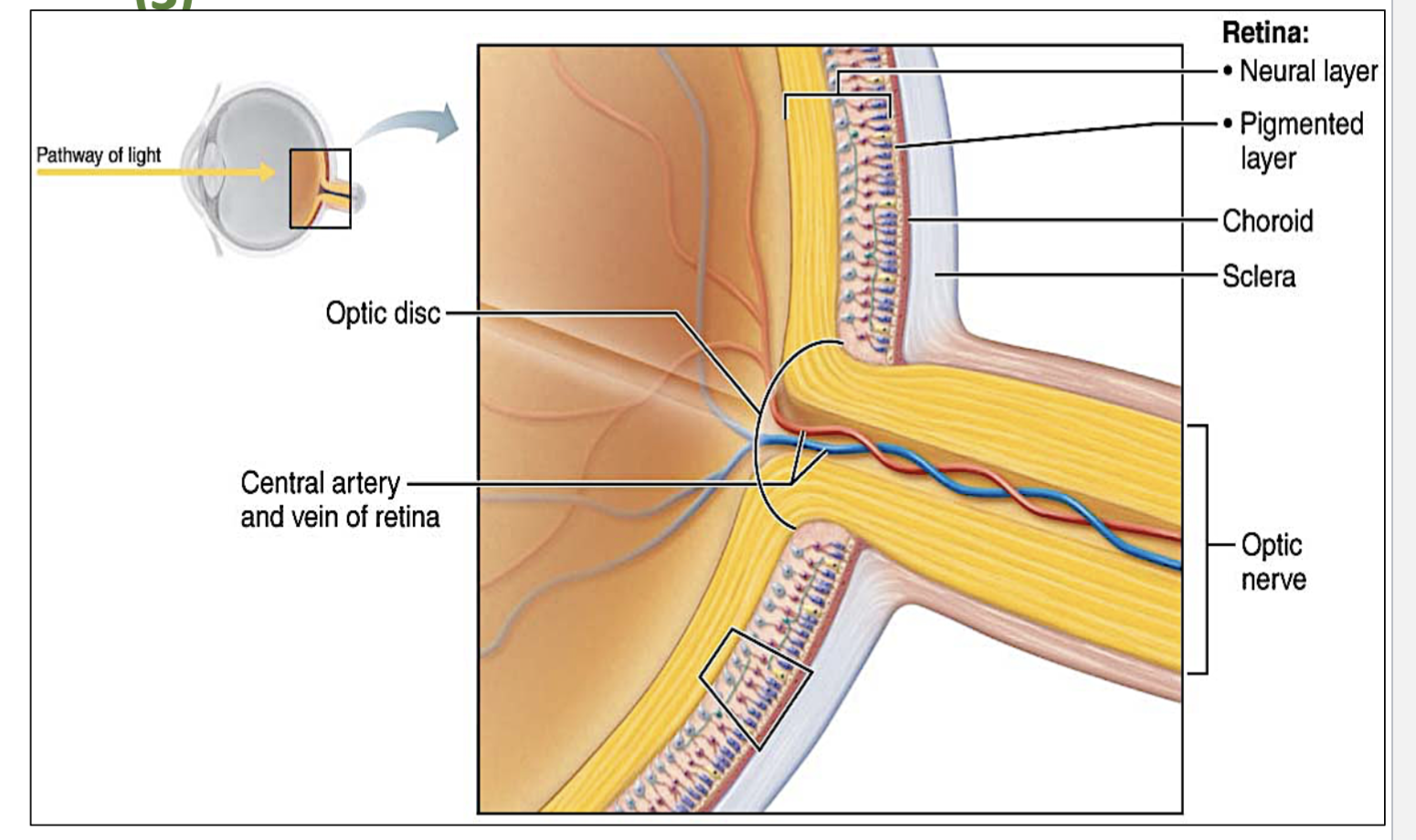
Sensory Tunic (retina)
pigmented layer (deepest part of the retina)
neural layer - becomes optic nerve
F.V.S
Tunic- layer/coat

What does the Fibrous Tunic layer of the eye contain?
this is the outermost layer of the eye, and it contains the Sclera & Cornea (transparent layer that let’s light in)

What does the Vascular Tunic layer of the eye contain?
this is the middle layer of the eye, and it contains the …
Choroid - under sclera
Ciliary body - secretes aqueous humor (in anterior & posterior chamber) and holds lens in place
Iris : Color, changes the pupil size
What does the Sensory Tunic layer of the eye contain?
this is the innermost layer of the eye, and it contains the ….
pigmented layer (deepest part of the retina)
neural layer - becomes optic nerve
What is the pupil?
the black hole in the iris.
What changes the size of the pupil depending on intensity of light?
the iris.

in which tunic layer of the eye is the retina located?
in the Sensory Tunic layer

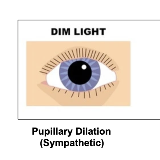
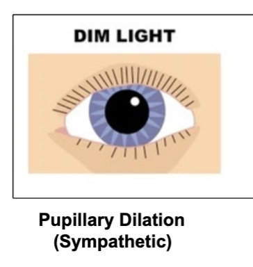
What happens to the pupil in dim light?
it dilates.
(photoreceptor rods are in charge of this)
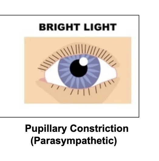
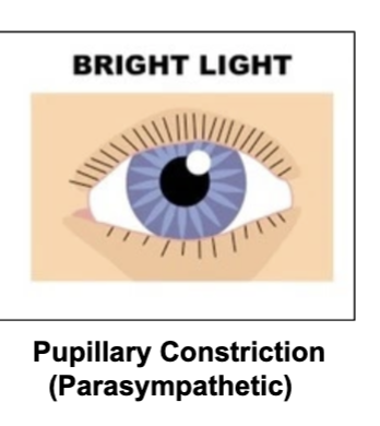
What happens to pupil in bright light?
it constricts
Is pupil dilation sympathetic or parasympathetic?
sympathetic
occurs when it is dark/dim outside to let more light in the eye and be able to see better.
(size of pupil is changed by the iris)
Is pupil constriction sympathetic or parasympathetic?
parasympathetic
occurs in bright light to reduce light entering the eye and improve visual acuity.
(size of pupil is changed by the iris)
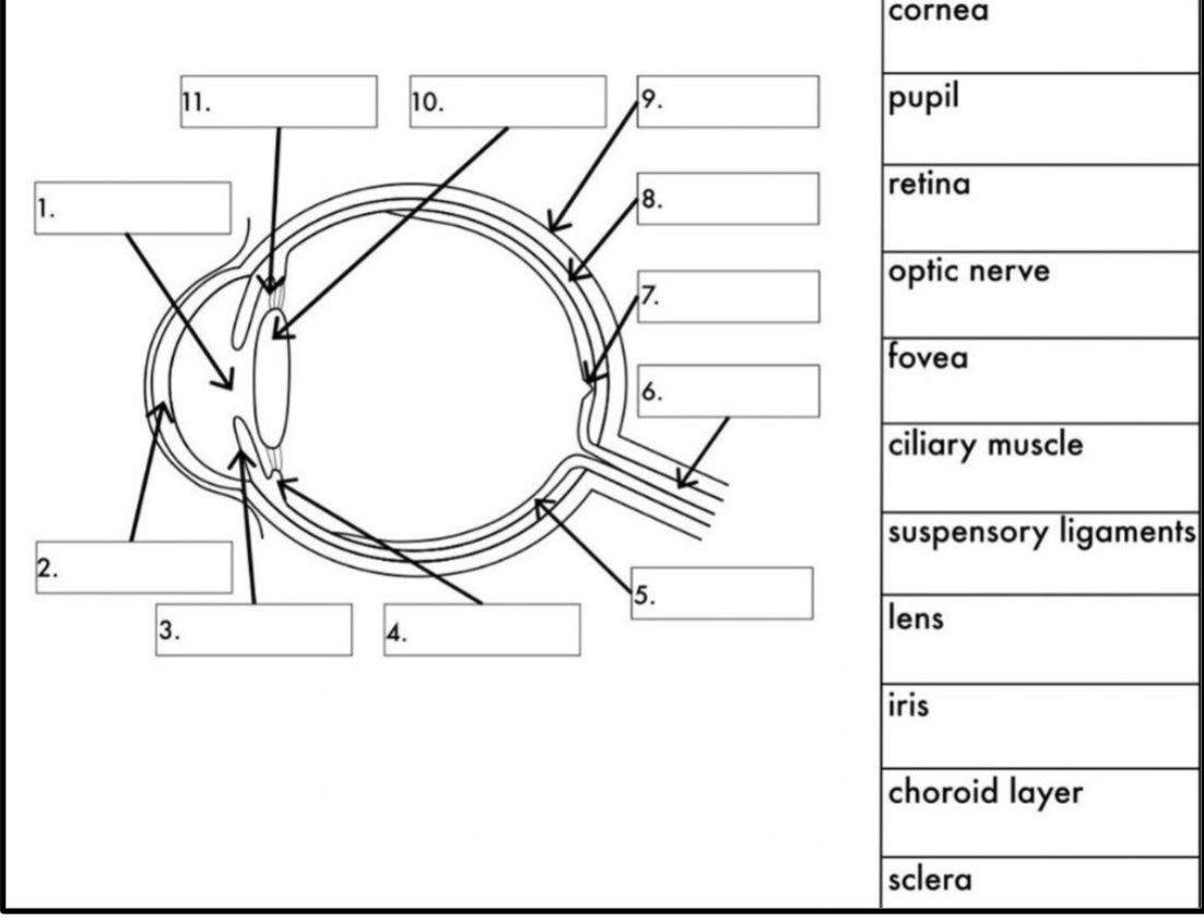
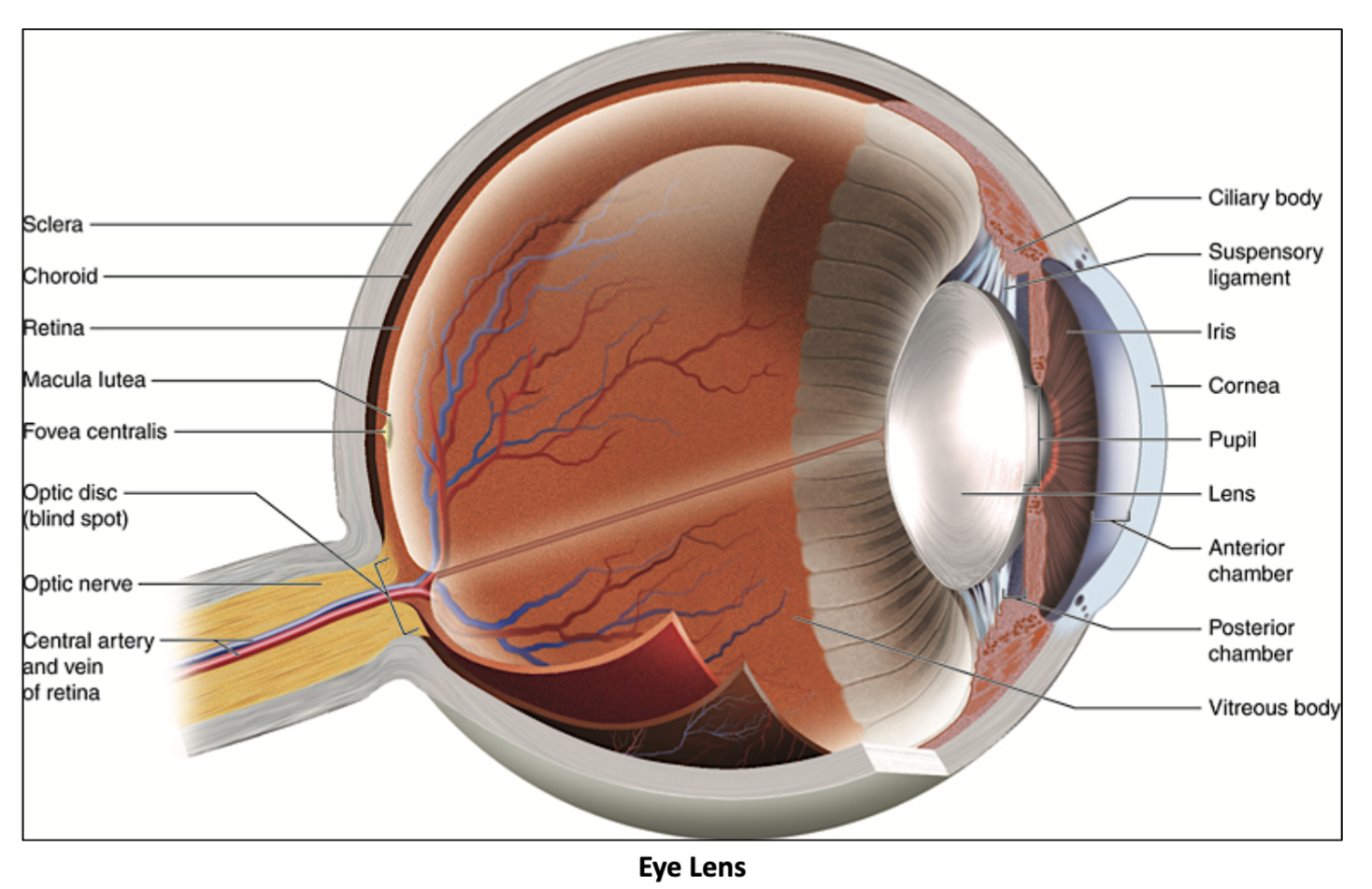
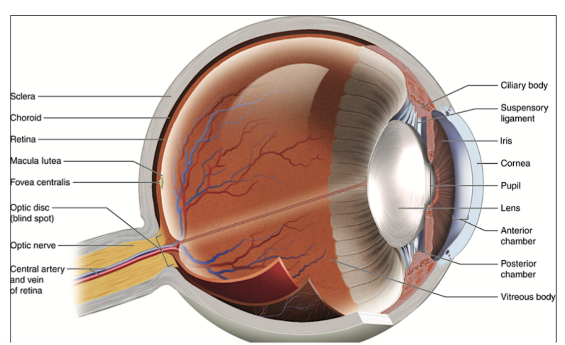
Label all the eye areas!
pupil
cornea
iris
ciliary muscle
retina
optic nerve
fovea
choroid (under sclera)
sclera
lens
suspensory ligaments
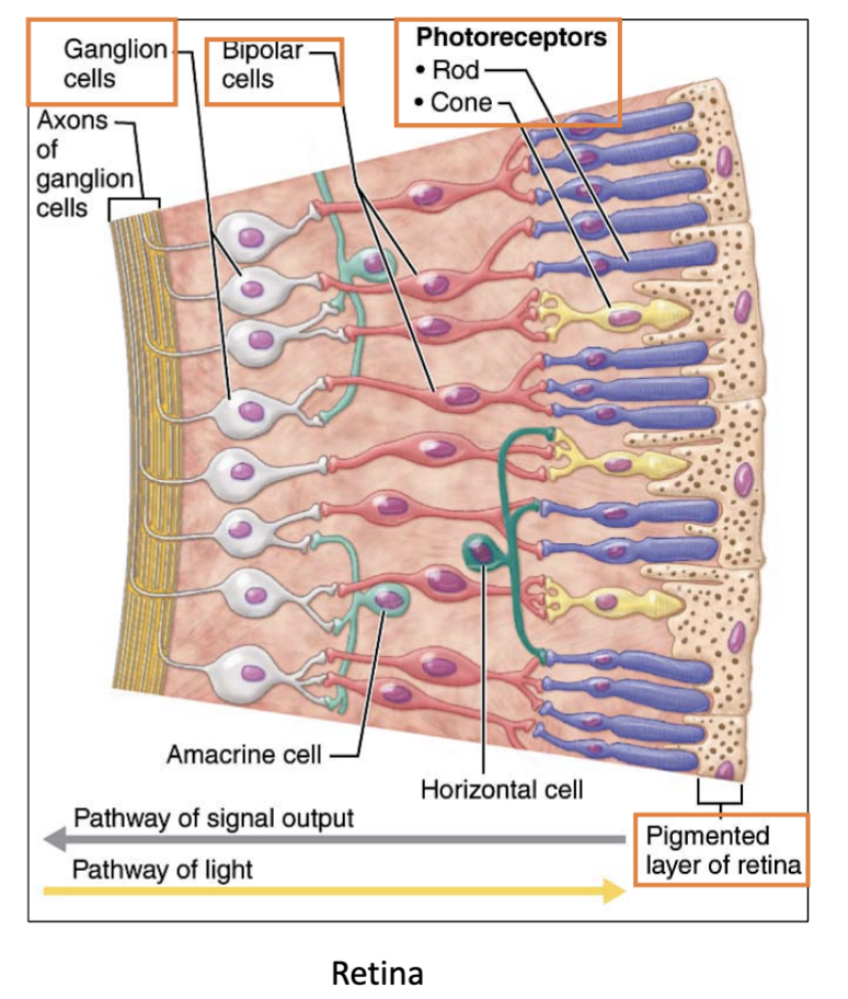
What happens at the Retina?
The retina converts light into neural signals (AP) for the brain to process images.

What’s the name of the layer that becomes the optic nerve?
The neural layer
→ part of the retina that contains the photoreceptors (rods and cones), bipolar cells, and ganglion cells (Axons of ganglion cells form the optic nerve)
What’s the name of the layer of the Retina that contains the photoreceptors (rods and cones), bipolar cells, and ganglion cells & also becomes the optic nerve?
The neural layer
(layer responsible for converting light into neural signals)

The axons of _____ cells all come together at the back of the eye, bundle up, and form the optic nerve, which carries the visual signals to the brain.
ganglion
Where is the pigmented layer in the retina & Whats it’s job?
The pigmented layer is located underneath the neural layer (innermost layer) of the retina, & it absorbs excess light

Conversion of light energy into (AP) action potentials occurs in the ______.
(a layer of the eye)
Retina

What are the 2 photoreceptors in the retina?
Rods & Cones
(there’s way more rods than cones)

What is the job of the Photoreceptor cells?
absorb light and convert it into electrical signals (AP) that can be processed by the bipolar cells to the Ganglion cells in the brain, allowing for vision.
The ______ layer of the Retina absorbs excess light.
Pigmented layer

Photoreceptor cells absorb light and generate electrical signals to the _____ cells & then to the _____ cells.
bipolar, ganglion

Axons of _____ cells form the optic nerve & then go to the primary ____ cortex in _____ lobe of the Cerebrum.
ganglion, visual, occipital

What cells receive signals from photoreceptors & where do they go after?
These signals go to bipolar cells, which pass them to ganglion cells.
The axons of ganglion cells bundle together to form the optic nerve, which carries the signals to the primary visual cortex in the occipital lobe for processing

What forms the optic nerve?
Axons of ganglion cells.

Where does the optic nerve send signals?
The optic nerve sends signals to the primary visual cortex in the occipital lobe for processing.

Where is the primary visual cortex located?
In the occipital lobe of the cerebrum.
Which layer does light reach first in the retina?
the deepest layer (the pigmented layer)
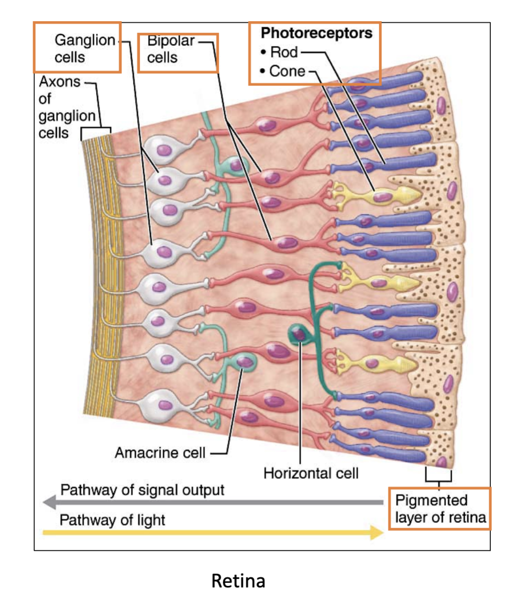
What is the correct order of the pathway of light in the retina? (5 steps)
(Light will reach the deepest layer first)
Pigmented layer → Photoreceptors (rod & cone) → Bipolar cells (pink) → Ganglion cells (white) → Axons of ganglion cells which form the optic nerve

What is the first layer light reaches in the retina?
Pigmented layer

After the pigmented layer, what cells does light hit next?
Photoreceptors (rod & cones)

What cells receive signals from photoreceptors?
Bipolar cells (long pink)

What cells receive input from bipolar cells?
Ganglion cells (white)

What do the axons of ganglion cells form?
The optic nerve.

What is the Fovea of the eye rich in?
Cones, which are responsible for sharp, detailed, and color vision.
What type of light activates rods?
rods are activated by dim light (night vision)
Which photoreceptor is in charge of night vision?
Rods are the photoreceptors responsible for night vision.
What type of light activates cones?
Bright light (day vision)
What kind of images do rods produce?
Fuzzy, indistinct images.
What kind of images do cones produce?
Sharp images with high acuity.
Do rods provide color vision?
No, only gray tones.
Do cones provide color vision?
Yes
Where are rods located in the retina?
Rods are primarily located in the peripheral regions of the retina, which enhances night vision and detects motion but does not contribute to color perception.
Where are cones located in the retina?
In the fovea centralis.
Which is more numerous: rods or cones?
Rods
What photopigment do rods contain?
Rhodopsin that needs Vitamin A to functions
There are ____ in the periphery of the retina.
(photoreceptors)
rods
There are more ____ than ____ in the fovea.
(photoreceptors)
Cones, rods
What vitamin is needed for rhodopsin to function?
Vitamin A.
( rhodopsin is the photopigment that rods contain)
What happens to your vision if there is a deficiency of vitamin A?
affects rods, so impaired night vision.
(won’t be able to see much in the dark)
What is the job of the Eye lens?
Focuses light and objects onto the retina
Which eye structure is flexible, and convex on both sides?
The eye lens
Which eye structure is made up oftTransparent protein fibers called crystallins?
The eye lens

Which Eye structure is Attached to ciliary body by suspensory ligament?
The eye lens
Which eye structure has the job of focusing light and objects onto the retina?
Eye lens

What is the shape of the eye lens?
Flexible and Convex (rounded) on both sides

What is the eye lens made of?
Transparent protein fibers called crystallins

How is the eye lens held in place?
By the suspensory ligament (ciliary zonule) attached to the ciliary body.

What structure helps adjust the shape of the lens?
The ciliary body, through the contraction and relaxation of its muscles, adjusts the shape of the lens for focusing.

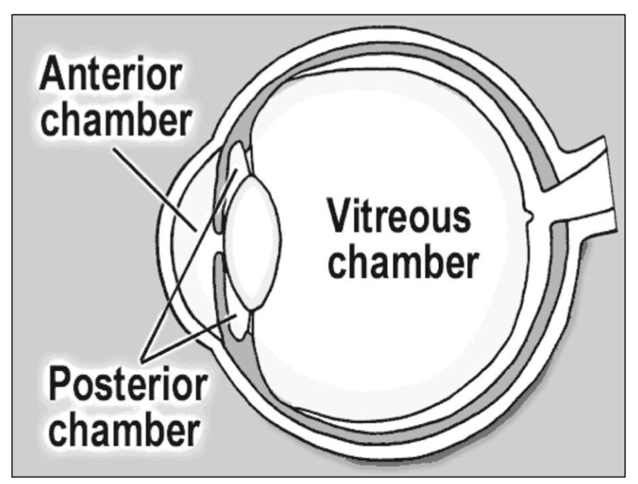
What are the 3 Chambers of the eye?
Anterior chamber is anterior (in front) to iris: filled with aqueous humor (watery transparent)
Posterior chamber is between iris and lens: aqueous humor (watery transparent)
Vitreous chamber is posterior (jn back) to lens: in vitreous humor (clear gelatinous)

What is the Aqueous humor?
A watery fluid that fills the anterior & posterior chambers of the eye, providing nutrients and maintaining intraocular pressure.
(formed by the ciliary body)
What is the Vitreous humor?
A clear gelatinous substance that fills the Vitreous chamber of the eye, providing structure and support.

What forms the Aqueous Humor?
primarily formed by the ciliary body, which secretes the fluid into the posterior chamber.
What does the Cilliary body form?
Aqueous Humor, which fills the anterior and posterior chambers.
anterior → in front of iris
posterior → between iris & lens
What is the Circulation of Aqueous Humor?
(5 steps)
Formed by ciliary body → posterior chamber → pupil → anterior chamber → reabsorbed by venous sinus

What reabsorbs the aqueous humor?
Venous sinus, or Schlemm's canal.
What is the Venous Sinus?
drains aqueous humor from the anterior chamber into the bloodstream, helping to regulate eye pressure.
What are Cataracts?
Clouding of the lens, which becomes opaque with age
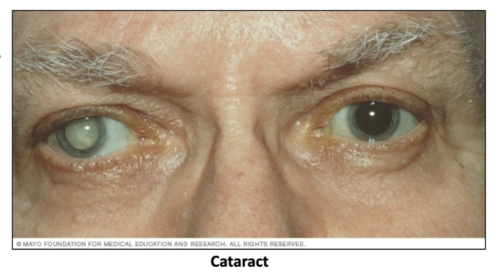
What can cause cataracts?
Diabetes, smoking, drugs, ultraviolet radiation, and certain viruses

How is a cataract treated?
By replacing the natural lens with a plastic one
What is glaucoma?
Elevated pressure within the eye due to improper drainage of aqueous humor