PSYC327 Test 1
 Call Kai
 Learn
Learn Practice Test
 Spaced Repetition
 Match
Match Flashcards
 Knowt Play
1/63

There's no tags or description
Looks like no tags are added yet.
Name | Mastery | Learn | Test | Matching | Spaced | Call with Kai | Chat |
|---|
No analytics yet
Send a link to your students to track their progress
64 Terms
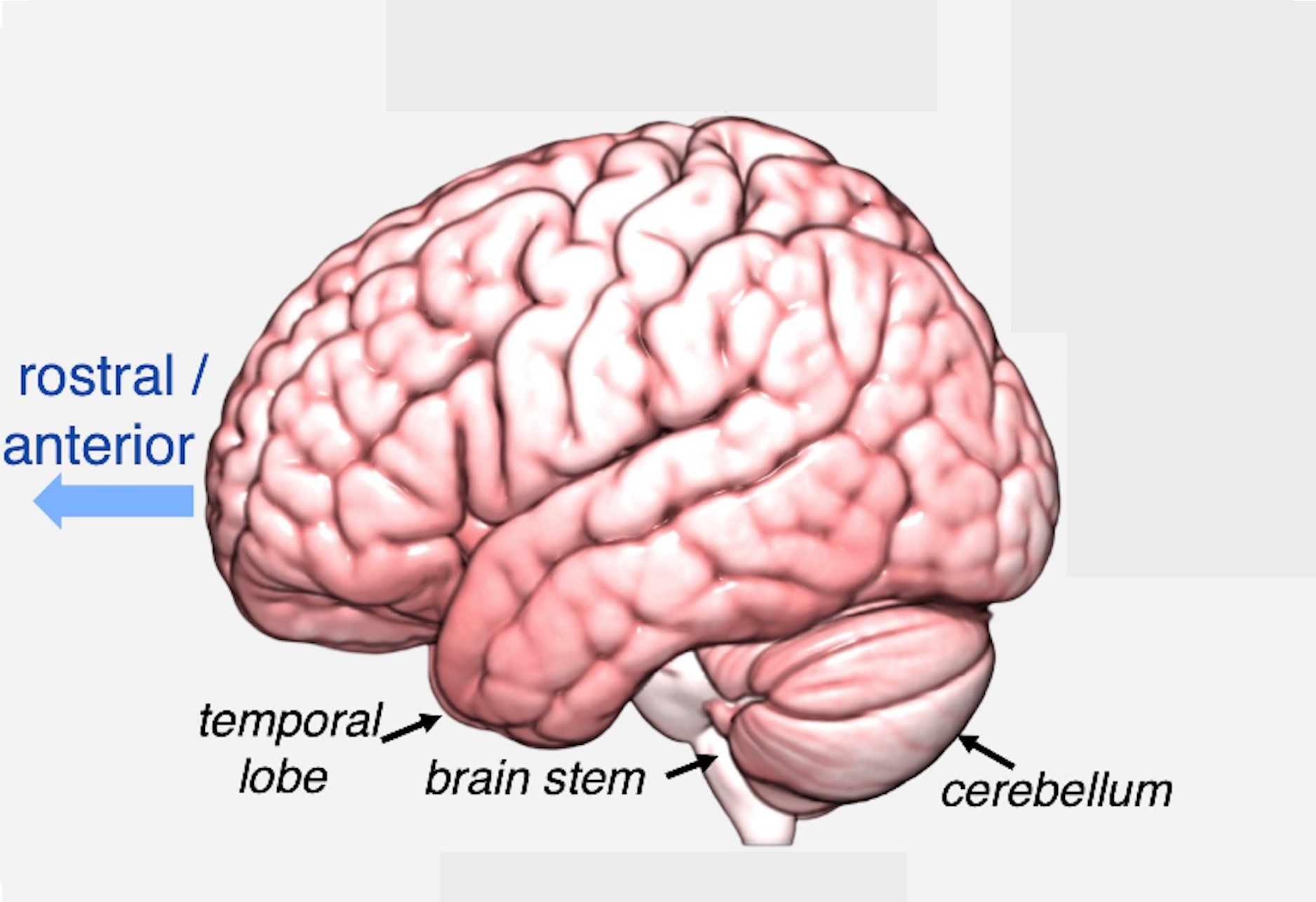
Anterior
The term rostral is sometimes used instead of anterior (for example, the rostral prefrontal cortex refers to the anterior tip of the frontal lobes). This is the forward part of the brain
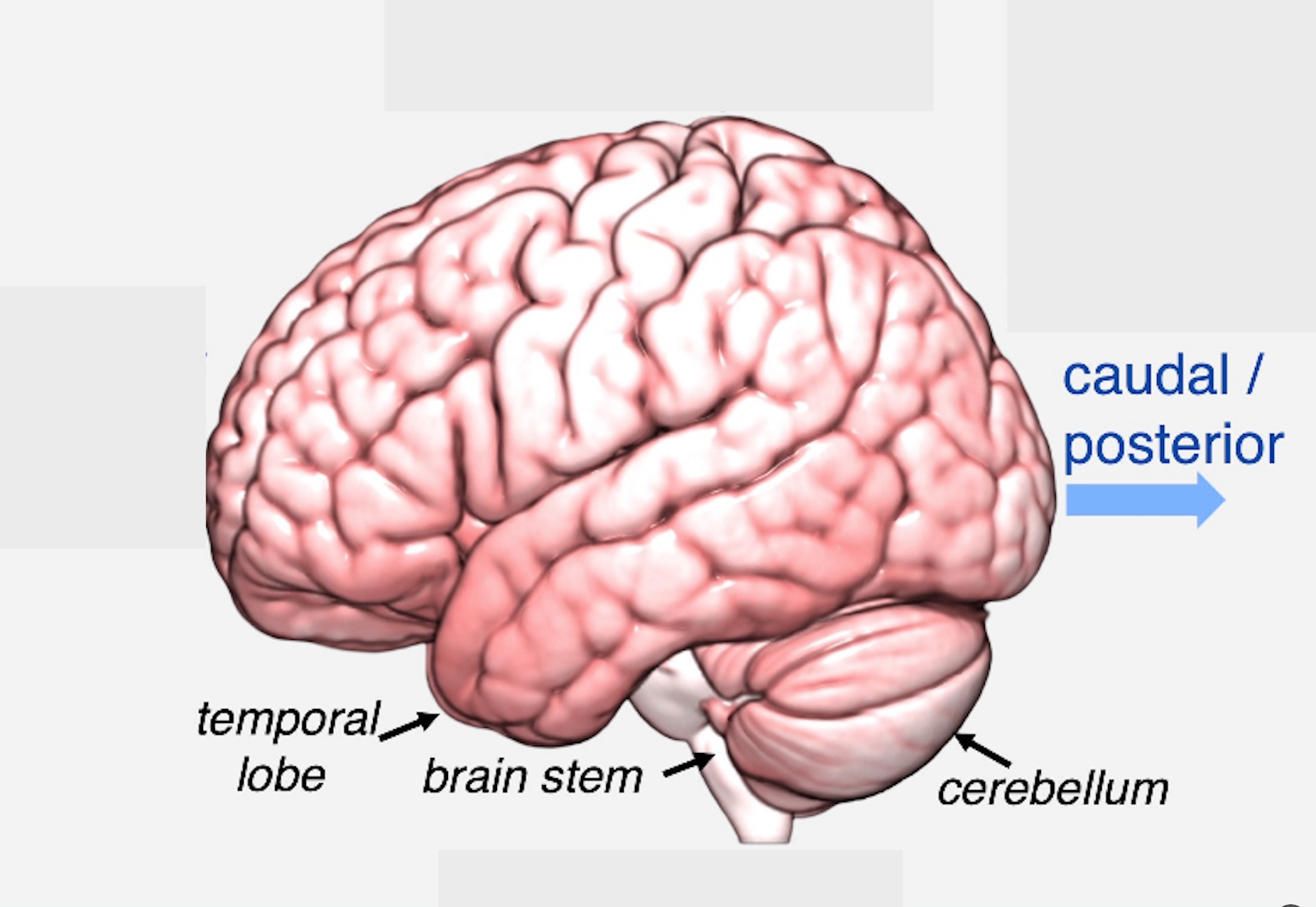
Posterior
The term caudal is sometimes used instead of posterior. This is the back part of the brain
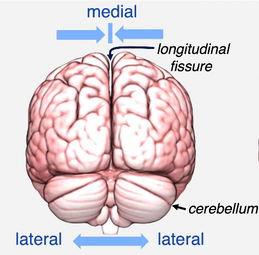
Posterior view
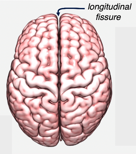
Dorsal view
What is the “seductive allure” of neuroscience explanations?
People tend to find explanations containing neuroscience information more satisfying, even when the neuroscience information does not actually improve the explanation. It can create an illusion of understanding.
What is the difference between structural MRI and fMRI?
Structural MRI (T1) examines the physical structure/anatomy of the brain. fMRI examines changes associated with brain activity, typically using the BOLD signal.
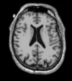
What does a T1-weighted MRI measure?
It uses magnetic properties of different tissues to produce detailed images of brain structure, allowing researchers to identify things such as tissue loss, tumours, stroke damage and enlarged ventricles.
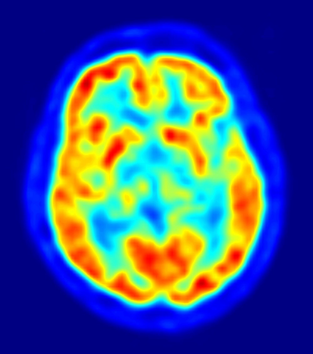
What is PET and what does it measure?
Positron Emission Tomography (PET) uses a radioactively tagged tracer to measure the uptake/metabolism of substances such as glucose. It can therefore provide information about brain function/metabolism.
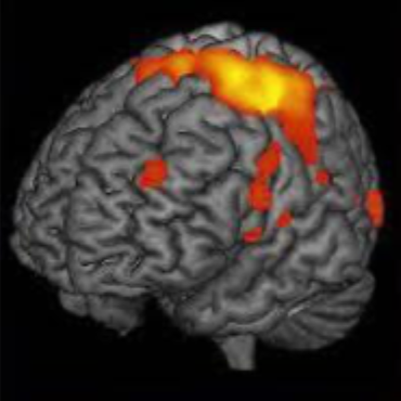
What is the BOLD signal?
Blood Oxygen Level Dependent signal. fMRI uses changes in blood oxygenation as an indirect proxy for neural activity. More active regions generally produce greater changes in oxygen use.
What is the reverse inference problem?
Inferring a particular mental process from activation in a particular brain region. This is problematic because brain regions generally support multiple functions. Knowing where activation occurs does not necessarily tell us what psychological process is occurring
Why can't fMRI establish causation?
fMRI shows that brain activity is associated with a task or behaviour. It does not demonstrate that activity in that region causes the behaviour or psychological process.
Why is task design so important in functional neuroimaging?
What brain activity researchers observe depends on the comparison between tasks/conditions. Carefully designed contrasts allow researchers to isolate the psychological process of interest and minimise unrelated processes.
What is the lesion method?
A method of studying brain function by examining people with brain damage. If damage to a particular region produces an impairment in a particular ability, this provides evidence that the region contributes to that function.
What is a double dissociation?
When two patients show opposite patterns of impairment across two tasks. For example, H.M. had impaired long-term memory but intact short-term memory, while E.E. showed the opposite pattern. This provides strong evidence for separate cognitive systems.
What did H.M.'s case demonstrate?
H.M. had severe difficulty forming new long-term memories after medial temporal lobe/hippocampal removal, while his short-term memory remained relatively intact. This provided evidence for separate memory systems.
What did E.E.'s case add to our understanding of memory?
E.E. had impaired digit span/short-term memory but relatively intact long-term memory. Combined with H.M., this created a double dissociation, strengthening evidence that short- and long-term memory rely on different systems.
What is a major limitation of naturally occurring brain lesions?
Brain damage is messy and non-random. Lesions can vary in size/location and may damage connections between regions. Patients can also differ in premorbid characteristics and compensatory changes.
What is lesion mapping/VLSM?
Voxel-based lesion-symptom mapping (VLSM) examines many patients with brain damage and determines whether damage to particular voxels/regions is associated with poorer performance on a task.
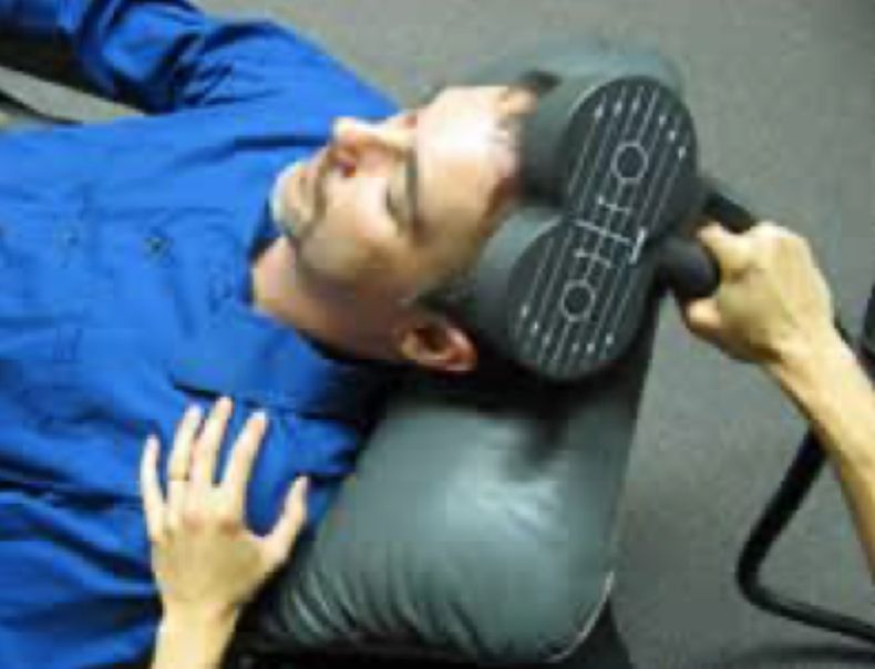
What is TMS?
Transcranial Magnetic Stimulation uses a powerful magnetic pulse applied over the scalp to temporarily disrupt activity in a relatively localised brain region, producing a temporary “lesion.”
What are the main strengths of TMS?
Researchers can randomly select healthy participants, temporarily disrupt a targeted region, stimulate areas that are rarely damaged naturally, and compare participants' performance with themselves.
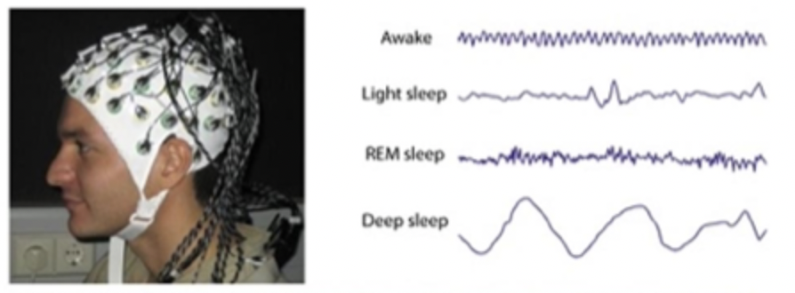
What does EEG measure?
EEG measures the summed electrical activity of large populations of neurons using electrodes placed on the scalp. It primarily reflects postsynaptic activity rather than individual neuronal action potentials.
What is EEG's major strength and weakness?
Strength: excellent temporal resolution — it can detect changes occurring within milliseconds. Weakness: relatively poor spatial resolution, making it difficult to determine exactly where activity originates.
What is an ERP?
An Event-Related Potential (ERP) is an averaged EEG response associated with a particular sensory, cognitive or motor event. Examples include P1/N1, P3, N2PC, MMN and ERN.
What is the method of converging operations?
Using multiple complementary methods to investigate the same psychological question. If different methods produce converging evidence, confidence in the conclusion increases because each method has different strengths and limitations.
What are the main differences between rods and cones?
Rods: ~120 million, concentrated in peripheral retina, highly sensitive to low light, support low-resolution/colourless vision. Cones: ~6 million, concentrated in the fovea, require more light, support high-acuity and colour vision.
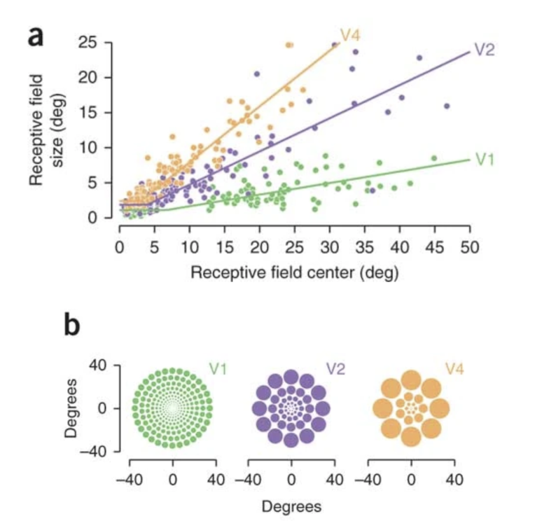
What is a receptive field?
The region of visual space in which stimulation produces a response in a neuron. Receptive fields allow researchers to determine what aspects of visual stimulation a neuron responds to.
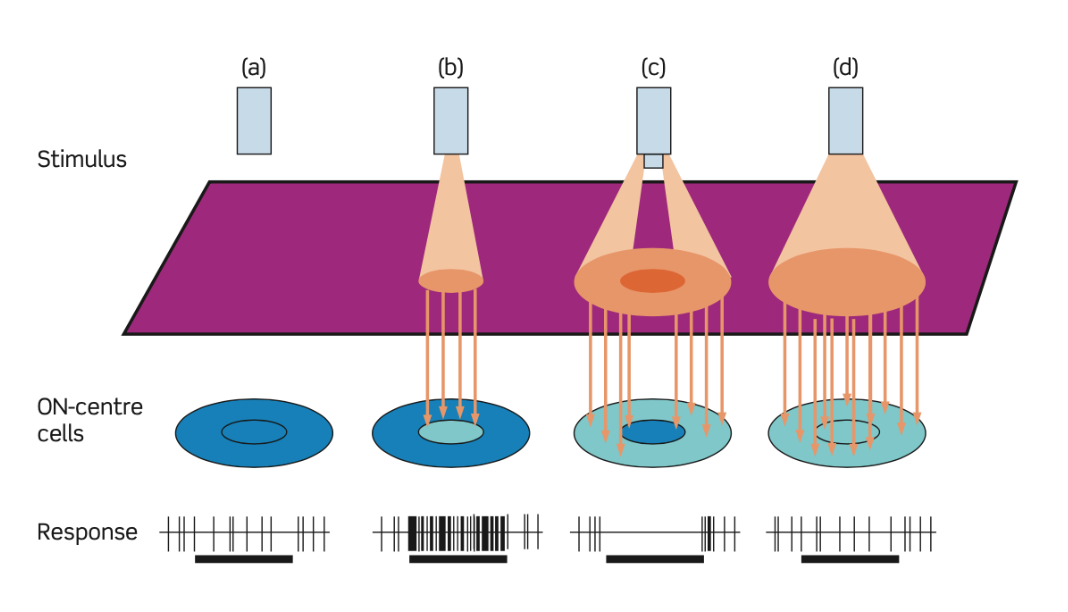
What is centre-surround organisation?
Retinal ganglion cells have receptive fields divided into a centre and surround with opposing effects. For an ON-centre/OFF-surround cell, light in the centre increases firing while light in the surround decreases firing.
What is lateral inhibition and why is it important?
Activity in one visual pathway can inhibit neighbouring pathways. This enhances differences in stimulation and helps the visual system detect edges and contrast rather than simply overall brightness.

What is retinotopy?
The organisation whereby neighbouring locations in the visual field are represented by neighbouring locations in visual cortex. V1 therefore contains a spatial map of the visual field.
What does contralateral organisation mean in V1?
Each hemisphere primarily processes the opposite visual field: left V1 → right visual field, while right V1 → left visual field. Importantly, this refers to visual fields, not simply the opposite eye.

What is cortical magnification?
The central visual field/fovea receives a disproportionately large representation in V1. This helps explain why we have much greater visual acuity in central than peripheral vision
What are simple cells?
V1 neurons that respond strongly to an oriented edge or bar at a particular position within their receptive field. They are constructed by pooling aligned inputs from LGN cells.
What are complex cells?
V1 neurons that prefer a particular orientation but can respond when the stimulus occurs at different positions within their receptive field. This provides early position invariance.
What is a tuning curve?
A graph showing how strongly a neuron responds to different stimulus values. For V1 orientation-selective cells, responses are strongest at a preferred orientation and gradually decrease as orientation deviates from that preference.
What is blindsight?
A condition in which a person has no conscious visual awareness in part of their visual field but can nevertheless perform certain visual tasks above chance. It demonstrates that visual processing can occur without conscious awareness.
What does blindsight tell us about V1?
V1 appears to be particularly important for conscious visual awareness, but it is not necessary for all visual processing. Other visual pathways can bypass V1 and support some visual behaviours.
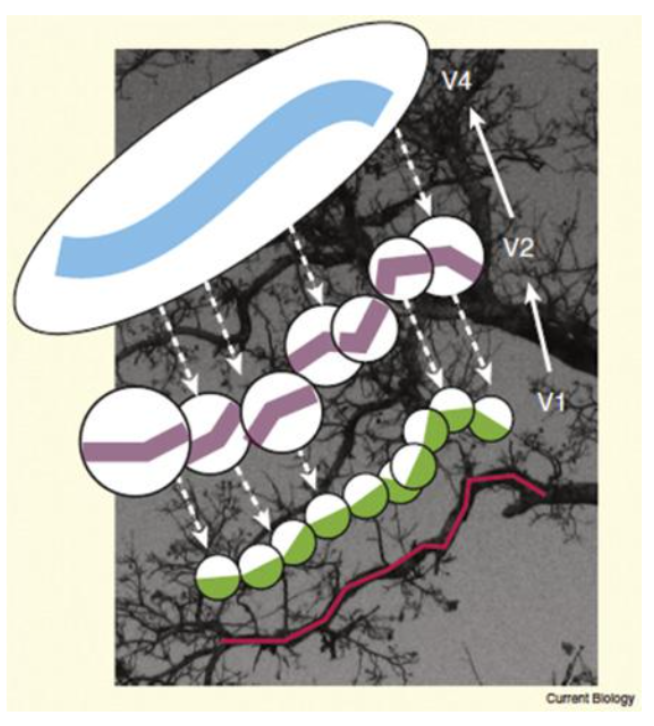
What is hierarchical processing in vision?
Visual processing becomes increasingly complex and integrated as information moves from early areas such as V1 to higher visual areas. Receptive fields generally become larger and neurons respond to increasingly complex stimulus properties.
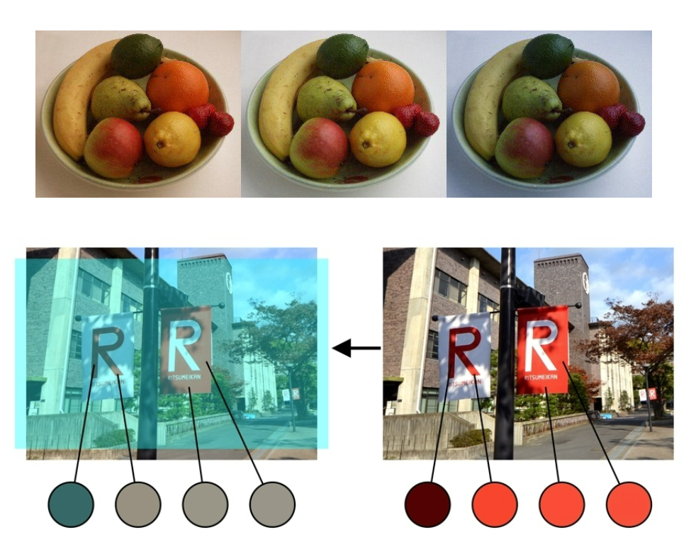
What is colour constancy?
The tendency to perceive an object's colour as relatively stable despite changes in illumination. Higher visual areas such as V4 are more sensitive to perceived colour than simply the physical wavelength of light.
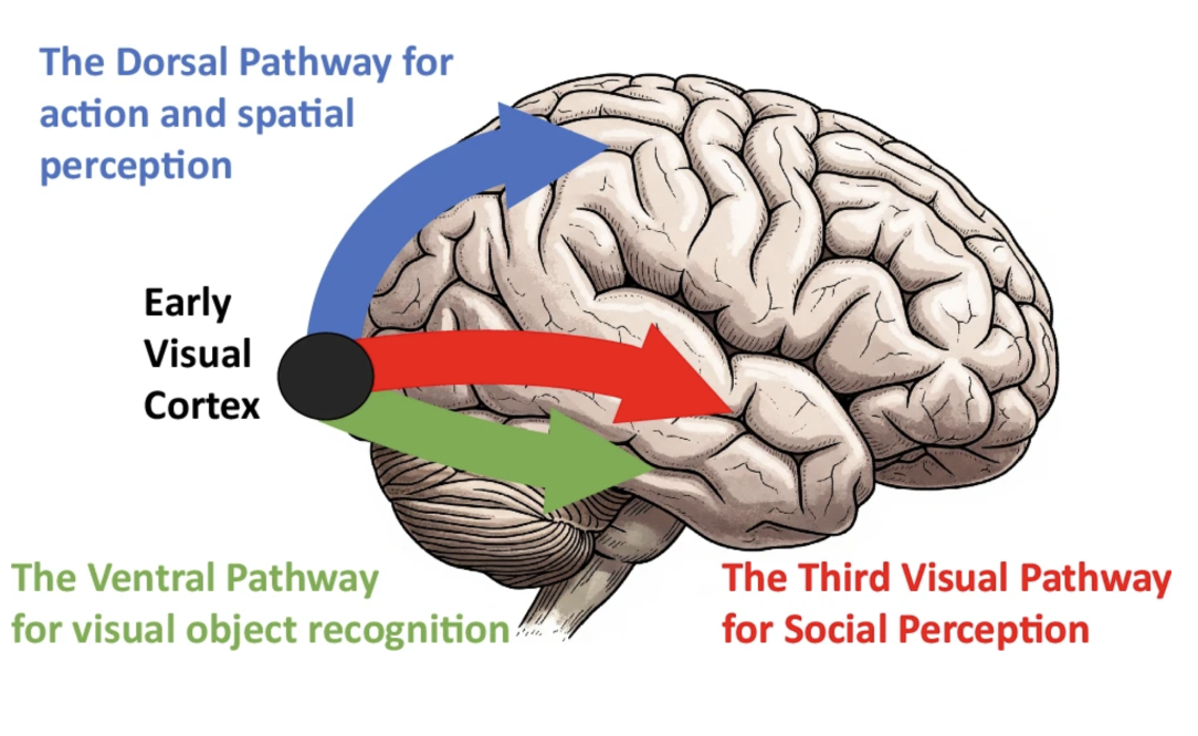
What are the ventral and dorsal visual streams?
Ventral/"what" stream: involved in identifying and recognising objects. Dorsal/"where/how" stream: involved in spatial processing and visually guided actions such as reaching.
What is visual agnosia?
Difficulty recognising or identifying objects despite relatively intact basic visual abilities. It can result from damage to higher-level visual processing, particularly the ventral pathway.
What is the difference between apperceptive and associative visual agnosia?
Apperceptive: difficulty constructing/perceiving the object's visual form. Associative: visual perception of the object is relatively intact, but the person cannot connect it with stored knowledge/meaning.
What is optic ataxia?
Difficulty using visual information to guide movements toward objects, despite relatively intact visual perception. It is associated with disruption of dorsal-stream processing.
What is prosopagnosia and what does it tell us about face perception?
Prosopagnosia is impaired face recognition despite relatively intact basic vision. Its selective nature suggests that face processing involves specialised neural systems, including the face-selective network/FFA.
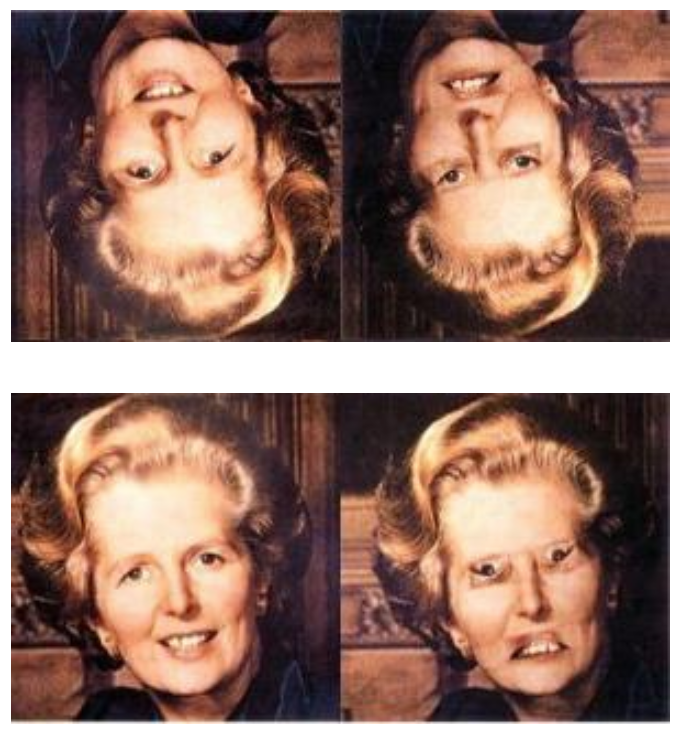
What is holistic/configural face processing?
Faces tend to be processed as an integrated whole, with relationships between features such as the eyes, nose and mouth being particularly important. This differs from how many ordinary objects are processed and helps explain the distinctive characteristics of face perception.
What is the primary function of the dorsal visual stream?
Processing spatial information, including object location, spatial relationships, movement, navigation, and visually guided actions.
What is the key difference between the dorsal and ventral visual streams?
Dorsal stream: where/how — location, movement, spatial relationships and action.
Ventral stream: what — colour, form and object identity.
What is an egocentric spatial representation?
A representation of an object's location relative to oneself (e.g., "the cup is to my left").
What is an allocentric spatial representation?
A representation of spatial relationships between objects, independent of one's own position (e.g., "the cup is beside the laptop").
Why is spatial processing necessary when we move?
Movement changes the position of objects on the retina. The brain must integrate visual information with eye, head and body position to maintain a stable representation of the environment.
What is optic ataxia?
A disorder involving difficulty reaching accurately toward visually perceived objects, despite relatively intact visual perception.
What does the case of optic ataxia tell us about the dorsal stream?
It demonstrates that the dorsal stream contributes to visually guided actions, particularly transforming visual information into appropriate movements.
What is mental rotation?
The ability to mentally manipulate or rotate an object to determine whether it matches another object.
What happens to reaction time as the amount of rotation increases in mental rotation tasks?
Reaction time generally increases as angular disparity increases, suggesting that participants mentally rotate the object.
Which hemisphere is particularly important for complex spatial reasoning?
The right hemisphere, particularly right parietal regions, shows greater involvement in complex spatial reasoning tasks.
What is hMT+/MT (V5)?
A visual area involved in motion perception. It responds strongly to moving visual stimuli and is part of the dorsal visual system.
What is akinetopsia?
A disorder involving impaired visual motion perception. Moving objects may appear as a series of disconnected snapshots rather than continuous movement.
Why does severe akinetopsia usually require bilateral damage?
Motion processing is represented across both hemispheres. Damage to only one side can leave the other hemisphere able to support motion perception.
What is attention?
The process of selecting certain information for further processing, allowing limited cognitive resources to be directed toward relevant stimuli.
What is a salience map?
A preattentive representation of visual space that identifies locations containing potentially behaviourally relevant or salient stimuli.
What is exogenous attention?
Stimulus-driven, bottom-up attention that is automatically captured by a salient or unexpected stimulus, such as a sudden flash.
What is endogenous attention?
Goal-directed, top-down attention that is voluntarily directed toward a location based on expectations or current goals.
What is Bálint's syndrome?
A disorder caused by bilateral parietal damage, typically involving:
Simultanagnosia — difficulty perceiving multiple objects simultaneously
Optic ataxia — difficulty visually guided reaching
Abnormal/difficult eye movements
What is unilateral neglect?
A disorder of spatial attention in which a person fails to attend to information on the side of space opposite their brain lesion. Right-hemisphere damage commonly produces left-sided neglect.
What is extinction, and what does it tell us about neglect?
Extinction occurs when a patient detects a stimulus on the neglected side when it is presented alone but fails to detect it when a competing stimulus appears simultaneously on the intact side.
This demonstrates that neglect involves competition for attentional resources, rather than simply an inability to see the neglected side.