Physiology 2130 Unit 3: Sensory System
1/98
There's no tags or description
Looks like no tags are added yet.
Name | Mastery | Learn | Test | Matching | Spaced | Call with Kai |
|---|
No analytics yet
Send a link to your students to track their progress
99 Terms
how does the body detect external changes rapidly?
many sensory systems → help keep homeostasis
detect external stimuli to keep internal enviro
Somatosensory (touch) system
Visual system
Auditory system
Vestibular system
Olfactory (smell) system
Gustatory (taste) system
sensory receptor
specialized cell or structure
detects internal + external stimuli → converts it into electrical signals the NS can interpret
how does stimuli become AP?
transduction of enviro info → how info from external envrio become info brain uses w/ APs
sensory receptors 1st take info from external stimuli & convert to APs
what are sensory receptors stimulated by?
external stimuli detected by sensory receptor 4 conscious perception
need adequate stimulus
Mechanical stimuli
Chemical stimuli
Electromagnetic stimuli
Other stimuli
mechanical stimuli
stretch sensory receptors in skin → open ion channels, depolarization of sensory neuron making AP
EX → touch, pressure, vibration, proprioception, sound
chemical stimuli
stimuli bind w receptor, depolarize cell & cause AP
EX → taste, odours, pain
electromagnetic stimuli
light E absorbed by photoreceptors of eye (rod & cone in retina) → AP
EX → light
other stimuli
detected by hairs in vestibular system → convert it into AP
EX → gravity, motion, acceleration, heat
adequate stimulus
need specfic envrio stimulus that sensory receptor most sensitive
GOAL → optimize sensory detection
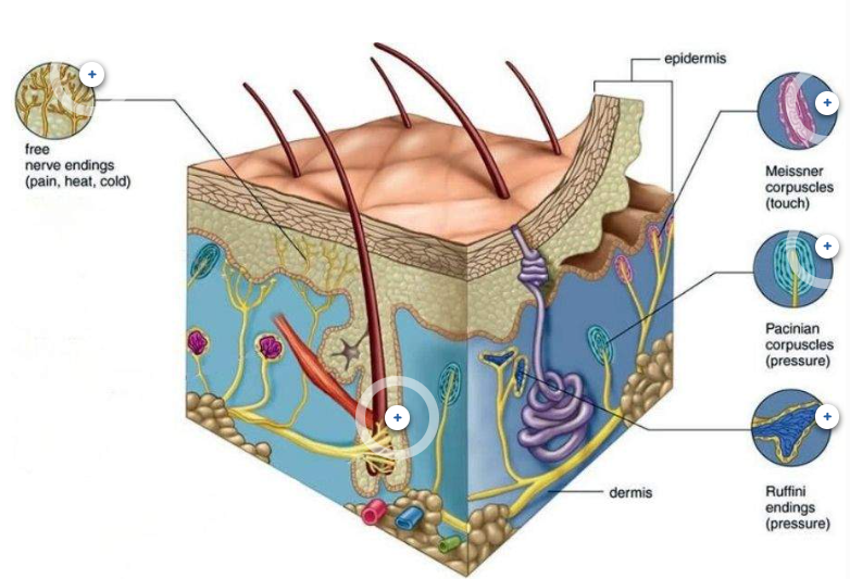
somatosensory system
detects & processes sensations of → touch, vibration, T, pain
each need many sensory receptors in skin 4 adequate stimulus
most sensations found from skin
cutaneous receptor
receptors in skin
free nerve endings
tactile (meissner) corpuscles
lammilar (Pacinian) corpuscles
bulbous (ruffini) corpuscles
hair follicles
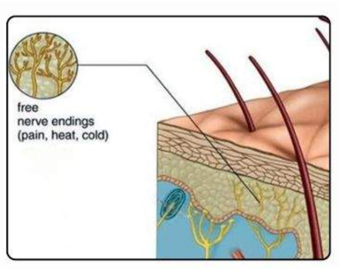
free nerve endings
type of cutaneous receptor
LOCATION→ thru out (skin, mucous membranes, muscles, internal organs)
FUNCTION → diff sensory stimuli & protect (sense harmful conditions)
pain (nociception)
temperature (thermoreception)
touch (mechanoreception) types
SENSITIVTY → wide range stimuli b/c unspecified, diff lvls based on location & f(x)
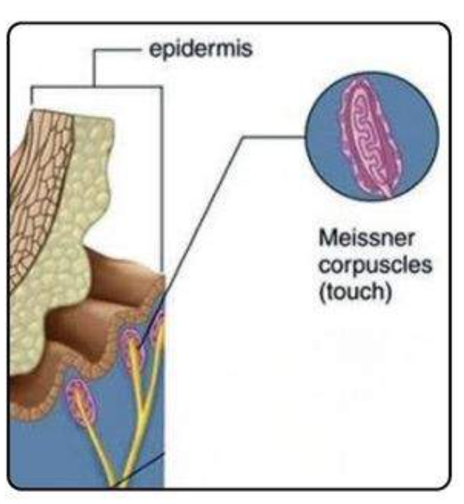
tactile (meissner) corpuscles
type of cutaneous receptor
LOCATION → glabrous skin (hairless)
fingertip, palm, feet soles, lip, tongue tip
FUNCTION → touch 4 detailed info, light & low frequency vibration (30-50 hz)
adaptive receptors → then DEC response if constant stimuli
SENSITIVTY → VVV & esp texture/fine touch
good 4 fine motor, specfic manipulation & recog. small object
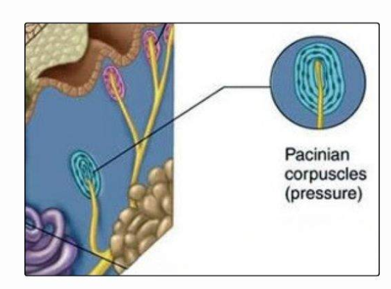
lammilar (Pacinian) corpuscles
type of cutaneous receptor
LOCATION → DEEP dermis & hypodermis (subcutaneous tissue)
FUNCTION → deep pressure & high frequency vibrations (250-350 hz)
adaptive receptors → then DEC response if constant stimuli
SENSITIVTY →
VV mechanical changes → deep pressure, stretch, INC frequency
DEC light touch & low frequency vibrations
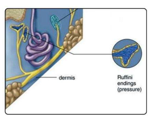
bulbous (ruffini) corpuscles
type of cutaneous receptor
LOCATION →
dermis & subcutaneous tissue
joint capsule, tendon, ligament
FUNCTION →
info abt sustained pressure, skin stretch → pov object manipulate & grip
know shape & move of objects
proprioception MAINLY → body position & moves
DEC adaptive receptors → respond as long as stimuli exist
SENSITIVTY → cont. pressure, stretch skin
DEC light, touch, rapid stimuli change
hair follicles
type of cutaneous receptor
LOCATION → all over except palm & soles
FUNCTION →
make hair w proliferate keratocytes in hair bulb
anchor points 4 hair shaft
form sense of touch & aware of hair moves
SENSITIVTY → X sensory cell
moves of hair follicle stimulate hair follicle receptors/hair root plexuses
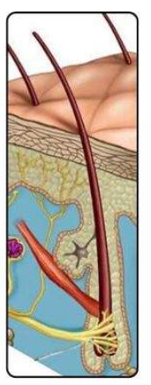
hair root plexuses
AKA hair follicle receptors
associated nerve endings near hair follicles
USE → sense movement of hair follicle
impt to detect insects & object contact skin/hair
adapt to cont. stimuli & keep sensitivity to changes in hair position over time
When are sensory receptor stimulated?
Ion channels open
Membrane permeability changes
A receptor-generated potential forms
If threshold is reached, action potentials are produced
when sensors sense something, where does info go?
senses make receptor generated potential
receptor pressed = skin receptor cells respond to stimuli, release neurotransmitters
movement detected in receptive field
neurotransmitters stimulate dendrites of sensory neuron & make receptor generated potential
receptor generated potential move down neurons & make AP @ axon terminals
axon terminals send info to another neuron take info to brain
receptor generated potential
changes in the membrane potential of sensory receptor cells in response to a stimulus
graded potentials that generate APs +send sensory info to CNS
X APs
X neurons
POTENTIAL DEPOLARIZING
release neurotransmitters
how are receptor generated potential similar to EPSP & IPSP
depolarize main but can hyper polarize
INC cell membrane permeability to Na/K
local & X propagate DOWN neuron → spread like EPSP
DEC over time & space
proportionate to stimuli (X all or nothing)
neural coding
how NS covert frequency info of neural activity 4 brain to understand
interpret changes of stimuli in waves & respond proportional to stimuli
MOSTLY → sensory cell release neurotransmitters proportional to stimuli
RESULT → make DEC/INC AP based on object weight
INC object weight = INC AP = brain sense INC AP frequency
What are the 2 paths a sensory signal takes to reach the brain?
Spinothalamic tract
Dorsal column-medial lemniscus pathway
Spinothalamic tract
USE →
(1) pain
(2) T
(3) crude touch
free nerve endings signal this path
—
1st order nerve (sensory nerve) detects info
send info ASAP & contralaterally to signals to 2nd order nerves
info sent to thalamus → signal 3rd order nerve
info stimulates sensory cortex & sent to correct places in brain
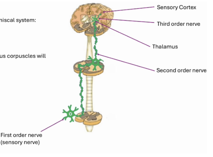
Dorsal column-medial lemniscus pathway
USE →
(1) fine detail
(2) vibration
(3) proprioception
tactile lammilar, bulbous corpuses signal this path
USE → detailed sensory discrimination & sense body position
—
1st order nerve (sensory nerve) detects info
send info to second order nerve and synapse contralaterally (longer time taken)
info sent to thalamus → signal 3rd order nerve
info stimulates sensory cortex & sent to correct places in brain
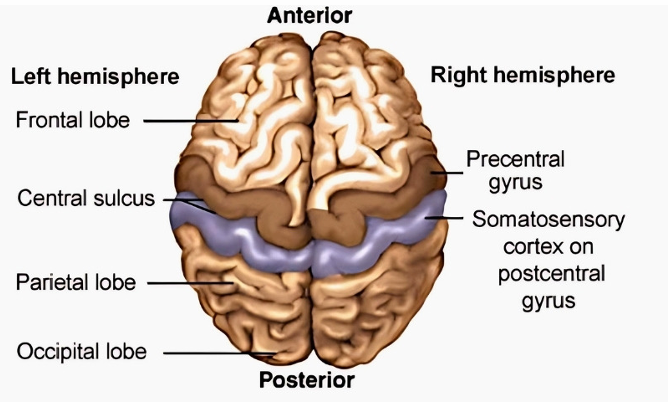
precentral gyrus
used 4 motor control
get info from frontal lobe
central sulcus
btwn precentral gyrus & postcentral gyrus
central ditch/divot
anterior = motor control
posterior central = somatosensory cortex
what describes the somatotopic organization of the postcentral gyrus?
postcentral gyrus has primary somatosensory cortex
somatotopic organization into a sensory homunculus
diff regions to specific cortical region
adjacent body part adjacent on cortical area
feet + leg = media
face + tongue = lateral
somatosensory cortex
in parietal lobe + within postcentral gyrus
USES →
interpret sensory data from receptors onto homunculus
pain, touch, T
perceive tactile sensation
proprioception → sense body position
localize stimuli 4 proper response & interact w enviro
where all info from sensory receptors sent & interpreted on homunculus map
How does the somatosensory cortex relate to the motor cortex?
somatosensory cortex lies directly posterior to motor cortex
sensory feedback from cortex helps guide & refine movement
both cortices organized somatotopically with corresponding body maps
Importance of the relationship between somatosensory cortex and motor cortex
sense body position
coordinate voluntary movement
adjust motor output based on sensory input
visual system
USE →
(1) detect light → convert to APs
(2) send info to primary visual area to process
(3) process & aware of visual world
What structures make up the visual system?
EYE
has photoreceptors that detect light & convert to electrical signals
VISUAL PATHWAY
carry APs from retina → brain
PRIMARY VISUAL CORTEX
on occipital lobe & process incoming visual info
what are the structures of the eye?
cornea
iris
lens
retina
fovea
optic nerve
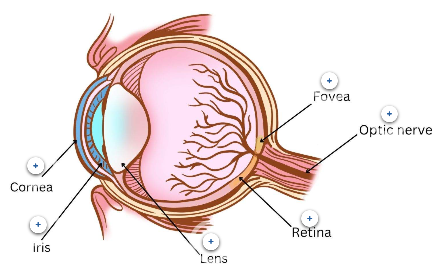
Cornea
USE →
main light focusing structure
bends/focuses incoming light rays to make clear image on retina
protect eye
ANATOMY →
transparent
dome shaped
cover iris, pupil, anterior of eye
avascular (X blood vessels) & get nourishment from tears
Iris
USES →
regulates amt of light entering eye → control pupil size w/ muscles
EX → bright = constrict, dim = dilate
helps eye look & colour → based on pigments it has
ANATOMY →
colored part of eye surround pupil
Lens
USES →
focuses light onto retina by ACCOMODATION
send info to retina & fovea
ANATOMY →
transparent + flexible
behind iris & pupil
accommodation of lens
contract & relax muscles in lens 4 visual acuity & distances
Retina
USES →
photoreceptors (rods + cones) + other cells → convert light into electrical signals → transmit to brain w optic nerve
vision &→ help see detailed image & colour
ANATOMY →
thin light sensitive tissue layer
back of eye
Fovea
USES →
highest lvl visual acuity
dense w cone photoreceptors (X rods) → good 4 detailed, colour vision in BRIGHT LIGHT
sharp central vision → read & drive
ANATOMY →
small central pit in retina
Optic nerve
USES →
send visual info to brain
get info from fovea & retina
ANATOMY →
nerve run from optic disc → brain
retinal ganglion cell axons + supportive cells
What cell types in the retina and how does their organization contribute to their function?
organized cells in layers to process visual info progressively
Rod cells
Cone cells
Bipolar cells
Ganglion cells
Amacrine cells
Pigment layer
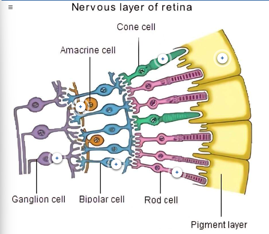
what retina cells can make AP + graded potentials
bipolar = graded & AP
specialized so spontaneously make potentials
inhibited by rod & cone cells to stop sending signals
ganglion = graded & AP
graded potential = allow fine & cont changes in signaling to reflect diff light intensities + better for visual info in retina
AP = send info over long distance, & all or nothing, better to travel signal via optic nerve to brain
photoreceptor & photopigment
receptor = respond to light
pigment = chem sensitive to light
How do rods and cones function?
in retina
X axon → X neuron, X APs make itself
receptor cells → release neurotransmitters & make graded receptor potentials to pass along info
can add or subtract signals → modulate by amacrine & bipolar cells to send ganglion cells proper info
FULL DARK→
ROD & CONE depolarize = release NTM inhibit bipolar cell = X make graded receptor potentials = X info sent to visual cortex w ganglion cells = X see much
depolarize → Na in & K out of cell lead to APs & inhibit NTMs
DIM LIGHT→
ROD hyperpolarize = X release NTM inhibit bipolar cell = make graded receptor potentials = info sent to visual cortex w ganglion cells = see B/W + objects w/o fine detail & colour
hyperpolarize → Na channel close bc light hit photopigments & change shape (Na permeability DEC & channel close) BUT K channel still open
THUS → cell hyperpolarize b/c Na DEC in & K INC out
CONE depolarize little = release NTM inhibit bipolar cells = X see much (X colour, X info sent to visual cortex)
BRIGHT LIGHT →
CONE hyperpolarize more = X release NTM inhibit bipolar cells = make graded receptor potentials = info sent to visual cortex w ganglion cells = see colour & fine details
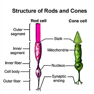
Rod cells
USE →
low light, night vision, peripheral vision
XXXX detail
dark = rod cells depolarizes & release neurotransmitter inhibit bipolar cells
ANATOMY →
mainly in retina (out & around fovea)
VV sensitive to light
FAIL → detail, colour (1 photopigment only)
Cone cells
USE →
bright light, high detail, colour
dark = cone cells depolarizes & release neurotransmitter inhibit bipolar cells
ANATOMY →
3 photopigments of wavelength sensitivity
S → short like BLUE
M → medium like GREEN
L → long like RED
mainly in fovea area (#1 [ ] in this area)
Bipolar cells
integrate visual signals BEFORE send forward
each cell gets info from diff rods + cones → collect visual info & INC sensitivity to light & colour
depolarized by cone & rod cell’s neurotransmitters in the dark
Ganglion cells
LAST output neurons of retina → get processed visual info from bipolar & amacrine cells
send info w axon (form optic nerve) → brain
Amacrine cells
modify signals btwn bipolar & ganglion cells → synapse both & affect visual signal before get to ganglion
Pigment layer
cell layer in retinal support photoreceptors → absorb excess light
RESULT → DEC scattering & INC photoreceptor health
how does the the eye process info?
light pass thru CORNEA of eye
IRIS regulates light passed in eye
LENS focus light on retina (back of eye)
RETINA uses photoreceptors to convert light as electrical signals → see image
uses graded potentials to transfer APs along visual path to primary visual area
signals transmit to brain w OPTIC NERVE
vision focused on FOVEA
Visual information flows ?
Photoreceptors → Bipolar cells → Ganglion cells → Brain
how does the retina convert light into electrical signals?
use graded potentials
which cells make graded potentials
bulbous + rod
both release NTMs cause graded potentials down line
sum OR subtract potentials to be proportional to stimuli
How is light transduced to action potentials?
Ganglion cell axons form the optic nerve and send APs to the visual cortex.
types of eye movements
focus on object = ensure image focused on fovea b/c most cone cells present here
1. Saccades
2. Smooth pursuit
3. Vestibular ocular reflex (VOR)
4. Vergences
Saccades
rapid jerky eye movements
quick move eye to object
EX → reading or gazing around a room while keeping head still
Smooth pursuit
smooth movement of eyes to follow path & keep moving object focused on fovea
EX → the butterfly moving from one flower to the next with just your eyes if you are able
vestibular ocular reflex (VOR)
eye movement when focus attention on object + shake up/down or back/froth
EX → shake your head, yes or no
Vergences
movements made when object approach/move away → eyes converge or diverge
EX → staring at a pencil and moving it away or towards your face
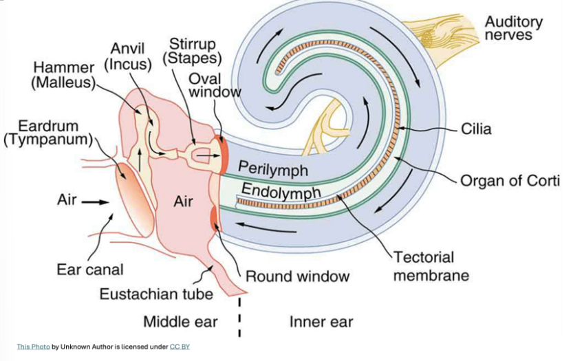
what are the structures of the ear?
OUTER EAR
auricle
external auditory canal (ear canal)
tympanic membrane (ear drum)
MIDDLE EAR
eustachian/auditory tube
auditory ossicles
incus
malleus
stapes
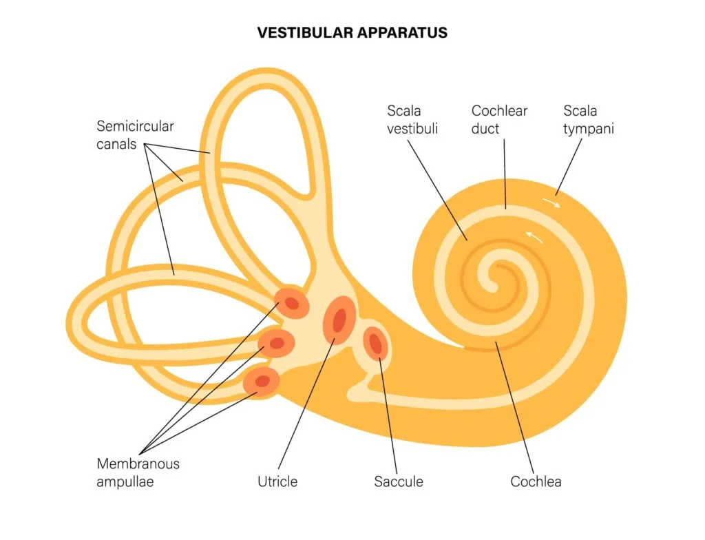
INNER EAR
oval window
round window
semi-circular canals
cochlea
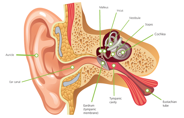
auricle
collect & amplify sound by capture sound wave & direct → ear canal
localize sound → tell sound direction & distance
external auditory canal
AKA ear canal
(1) sound transmission
carry sound waves from external envrio to eardrum
S shape curve → amplify & enhance frequences & help hearing process
(2) protect eardrum & middle ear
canal curve → protect eardrum from foreign objects
cermen (earwax) → trap dust, debris, antimicrobe & stop infection
cermen
AKA earwax
made from glands on cartilaginous part of canal
tympanic membrane (eardrum)
thin cone membrane
(1) sound transmission
convert sound waves in air → mechanical vibrations
sound wave strike eardrum = eardrum vibrate = vibration sent to malleus (on inner eardrum)
(2) protection
barrier separate external ear & protect middle ear
keep sterile envrio of middle ear
Eustachian/auditory tube
part of middle ear
narrow canal → connect middle ear to nasopharynx (upper part of throat behind nose)
USE →
equalize air pressure on eardrum sides
drain fluid from middle ear
auditory ossicles
part of middle ear
connect to oval window via stapes
3 tiny bones
incus → anvil
malleus → hammer
stapes → stirrup
USE → transmit sound vibration from eardrum to inner ear
sound waves strike tympanic membrane → membrane vibrate → malleus move → movement send to incus → sent to stapes → inner ear
oval window
part of inner ear
USE →
control movement of fluid in cochlea
activate receptors for hearing
stapes vibrate on oval window → standing waves in cochlea → detected by specialized hairs on nerve cells → cells carry APs to auditory centre of brain
round window
part of inner ear
USE → pressure-relief valve from cochlea
after sound travel thru cochlea → stops waves spread thru round window = X vibrate in cochlea
semicircular canals
part of inner ear
3 look shapes
USE → keep balance + spatial orientation
each canal used for specfic movement plane → horizontal, anterior/superior, posterior (X/Y/Z)
cochlea
part of inner ear
spiral shape → 3 divisions
1. scala vestibili
2. cochlear duct
3. scala tympani
fluid filled → transform sound vibration to neural signals & send to brain (AP made & send via cochlear nerve)
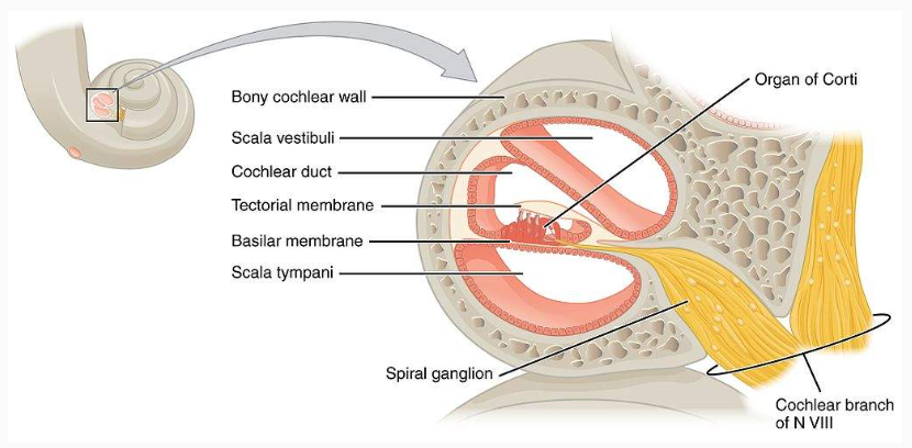
divisions of the cochlea
upper → scala vestibili (vestibular duct)
middle → cochlear duct
lower → scala tympani
tectorial membrane
basilar membrane
organ of corti
scala tympani & vestibli filled w perilymph
cochlea duct filled w endolymph
perilymph
ionic sol’n → INC [Na], DEC [K]
like extracellular fluid
fills scala tympani & vestibli
endolymph
sol’n → INC [K], DEC [Na]
like intracellular fluid
basilar membrane
separate cochlear duct & tympanic duct
has organ of corti
organ of corti
has special hair cels turn sound waves → AP
hairs embedded in tectorial membrane
sound waves = basilar membrane vibrates → fixed hair cells in tectorial membrane bend
what is sound?
series of pressure WAVES emitted, travel thru air, collected in ear
wave features
amplitude → V & loudness of sound
frequency → distant of waves & sound pitch
pressure wave moves thru air & hit ear → transmit to tympanic membrane → membrane vibrates = ossicles vibrate → oval window vibrates, make standing wave in cochlea → vibrates basilar membrane
how does the ear detect different frequencies and how is sound heard?
WAVE IN AIR
sound waves rep alternating areas of high & low volumes
pressure waves from stimuli measured for specfic wavelength
sound waves funneled through outer ear
auricle → ear canal → tympanic membrane
tympanic membrane vibrates to respond to sound waves
transfer pressure wave to middle ear to ossicles
vibrations amplified across ossicles
pass thru malleus, incus, stapes
3 bones connect tympanic membrane to oval window (inner ear)
(1) malleus → attach tympanic membrane, move based on vibration of eardrum → sent to incus & stapes
(2) stapes → connect oval window & move to create pressure waves in perilymph fluid of cochlea
WAVE IN FLUID
vibrations against oval window create standing wave in endolymph OR vestibuli
oval windows vibrate
cochlea fluid vibrates → pressure wave travelling thru perilymph OG & displace endolymph fluid
create standing waves when waves resonate @ specfic frequence of sound wave → standing wave in fluid bending of cochler duct me
standing wave in basilar membrane → go to cochlear duct
pressure bend cochlear duct membrane @ resonance
basilar membrane airs vibrate at specfic point → connect to specfic sound frequency
sound frequency is determined
sound resonates throughout whole cochlea & return back out round window → stop vibration & resonance forever
resonance
bending of cochlear duct membrane @ max vibration of a frequency
how are standing waves in the cochlea formed?
USE OF WAVES →
part of process to make cochlea’s fluid filled structure convert mechanical E → neural signals
PROCESS →
standing wave = when incoming pressure wave & its reflection in cochlear duct interfere
RESULT → regions of max & min displacement occur as the frequency of sound tries to match natural frequency of a region on basilar membrane
basilar membrane has variations for stiffness & width = resonance occur at diff frequences
how are different frequencies detected by the ear?
tonotopic organization → based on location in cochlea
CAUSE → diff thickness & flexibility of material
max vibrations found at →
BASE of basilar membrane = DEC frequency heard
thicker narrow & DEC flexible membrane
APEX of basilar membrane= INC frequency heard
thinner wider & INC flexible membrane
stereocilia
hair cells embedded on tectorial membrane
tectorial X move BUT basilar membrane moves
how does the cochlea create APs?
fluid waves move through cochlea → moves oval window & create standing wave in cochlear fluid
basilar membrane vibrates & hair cells bend
basilar membrane fluid movement = bend hair cell’s stereocilia as response to vibrations
hair cells = organ of corti
mechanically gated ion channels open
K⁺ enters hair cells & depolarize
neurotransmitter release
depolarized hair cells release neurotransmitter onto auditory neurons
auditory nerve fibers generate APs
AP travel to auditory cortex
cochlear nerve used to take info to brain
What transforms sound vibrations into neural signals that are sent to the brain?
the cochlea
What would happen if the basilar membrane was damaged at the apex?
hard time detecting low pitch sounds
what is damaged if a person is having trouble hearing quieter sound?
affect structures needed to make vibrations in cochlea to detect sound
malleus + incus → amplify sound by causing stapes to vibrate in the oval window
tympanic membrane → vibrate ossicles as sound pressure waves hit it
T/F. The tympanic membrane, or eardrum, vibrates when sound pressure waves strike it, and these vibrations are passed through the ossicles
T
vestibular system
USE → balance + spatial orientation
coordinate w other sensory systems to share info → semicircular canal + otolith organ inputs + visual & proprioceptive system
semicircular canals sed in VOR → stabilize vision w eye movements counteract head moves
FOUND → inner ear
PROCESS → detect head move, change in position & sends sensory info to brain to keep balance, posture, eye movement
structures of vestibular system
vestibular apparatus →
3 semicircular canals/ducts
anterior
lateral
posterior
all looped tubulars in inner ear
ampullae
utricle
saccule
semicircular canals
USE → balance + spatial orientation
ANATOMY →
inner ear
3 looped canal tublar structures
anterior (superior) → vertical
ROLE → front/back moves
posterior → vertically BUT perpendicular to anterior
ROLE → head tilts to shoulders
lateral (horizontal) → horizontal
ROLE → detect horizontal head moves
ampullae
sensors detect where body is in space w/ sensory hair cells
@ end of each semicircular duct
filled w/ endolymph & pushes cupula → hair cells in cupula move as endolymph moves → hair cells release NTMs (constant) → fire APs along sensory nerve toward head
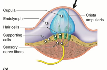
how does the body detect angular & rotational moves?
use semicircular canals’ AMPULLAE
based on move, cause change in hair cells of cupula inside ampullae → change release of NTMs that affect AP firing frequency
EX → stand still
baseline amt of NTMs released
NTMS stimulate sensory nerve fibers & cause APs
AP frequency (based on amt of NTMs released) interpreted by brain
EX → dancing
inertia of endolymph lag after canal wall moves
lag push against cupola & stimulate hair cells
hair cells bent & based on direction, release INC/DEC NTMs → change AP frequency along sensory nerve
info sent to brain w vestibular nerve & brain interprets it as movement of heads
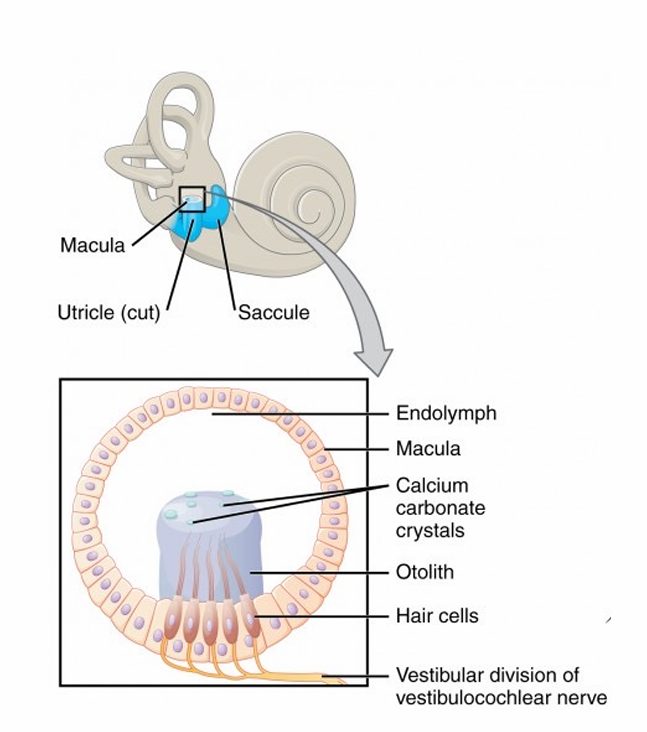
utricle
USE → balance + spatial orientation (horizontal linear acceleration)
ANATOMY →
inner ear
filled w endolymph
bigger + horizontal position
has hair cells in gelatinous layer w/ otoliths
saccule
USE → balance + spatial orientation (vertical linear acceleration)
ANATOMY →
inner ear & closer to cochlea
filled w endolymph
smaller + vertical position
has hair cells in gelatinous layer w/ otoliths
compare contrast utricle & saccule
(+) →
both have hair cells w/ otoliths
detect linear acceleration & head position change
send info to brain w vestibular nerve
work for balance + spatial orientation
(-) →
U → horizontal
S → vertical
how does the body detect up and down moves?
UTRICLE + SACCULE
MACULA → tissue & structure
based on head move, otolith crystals move → affect hair cell direction & change NTM release & ATP pxdtn on neuron (still release NTMs during rest)
STRUCTURE →
vestibular division of vestibular cochlear nerve nerves in tissue → connect to hair cells in gelatinous membrane (like cupula) that release NTMs
membrane connected to otolith & has Ca carbonate crystals
EX → look ahead & upright
all structures upright, hair cells release NTMs → cause frequency of APs in nerve → brain detect head stable & align
EX → head tilt forward
gravity move otolith crystals forward, pulling on hair cells → change amt NTMs released → affect amt APs along neuron → brain interpret & know head move forward
head move forward so lag of hair cells & they bend back as they slowly move forward
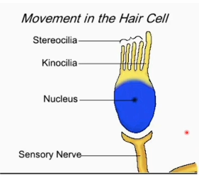
how does a hair cell of the vestibular apparatus tell how much neurotransmitters are released?
based on bending of hair cells
USE →
stereocilia + kinocilia vertical = AP steady
ACCELERATE
stereocilia bend → kinocilia = INC AP w NTM INC
DECELRATE
stereocilia bend ← kinocilia = DEC AP w NTM DEC
ANATOMY →
stereocilia = shorter hair
kinocilia = 1 long hair
stereocilia + kinocilia connect to cell body at base in nucleus
nucleus connected to sensory nerve
what is the clinical significance of the vestibular system?
semicircular canal pxb = vertigo, dizziness, balance issue
EX → benign paroxysmal positional vertigo (BPPV)
dislodged otoliths from utricle enter a semicircular canal
RESULT → disrupt function & cause vertigo
SOLVE → series of head positions help restore position of otolith crystals, & DEC symptoms