Lecture 1 & 2: Glands & Integumentary System
 Call Kai
 Learn
Learn Practice Test
 Spaced Repetition
 Match
Match Flashcards
 Knowt Play
1/112

There's no tags or description
Looks like no tags are added yet.
Name | Mastery | Learn | Test | Matching | Spaced | Call with Kai | Chat |
|---|
No analytics yet
Send a link to your students to track their progress
113 Terms
Glands are what type of organs?
Secretory organs:
make and release chemical substances
they are made of epithelium & supportive connective tissue
Two Types of Glands formed by Infolding of Epithelium:
Endocrine
Exocrine
Endocrine Glands
Ductless glands that secrete hormones directly into blood or interstitial fluid. (No ducts! Extensive capillary supply)
Exocrine Glands
Glands that release secretions onto epithelial surfaces through ducts. (YES DUCTS! ie. saliva & sweat)
- classified by mode of secretion
Exocrine Duct Structure & Type
Structure: Unicellular vs Multicellular
Duct Type: Simple vs Compound branching
Three Modes of Secretion by _____ Glands
Exocrine Glands mode of secretion:
- Merocrine
- Apocrine
- Holocrine
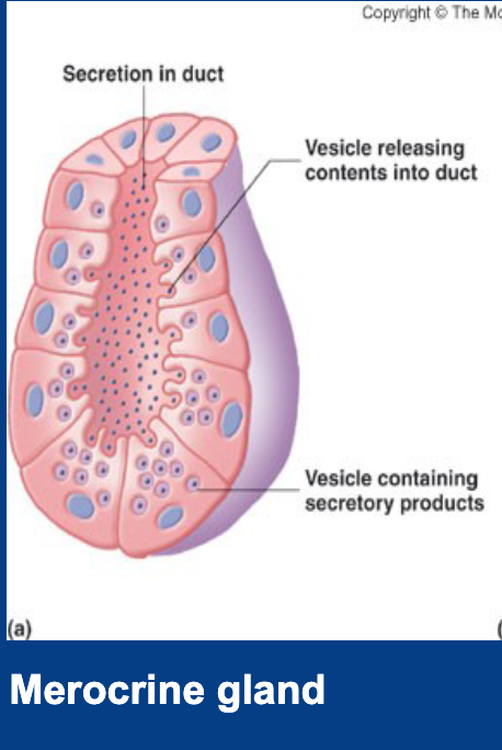
Merocrine (Eccrine) Secretion
Secretion mechanism where contents are released via exocytosis or active transport (w/ ATP) without cell damage (loss of cytoplasm).
- ie. sweat glands
Apocrine Secretion
- Secretion mechanism where the apical portion of the cytoplasm is shed with product.
- APO- A PART OF cell & cytoplasm pinches off and is released.
ie. Mammary gland

Holocrine Secretion
- Secretion mechanism where entire cell ruptures, dies, and forms the secretory product.
- ie. sebaceous gland

Unicellular Exocrine Glands
Single isolated secretory cells found in an epithelium (e.g., goblet cells).

Multicellular Exocrine Glands
Glands composed of multiple secretory cells and supportive connective tissue.
many cells acting as one unit
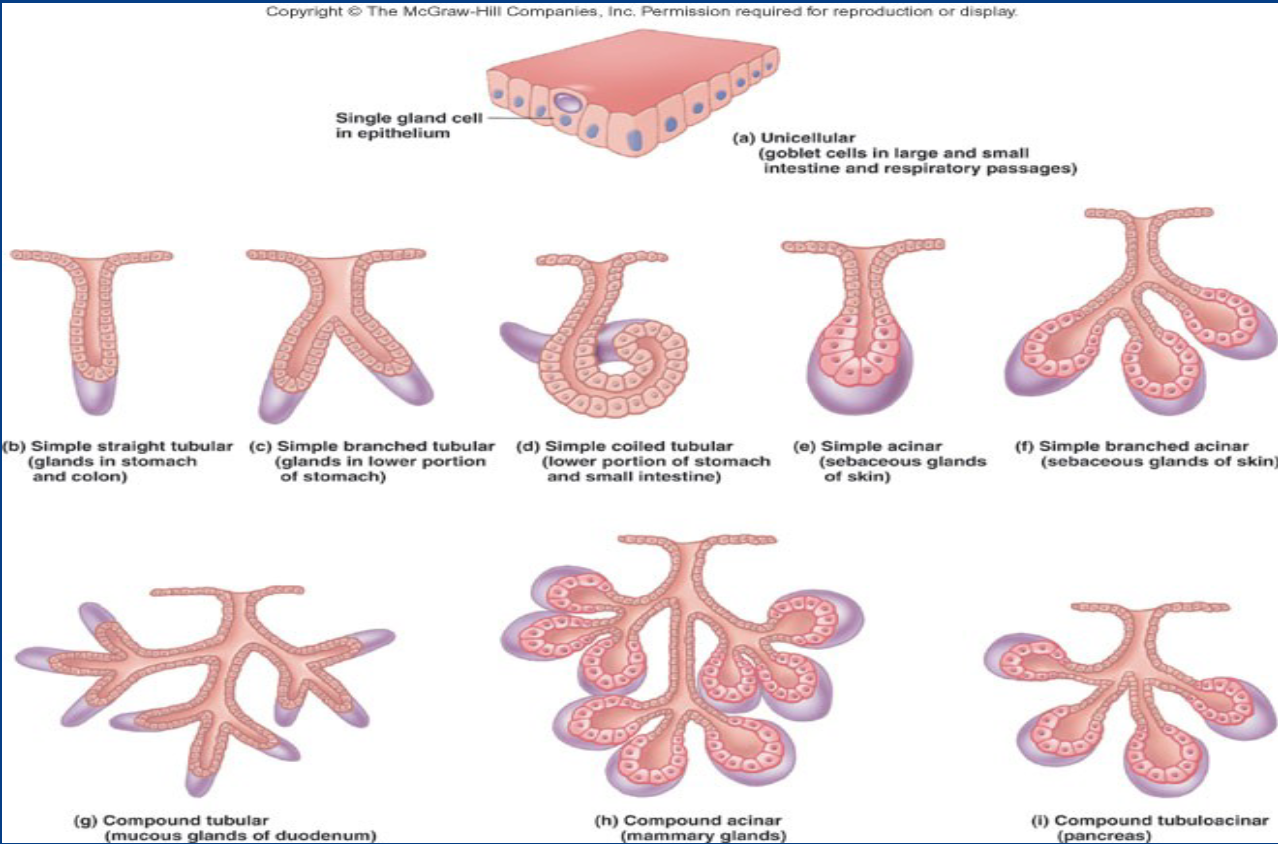
Simple Exocrine Glands
Glands with a single unbranched duct leading to secretory units.

Compound Exocrine Glands
Glands with a branched duct system leading to multiple secretory units.

Acini / Acinar (Alveolar)
- Sac-like or bulbous clusters that produce or hold secretions
Integumentary System Consist of
Skin
Accessory organs: hair, nails, cutaneous glands
T/F - Skin is the most vulnerable organ
True: skin is exposed to radiation, trauma, infection, and toxic chemicals
- receives more medical treatment than other organ systems.
General Functions of the Skin
- Resistance (Trauma & Infection)
- Barrier (Waterproofing, UV Light (melanin), Chemicals)
- First Step for Vitamin D Synthesis
- Sensation & Thermoregulation (vasodilation/constriction)
- Nonverbal communication (acne, birthmark,scar)
- Transdermal absorption (drug patch)
What makes this skin resistant to infection
Keratinized epidermis does not contain live cells that can act as hosts fo pathogens
Acid mantle…
Three Layers of Integumentary System (which is not a part of the skin?)
Skin Layers: Epidermis & Dermis
Non Skin, Fat & Neuro-Vascular Layer below Dermis: Hypodermis

Thick vs Thin Skin & Glands
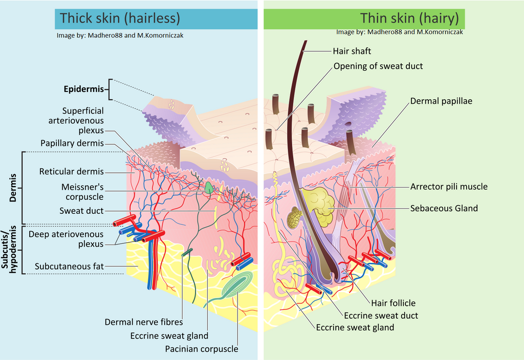
Thick Skin =
- on palms and sole & digits (epidermis) 0.5mm thick
- has sweat glands, no hair follicles or sebaceous (oil) glands
Thin Skin =
- covers rest of body (epidermis) 0.1 mm thick ;
- has sweat glands, hair follicles, and sebaceous glands
continuously shedding & regenerating
What type of epithelium does the Epidermis have & general characteristics
- keratinized stratified squamous epithelium:
- dead cells packed with keratin (tough protein)
- lacks blood vessels
- sparse nerve ending for touch & pain

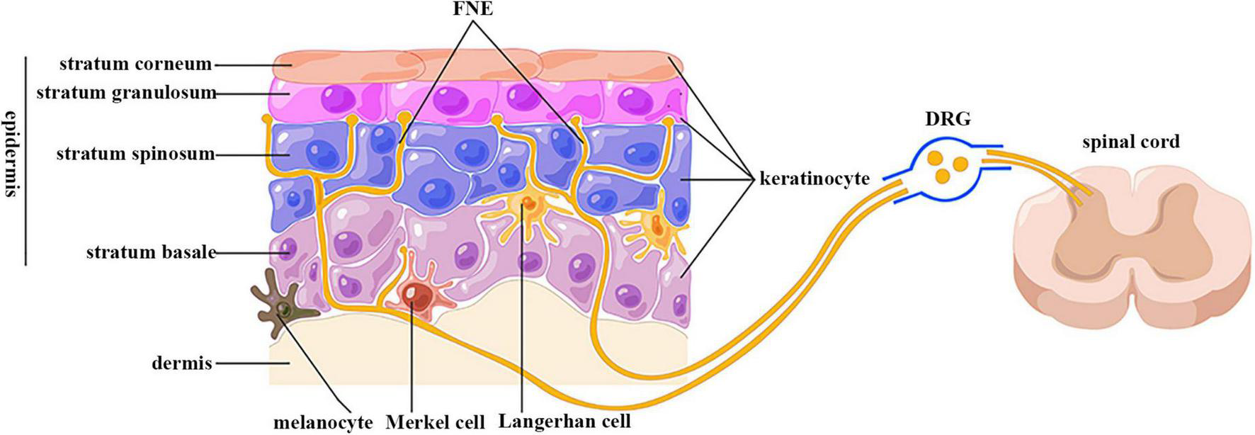
5 Types of Cells in Epidermis
- stem cells
- keratinocytes
- melanocytes
- tactile (merkel) cells
- dendritic (langerhans) cells

Stratum Basale
Deepest epidermal layer containing stem cells (which keratinizes cells), melanocytes, and tactile (Merkel) cells.
above basement membrane where nutrients diffuse..*

Stratum Spinosum
Above Basale
Epidermal layer of thick keratinocytes attached by desmosomes; contains dendritic cells.

Stratum Granulosum
Epidermal layer containing keratohyalin granules and lamellar granules for water barrier.
Stratum Lucidum
Thin, Clear epidermal layer containing eleidin;
found exclusively in thick skin (palm & soles) ; cells are dead

Stratum Corneum
Outermost epidermal layer of dead, fully keratinized, highly resistant squames.

Keratinocytes
Principal epidermal cell type synthesizing keratin for structural protection.
Melanocytes
Cells in stratum basale that produce melanin to shield nuclear DNA from UV.
Dendritic (Langerhans) Cells
Macrophage-like immune cells found mainly in stratum spinosum and granulosum.
Tactile (Merkel) Cells
Sensory receptors located in stratum basale associated with nerve fibers for touch.
Epidermal Stem Cells
Undifferentiated mitotic cells in stratum basale that give rise to keratinocytes.
Keratinocyte Life Cycle
30 to 40 day transition of keratinocytes from stratum basale to surface exfoliation.
Epidermal Water Barrier
Hydrophobic barrier between granulosum and corneum formed by lipids and tight junctions.
Filaggrin
Protein from keratohyalin granules that aggregates keratin filaments into tough bundles.
FLG Gene
Gene located on chromosome 1q21 encoding profilaggrin.
Profilaggrin
Large precursor protein stored in keratohyalin granules that cleaves into filaggrin.
Natural Moisturizing Factor (NMF)
Breakdown products of filaggrin (PCA, UCA) that maintain skin hydration and pH.
trans-Urocanic Acid (trans-UCA)
Filaggrin breakdown product in stratum corneum that absorbs UV-B radiation.
Pyrrolidone Carboxylic Acid (PCA)
Filaggrin breakdown product that serves as a potent hygroscopic humectant in skin.
Ichthyosis Vulgaris
Autosomal semi-dominant skin disorder caused by FLG loss-of-function mutations.
Atopic Dermatitis (Eczema)
Chronic inflammatory skin disease associated with filaggrin deficiency and skin barrier loss.
Atopic March
Progression from eczema in infants to asthma, allergic rhinitis, and food allergies.
Thick Skin
Epidermis (~0.5 mm) with stratum lucidum and prominent ridges; lacks hair and oil glands.
Thin Skin
Epidermis (~0.1 mm) covering most of body; lacks stratum lucidum; contains hair and glands.
Papillary Layer of Dermis
Superficial dermal layer composed of areolar connective tissue and dermal papillae.
Reticular Layer of Dermis
Deep dermal layer composed of dense irregular connective tissue providing strength.
Dermal Papillae
Upward finger-like projections of dermis that interlock with epidermal ridges.
Epidermal Ridges
Downward projections of epidermis into dermis forming friction ridges (fingerprints).
Cleavage (Langer's) Lines
Predominant orientation of collagen bundle fibers within reticular layer of dermis.
Striae (Stretch Marks)
Dermal tears resulting from severe stretching of collagen fibers.
Hypodermis (Subcutaneous Layer)
Subepidermal layer of loose areolar and adipose tissue anchoring skin to fascia.
Subcutaneous Fat
Adipose deposit within hypodermis providing thermal insulation and energy storage.
Eumelanin
Dark brownish-black pigment produced by melanocytes.
Pheomelanin
Reddish-yellow sulfur-containing pigment produced by melanocytes.
Tyrosinase
Enzyme required for synthesis of melanin from tyrosine in melanocyte melanosomes.
Carotene
Yellow-orange dietary pigment accumulating in stratum corneum and subcutaneous fat.
Hemoglobin
Iron-bearing red pigment of red blood cells imparting pinkish skin tones.
Cyanosis
Blueness of skin resulting from oxygen deficiency in circulating blood.
Erythema
Abnormal skin redness due to dilated dermal cutaneous blood vessels.
Pallor
Pale or ashen skin shade from diminished cutaneous blood flow.
Albinism
Genetic deficiency of melanin production due to absent/defective tyrosinase.
Jaundice
Yellowish staining of skin and sclera from elevated serum bilirubin.
Hematoma
Localized collection of clotted blood outside blood vessels (bruise).
Friction Ridges
Markings on fingertips/soles created by dermal papillae enhancing grip.
Flexion Lines
Marks on skin where skin folds over joints during movement.
Freckles
Flat melanized patches of skin resulting from localized melanin aggregation.
Nevus (Mole)
Elevated melanized patch of skin, often containing hair.
Hemangioma
Benign tumor of cutaneous blood capillaries (birthmark).
Hair Shaft
Visible portion of hair extending above skin surface.
Hair Root
Portion of hair embedded beneath skin surface within hair follicle.
Hair Bulb
Swollen base of hair root situated in deep dermis or hypodermis.
Hair Papilla
Vascular connective tissue projection into hair bulb providing nutrients.
Hair Matrix
Mitotic layer of growth cells resting directly over hair papilla.
Hair Medulla
Innermost core layer of hair follicle containing soft keratin and air spaces.
Hair Cortex
Middle layer comprising bulk of hair tissue containing hard keratin.
Hair Cuticle
Outermost layer of hair shaft composed of overlapping scaly cells.
Epithelial Root Sheath
Extension of epidermal strata surrounding hair root.
Dermal Root Sheath
Derived from dermal connective tissue surrounding epithelial root sheath.
Arrector Pili Muscle
Smooth muscle bundle extending from dermal collagen to epithelial sheath.
Lanugo
Fine, unpigmented fetal hair present during last trimester.
Vellus
Fine, pale hair covering majority of children and female body surface.
Terminal Hair
Coarse, heavily pigmented hair on scalp, eyebrows, axillary, and pubic regions.
Anagen Phase
Active growth stage of the hair cycle where matrix cells undergo active mitosis.
Catagen Phase
Degenerative transition phase of hair cycle where follicle shrinks and matrix dies.
Telogen Phase
Resting phase of hair cycle prior to hair shedding and new growth re-entry.
Alopecia
Hair thinning or baldness resulting from follicle aging or disease.
Pattern Baldness
Genetic condition where hair loss occurs selectively in specific regions due to DHT.
Hirsutism
Excessive body hair growth in areas atypical for female sex due to androgen surge.
Nail Plate
Hard external portion of nail including free edge, body, and root.
Nail Body
Visible attached portion of nail plate overlying nail bed.
Nail Root
Proximal hidden portion of nail plate extending beneath skin fold.
Nail Bed
Skin layer situated directly beneath nail plate.
Nail Matrix
Thickened stratum basale at proximal nail fold responsible for nail growth.
Eponychium (Cuticle)
Narrow zone of dead skin overhanging proximal end of nail plate.
Hyponychium
Epidermis of skin underlying free edge of nail plate.
Lunule
White crescent-shaped area at proximal base of nail plate.
Sebaceous Glands
Holocrine glands associated with hair follicles that secrete oily sebum.
Sebum
Oily secretion rich in lipids produced by sebaceous glands.
Merocrine (Eccrine) Sweat Glands
Simple tubular cutaneous glands producing watery perspiration for thermoregulation.