Dental Sciences & Prev Dentistry- Ch 10/COMPLETED
1/28
Earn XP
Description and Tags
Name | Mastery | Learn | Test | Matching | Spaced | Call with Kai |
|---|
No study sessions yet.
29 Terms
Regions of the Face
Forehead: Extending from the eyebrows to the hairline
Temples: Lateral to the eyes
Orbital: Eye area that is covered by the eyelids
External nose
Zygomatic (malar): Prominence of the cheek
Mouth and lips
Cheeks
Chin
External ear
Features of the Face
Outer and inner canthus
of the eye
Ala of the nose
Philtrum
Tragus of the ear
Nasion
Glabella
Root or “bridge” of nose
Septum of the nasal cavity
Anterior naris of the nostril
Mental protuberance of the mandible
Angle of the mandible
Zygomatic arch
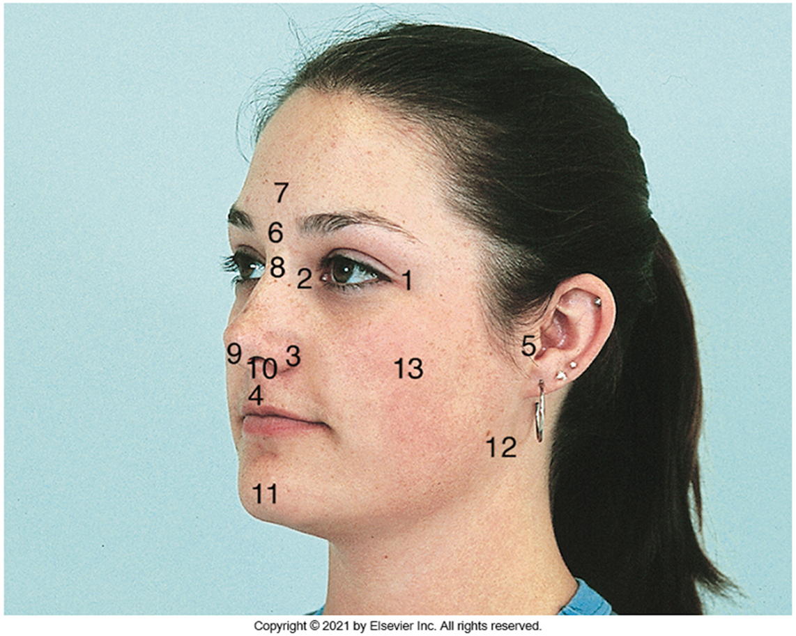
What are the facial numbers 1-13? What are they called? Where are they located?
Outer Canthus, located in the fold of tissue at the outer corner of the lip
Inner Canthus, located in the fold of tissue at the inner corner of the eye
Ala of the nose, located on either side of the nostrils
Philtrum, located in the middle area of the upper lip
Tragus, located on the outer ear, in front of the ear canal
Nasion, located at the bridge of the nose, between the eyes (just below the eyebrows)
Glabella, located between the eyebrows on the forehead
Root of the nose, located at the top of the nose, where it meets the forehead (bridge of the nose)
Septum, located in the middle of the nasal cavity, dividing the nostrils
Anterior naris, located at the nostril openings
Mental protuberance, located at the front of the chin
Angle of the mandible, located where the lower jaw turns upwards
Zygomatic arch, located as the bony structure of the cheek (cheek bone)
Skin
The skin of the face is thin to medium in relative thickness
It is soft and movable over a layer of loose connective tissue
The skin around the external ear and the ala of the nose is fixed to underlying cartilage
Facial skin contains many sweat and sebaceous glands
Lips
The lips are also known as labia
The lips are outlined by the vermilion border
The labial commissure is the angle at the corner of the mouth where the upper and lower lips join
The nasolabial sulcus is the groove extending upward between each labial commissure and the ala of the nose
The oral cavity
Lined with mucous membrane tissue
Consists of two areas
The vestibule is the space between the teeth and the inner mucosal lining of the lips and cheeks
The oral cavity proper is the space contained within the upper and lower dental arches
The Vestibule
The intraoral vestibule begins on the inside of the lips and then extends from the lips onto the alveolar process of both arches
The vestibular mucosa is thin, red, and loosely bound to underlying alveolar bone
The base of each vestibule, where the buccal mucosa meets the alveolar mucosa, is called the mucobuccal fold
The mucogingival junction is a distinct line of color change where the alveolar membrane meets with attached gingiva
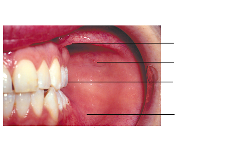
Label from 1-4
Buccal Frenulum
Papilla and Orifice of Parotid Duct
Crown of 2nd Maxillary Molar
Mucobuccal Fold
Labial and Other Frenula
A frenum is a narrow band of tissue that connects two structures
The labial frenum passes from the midline of the maxillary or mandibular arch to the midline of the inner surface of the lip
The buccal frenum passes from the oral mucosa near the maxillary or mandibular first molars to the inner surface of the cheek
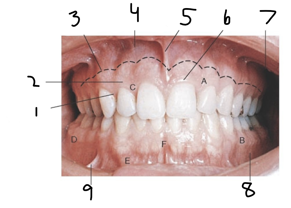
Label 1-9
Interdental Gingiva
Attached Gingiva
Mucogingival Junction
Alveolar Mucosa
Maxillary Labial Frenum
Marginal Gingiva
Maxillary Vestibule
Mandibular Vestibule
Mandibular Buccal Frenum
What is gingiva?
Gums, they surround the teeth and are self cleaning
how does normal gingiva look like? what are it’s characteristics?
Normal gingiva surround the teeth and are self cleaning. They are firm, resistant, and are tight around the tooth and bone. Surface color varies according to the individuals pigment. The surfaces of the attached gingivae and interdental papillae are stippled and similar in appearance to the rind of an orange.
Unattached Gingiva
Also known as marginal gingiva and free gingiva. The unattached gingiva is usually about 1 mm wide and forms the soft wall of the gingival sulcus.
In gingivitis, unattached gingiva is the first tissue to become inflamed.
Interdental gingiva (also called gingival papilla)
Extension of the free gingiva that fills the interproximal embrasure between two adjacent teeth
Gingival groove
The gingival groove is a shallow groove that runs parallel to the margin of the unattached gingiva and marks the beginning of the attached gingiva
Attached gingiva
The attached gingiva extends from the base of the sulcus to the mucogingival junction
What is the oral cavity proper?
It is the area inside the dental arches, in the back of the last moral on each side is a space that links the vestibule and the oral cavity proper
Hard palate
The hard palate separates the nasal cavity above from the oral cavity below. The nasal surfaces are covered with respiratory mucosa, and the oral surfaces are covered with oral mucosa. The mucosa of the hard palate is tightly bound to the underlying bone, and therefore submucosal injections into the palatal area can be extremely painful.
What is another term used to refer to the hard palate?
The roof of the mouth
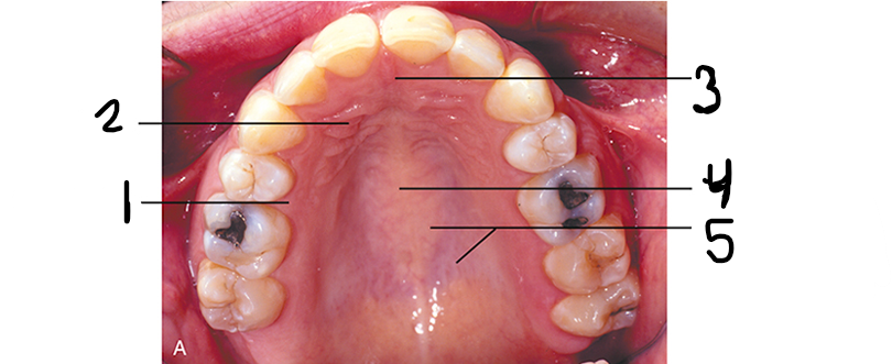
Label the land marks of the hard palate (roof of mouth) from 1-5
Lingual (palatal) gingiva
Palatal Rugae
Incisive Papilla
Median Palatine Raphe
Ducts for Palatal Glands
Soft palate
The soft palate is the movable posterior third of the palate. It has no bony skeleton and hangs like a limp curtain into the pharynx behind it. The soft palate ends posteriorly as a free edge with a hanging projection called the uvula.
The soft palate is supported posteriorly by two arches, the fauces:
The anterior arch runs from the soft palate down to the lateral aspects of the tongue as the palatoglossal arch
The posterior arch, the free posterior border of the soft palate, is called the palatopharyngeal arch
The opening between the two arches is called the isthmus of fauces and contains the palatine tonsil
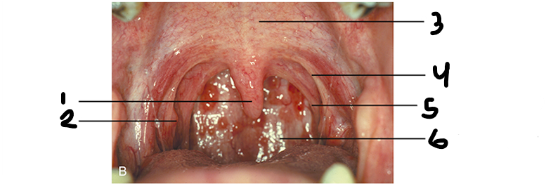
Label the soft palate surfaces from 1-6
Uvula
Palatine Tonsil
Soft Palate
Anterior Faucial Pillar
Posterior Faucial Pillar
Posterior Wall of Pharynx
What is the soft palate?
the back of the mouth/throat
Tongue
The tongue is an important organ, responsible for several functions:
Speech
Manipulation and positioning of food
Sense of taste
Swallowing
Cleansing of the oral cavity
Parts and Surface of the tongue
Body: Anterior two thirds of the tongue
Root: Posterior portion that turns downward toward the pharynx
Dorsum: Upper and posterior roughened surface
Sublingual surface: Covered with smooth, transparent mucosa
Lingual frenulum: Thin fold of mucous membrane that extends from the floor of the mouth to the underside of the tongue
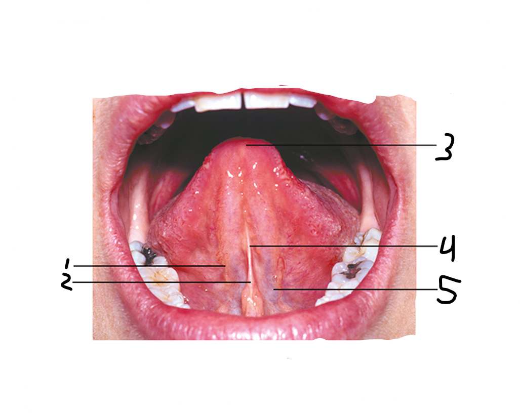
Label the parts of the tongue from 1-5
Fimbriated Fold
Lingual Frenum
Raised Tip of Tongue
Sublingual Caruncle
Lingual Veins
Taste buds
Located on the fungiform papillae and in the trough of the large vallate papillae, which form a V on the posterior portion of the tongue
The sense of touch is provided by numerous filiform papillae that cover the entire surface of the tongue
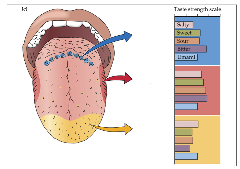
Label the tongue surface from top to bottom (Blue, red, yellow)
Page 38 (last page)
1 (Blue): Circumvallate Papillae
2 (Red): Foliate Papillae
3 (Yellow): Fungiform Papillae
Teeth
Teeth are either single-rooted or multirooted
The teeth sit in bony sockets, or alveoli, within the alveolar process of the maxilla and mandible
In the mouth, a cuff of gingival tissue surrounds each tooth
The portion of the tooth that is visible in the oral cavity is called the crown