A&P Lab 4- Appendicular Skeleton
 Call Kai
 Learn
Learn Practice Test
 Spaced Repetition
 Match
Match Flashcards
1/131

There's no tags or description
Looks like no tags are added yet.
Name | Mastery | Learn | Test | Matching | Spaced | Call with Kai | Chat |
|---|
No analytics yet
Send a link to your students to track their progress
132 Terms
What are the 3 types of cartilage
Hyaline cartilage, elastic cartilage, and fibrocartilage
Where is Hyaline Cartilage found?
Hyaline cartilage is found in the ribs, nose, trachea, and at the ends of long bones, providing support and flexibility.
Where is Fibrocartilage found
Fibrocartilage is found in intervertebral discs, the pubic symphysis, and menisci of the knee, providing tensile strength and support.
Where is elastic cartilage found
Elastic cartilage is found in the external ear, epiglottis, and parts of the larynx, providing flexibility and structure.
Epiphyseal Plate
layer of hyaline cartilage located between the epiphysis and diaphysis of long bones, allowing for longitudinal growth during development.


Long bone anatomy
Articular cartilage, medullary cavity on inside, bone marrow, periosteum (outside sheath), endosteum (inside lining), diaphysis, and epiphysis
What is within the appendicular skeleton?
pelvic girdle, shoulder girdle, and the bones of the limbs (arms and legs).

Name this structure
Sternoclavicular (SC) joint
-Only site of attachment to axial skeleton (between sternum and clavicle)

Name this Structure
Acromioclavicular joint
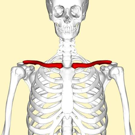
Name this bone
Clavicle

Name this portion of the clavicle
A= Lateral (Acromial End)
B=Middle
C= Medial (sternal end)


Name this bone
Scapula

Name this landmark on the scapula
Coracoid Process
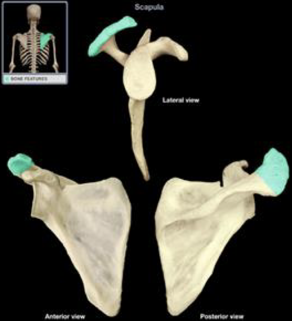
Name this landmark on the scapula
Acromion

Name this structure
Infraspinatus

Name this landmark on the scapula
Glenoid Cavity

Right or Left?
Right (remember the anterior is and smooth faces ribs)

Right or left?
Left
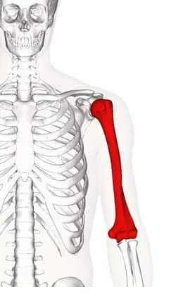
Name this bone
Humerus

Name this Landmark
Head

Name this Landmark
Greater Tubercle
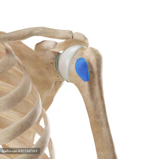
Name this landmark
Lesser Tubercle

Name this landmark
Intertubercular Sulcus (or groove)
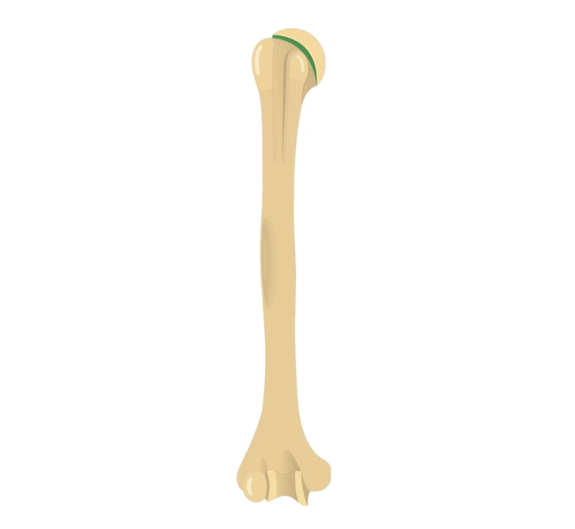
Name this landmark
anatomical neck of humerus

Name this landmark
Surgical Neck of humerus
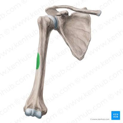
Name this landmark
Deltoid Tuberosity (on the lateral side of the arm)

Name this landmark
Olecranon Fossa (Posterior view)

Name this landmark
Coronoid Fossa (Anterior view of the humerus)

Name this landmark
Lateral Epicondyle

Name this landmark
Medial Epicondyle

Name this landmark
Trochlea of humerous
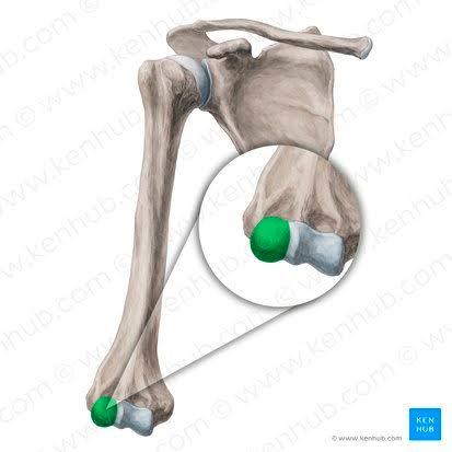
Name this landmark
Capitulum

Left or right?
Right Humerus

Left or right?
Right Humerus
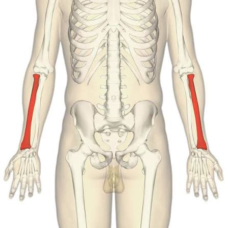
Name this bone
Radius
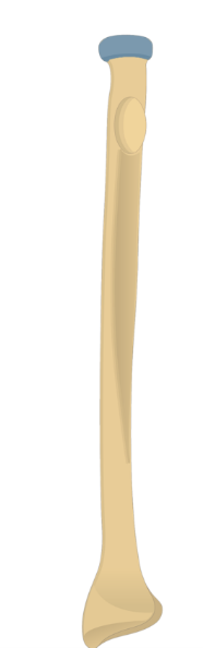
Name this structure
Head of radius

Name this structure
Neck of radius

Name this structure
Radial tuberosity

Name this structure
Styloid process of radius

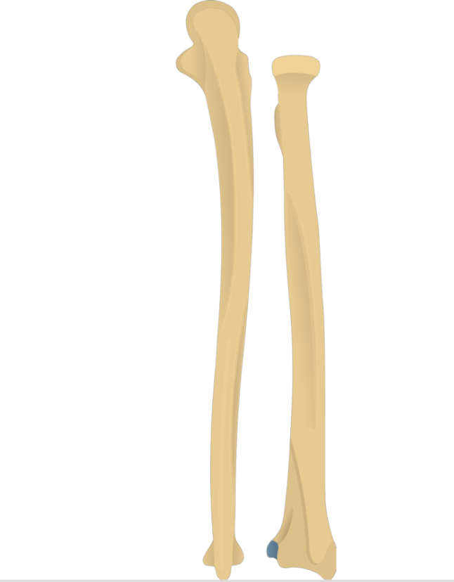
Name this structure
Ulnar notch

right or left?
right radius
-left = anterior view
-right= posterior image

Name this Bone
Ulna (more medial bone in anatomical position)

Name this structure
Olecranon process of ulna

Name this structure
Coronoid process of ulna

Name this structure
Ulnar tuberosity

Name this structure
Styloid process of ulna
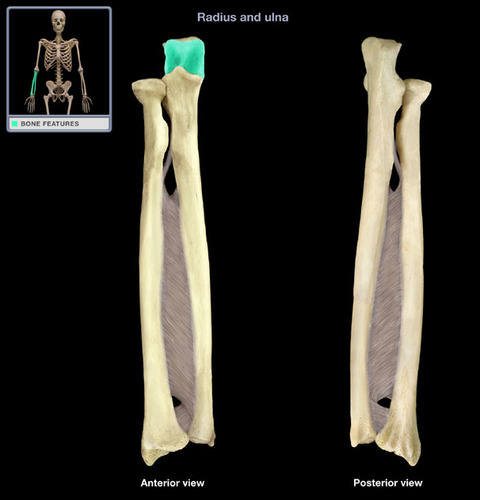
Name this structure
Trochlear notch of ulna

Name this structure
Radial notch of ulna
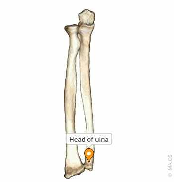
Name this structure
head of ulna

The anatomical name of the joint that occurs here is the middle radioulnar joint
Interosseous border

left or right?
Right
First view: Anterior
Second view: lateral
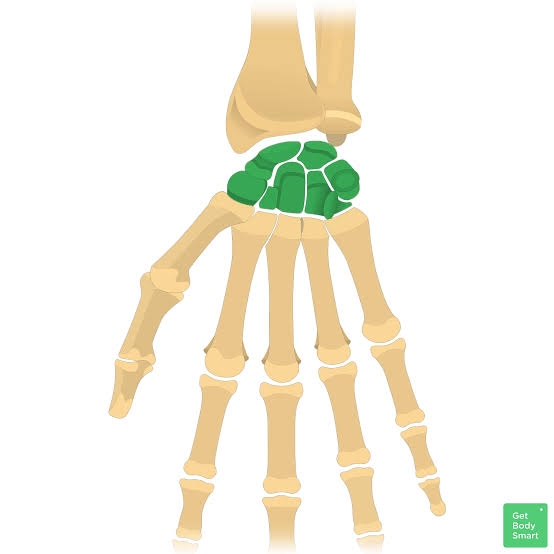
How many carpals in one hand
8
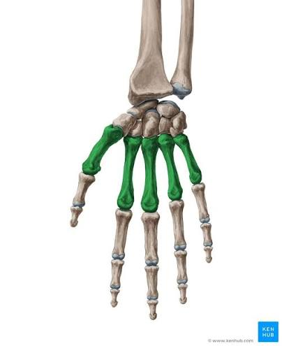
How many metacarpals in one hand
5
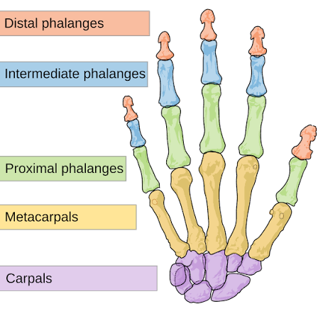
How many phalanges in one hand
14
(remember there are 56 total in the body)


Name the phalanges bottom up
Bottom: proximal phalanges
Middle: middle phalanges
Top: distal phalanges
How are the metacarpals numbered
thumb to pinky
thumb=1 pinky 5
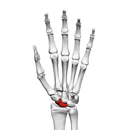
Name this Carpal
scaphoid carpal

Name this Carpal
Lunate
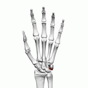
Name this Carpal
Trilateral

Name this Carpal
Pisiform
*cannot see from posterior view)

Name this Carpal
Hamate
hook shaped with hamate hook
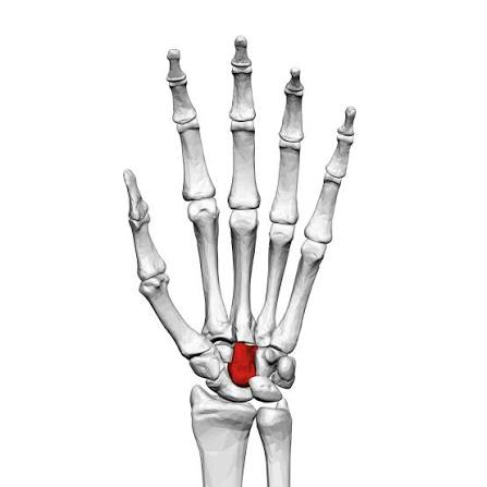
Name this Carpal
Capitate

Name this Carpal
Trapezoid

Name this Carpal
trapezium
How to remember the carpals
Mnemonic: "So Long Pinky Here Comes The Thumb”
Proximal Row (Thumb to Pinky)
So – Scaphoid
Long – Lunate
To – Triquetrum
Pinky – Pisiform
Distal Row (Pinky to Thumb)
Here – Hamate
Comes – Capitate
The – Trapezoid
Thumb – Trapezium:

Pelvic girdle
consisting of the hip (coxal) bones (ilium, ischium, pubis)

Name this bone
Ilium
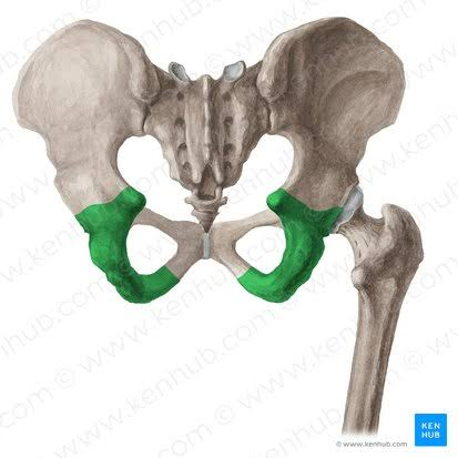
Name this bone
Ischium

Name this bone
Pubis

Name this structure
Pubic symphysis

Name this Structure
Iliac Crest

Name this structure
Sacroiliac joint

Name this structure
Iliac Fossa

Name this structure
Sacral promontory

Name this structure
Pelvic brim

Name this structure
Ischia spine

Name this structure
Acetabulum
fusion of all 3 bones
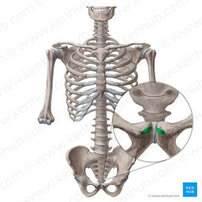
Name this structure
Pubic crest

Name this Structure
Ala of ilium

Name this structure
Ischial tuberosity

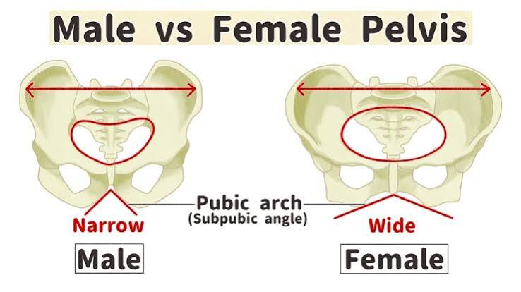
What are the structures that make the male and female pelvis different
Female: flared ilia, smaller ischia, >90° pubic arch, wider pelvic opening
Male: upright ilia, larger ischia, <90° pubic arch, narrower pelvic opening

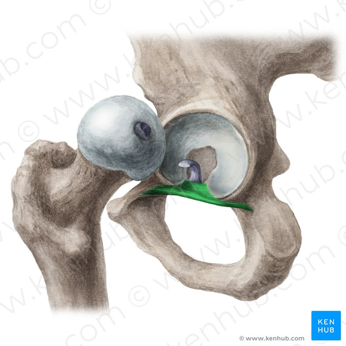
Name this structure
Acetabular labrum
fibrocartilaginious
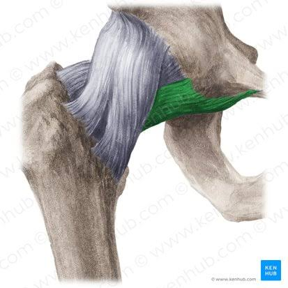
Name this ligament
Transverse acetabular ligament

Name this ligament
Ligamentum Teres

Name this ligament
iliofemoral ligament
Name this ligament
Pubofemoral ligament

Name this ligament
Ischlofemoral ligaments
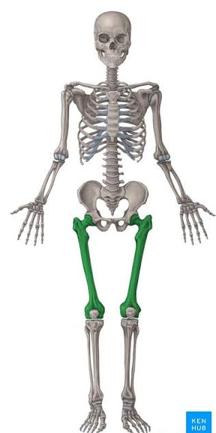
Name this bone
Femur

Name this structure
Fovea capitis


Name this structure
Head of femur
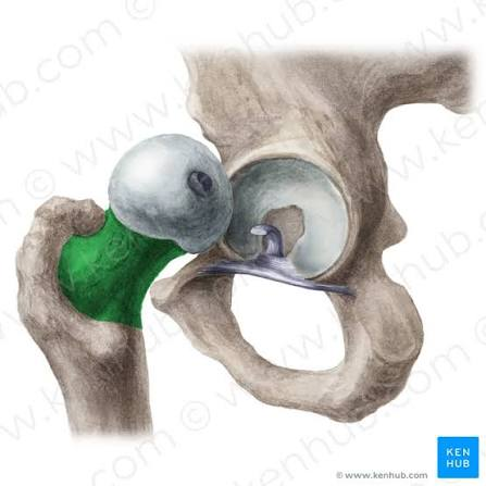
Name this structure
Neck of femur
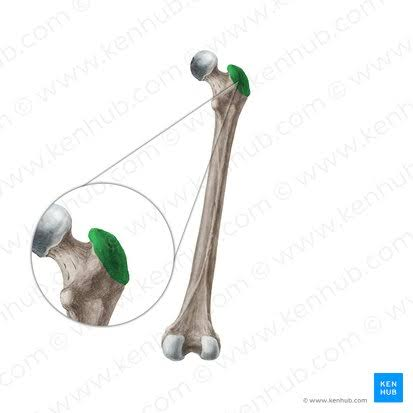
Name this structure
Greater trochanter of femur

Name this structure
Lesser trochanter of femur

Name this structure
intertrochanteric line (anterioir)

Name this structure
Intertrochanter Crest (posterioir)

Name this structure
Linea aspera of femur

Name this structure (only the part where it splits)
posterior view
medial and lateral supracondylar lines of femur
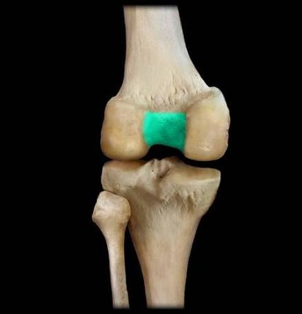
Name this structure
intercondylar notch of femur
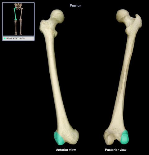
Name this structure
Lateral condyle of femur

Name this structure
Medial condyle of femur