Osteology
 Call Kai
 Learn
Learn Practice Test
 Spaced Repetition
 Match
Match Flashcards
 Knowt Play
1/93

There's no tags or description
Looks like no tags are added yet.
Name | Mastery | Learn | Test | Matching | Spaced | Call with Kai | Chat |
|---|
No analytics yet
Send a link to your students to track their progress
94 Terms
Anatomy
The term applied to the science of the structure of
the body
Physiology
he study of the function of the body organs
Osteology
The detailed study of the body of knowledge relating to the bones of the body
The adult human skeleton is composed of
206 primary bones
Bones provide the following:
1. Attachment for muscles
2. Mechanical basis for movement
3. Protection of internal organ
4. A frame to support the body
5. Storage for calcium, phosphorus, and other salts
6. Production of red and white blood cells
Correct Anatomic Position
Patient standing erect with the face and eyes directed forward, arms extended by the
sides with palms of the hands facing forward, heels together, and toes pointing
anteriorly
Four types of body planes
Sagittaal
Coronal
Horizontal
Oblique
The midsagittal plane and a horizontal plane intersect at the umbilicus and create the
boundaries.
This is useful for describing the location of the various abdominal organs.

Sthenic
• 50% of population
• Heart- moderately transverse
• Lungs- Moderate length
• Diaphragm- Moderately high
• Stomach- High, upper left
• Colon- spread evenly, slight dip in transverse
colon
• Gallbladder- Centered on right side, upper
abdomen
Hyposthenic
• 35% of population
• Organs and characteristics for this habitus are
intermediate between sthenic and asthenic
body habitus types; this habitus is the most
difficult to classify.
Asthenic
• 10% of population
• Heart- nearly vertical and at midline
• Lungs- long, apices above clavicles, may be
broader above base
• Diaphragm- Low
• Stomach- Low and medial, in the pelvis when
standing
• Colon- Low, folds on itself
• Gallbladder- Low and nearer the midline
Hypersthenic
• 5% of the population
• Heart- axis nearly transverse
• Lungs- Short, apices at or near clavicle
• Diaphragm- high
• Stomach- High, transverse, and in the middle
• Colon- Around periphery of abdomen
• Gallbladder- High, outside, lies more parallel
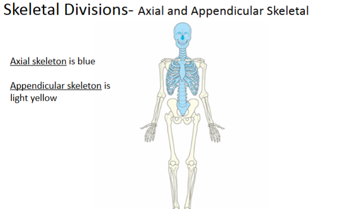
The 206 bones are divided into two main groups:
axial and appendicular
Axial Skeleton
Supports the skeleton and protects the head and trunk with 80 bones
Appendicular Skeleton
Allows the body to move in various positions and from place to place with its 126 bones
Compact bone
Strong, dense outer layer
Spongy bone
– Inner, less dense layer
– Contains a spiculated network called trabeculae
Trabeculae filled with
red and yellow marrow
Periosteum
tough fibrous connective tissue that covers bone, except at articular ends.
Compact bone
dense outer layer
Spongy/Cancellous bone
Less dense inner portion of bone
Trabeculae
Softer spongy bone contains a spiculated network of interconnecting spaces.
Medullary Cavity
contains trabeculae Filled with yellow marrow
Endosteum
The tissue lining the medullary cavity.
Bone Marrow
Red produces red and white blood cells found in ends of long bone. Yellow stores adipose fat cells.
Nutrient foramen
near the center of all long bones is an opening in the periosteum
Bone Development
• Ossification is the term that applies to the development and formation of bones
• Begins in the second month of embryonic life
• Two processes
– Intermembranous ossification
– Endochondral ossification
Intermembranous ossification
Flat bones- bone of skull, clavicles, mandible and sternum. Before birth these bones are not
joined.
Endochondral ossification
Short, irregular and long bones
Two distinct centers of development
Primary ossification- diaphysis and metaphysis
Secondary ossification- epiphysis
Classification of Bones
• Classified by shape
– Long
– Short
– Flat
– Irregular
– Sesamoid
Long Bones
Long bones found only in limbs
– Consist of body and two enlarged articular ends
– Consist of long cylindrical shaft called the body and
two enlarged, rounded ends that contain a smooth,
slippery articular surface.
– Examples: femur and humerus, the phalanges of the
fingers and toes are also considered long bones. A
primary function of long bones is to provide support.
Short bones
– Consist mainly of cancellous bone with a thin
outer layer of compact bone containing red
marrow.
– Example: carpal bones of
the wrist and tarsal bones of the
the ankle
Flat bones consist of two plates of compact
bones
– Middle layer of cancellous bone called diploë
– Examples: sternum, cranium and scapulae. The
flat surface provides protection, and their broad
surfaces allow muscle attachment.
Irregular bones are peculiarly shaped
– Examples: vertebrae, facial bones and pelvis. They
have compact bone on the exterior and cancellous
bone containing red marrow in the interior. They
serve for muscles, tendons and ligaments
attachment or they attach to other bones to
create joints.
Sesamoid bones
– Very small and oval
– Develop inside and beside tendons
– Protect the tendon from excessive wear
– Largest is patella
Three subdivisions based on mobility of joint
– Synarthroses = immovable
Sutures of the skull
– Amphiarthroses = slightly movable
Inferior tibiofibular joint
– Diarthroses = freely movable
Bones of most joints
Synarthroses
Synarthroses = immovable
Sutures of the skull
Amphiarthroses
slightly movable
Inferior tibiofibular joint
Diarthroses
freely movable Bones of most joints
Fibrous Joints
strongest joints in the body, virtually immovabel
Syndesmosis
an immovable joint or slightly
movable joint united by sheets of fibrous tissue.
Example- Inferior tibiofibular joint
Suture
An immovable joint occurring only in the
skull. Held together by strong connective tissue.
Example- sutures of the skull
Gomphosis
An immovable joint occurring only
in the roots of the teeth.
Cartilaginous Joints
similar to fibrous joints that
they do not have a joint cavity and they are virtually
immovable. Hyaline cartilage and fibrocartilage
unites these joints.
Symphysis
Slightly movable, example-pubic
symphysis or between each vertebral body
Synchondrosis
An immovable joint containing a
rigid cartilage that unites two bones.
Synovial Joints
permit a wide range of motion,
and they all are freely movable.
Gliding
uniaxial movement, example-
intercarpal and intertarsal joints of wrist and
foot
Hinge
uniaxial movement, permits only
flexion and extension. Example- elbow, knee
and ankle
Pivot
uniaxial movement, these joints allow
only rotation around a single axis. Example-
atlas and axis of cervical spine.
Ellipsoid
Biaxial movement, primary.
Movement in two directions at right angles to
each other. Examples- radiocarpal joint.
Saddle
Biaxial movement, this joint permits
movement in two axes. Example-
Carpometacarpal joint
Ball and Socket Join
multiaxial movement,
Example- hip joint and shoulder joint
Condyle
rounded process at an extremity.
Coracoid or Coronoid
crownlike process
Crest
ridgelike process
Epicondyle
projection above condyle
Facet
small, smooth-surfaced process for articulation with another structure.
Hamulus
hook-shaped process
Head
hornlike process on a bone
Horn
hornlike process on a bone
Line
less prominent ridge than a crest, a linear elevation.
Malleolus
club-shaped process
Protuberance
projecting part or prominence
Spine
sharp process
Styloid
long, pointed process
Trochanter
either of two large, rounded, and elevated processes.
Tubercle
small, rounded and elevated process.
Tuberosity
large, wounded and elevated process
Fissure
cleft or deep groove
Foramen
hole in a bone
Fossa
pit or hollow space
Groove
shallow linear channel
Meatus
ubelike passageway
Notch
an indentation into border of a bone
Sinus
recess, groove, cavity or hollow space
Sulcus
furrow or trench
Closed
fracture that does not break through
the skin
Open
Serious fracture in which broken bone
or bones project through the skin.
Nondisplaced
fracture in which bone retains
its normal alignment.
Displaced
serious fracture in which bones are
not in anatomic alignment.
Common classifications of fractures:
Compression
Open or compound
Simple
Greenstick
Transverse
Spiral or Oblique
Comminuted
Impacted
Radiation
is energy that is transmitted by waves through
space or through a medium (matter).
Ionization
is any process by which a neutral atom gains or
loses an electron, thus acquiring a net charge. This process
has the ability to disrupt the composition of matter.
Sound
Medical Sonography
Electrical
Electrocardiography
Heat (thermal)
Thermography
Magnetic
Magnetic resonance imaging
Electromagnetic
X-rays
Radio waves
Nuclear
Gamma radiation
Radiography
Uses electromagnetic energy
in the form of x-rays to create
medical images