Introduction to Mycology (Exam 3)
1/77
There's no tags or description
Looks like no tags are added yet.
Name | Mastery | Learn | Test | Matching | Spaced | Call with Kai | Chat |
|---|
No analytics yet
Send a link to your students to track their progress
78 Terms
Fungi are ______ and plant-like.
eukaryotic
Fungi are ______ and absorb nutrients from the enviroment.
heterotrophic
Reproduction of fungi can be ______ and ______.
sexual, asexual
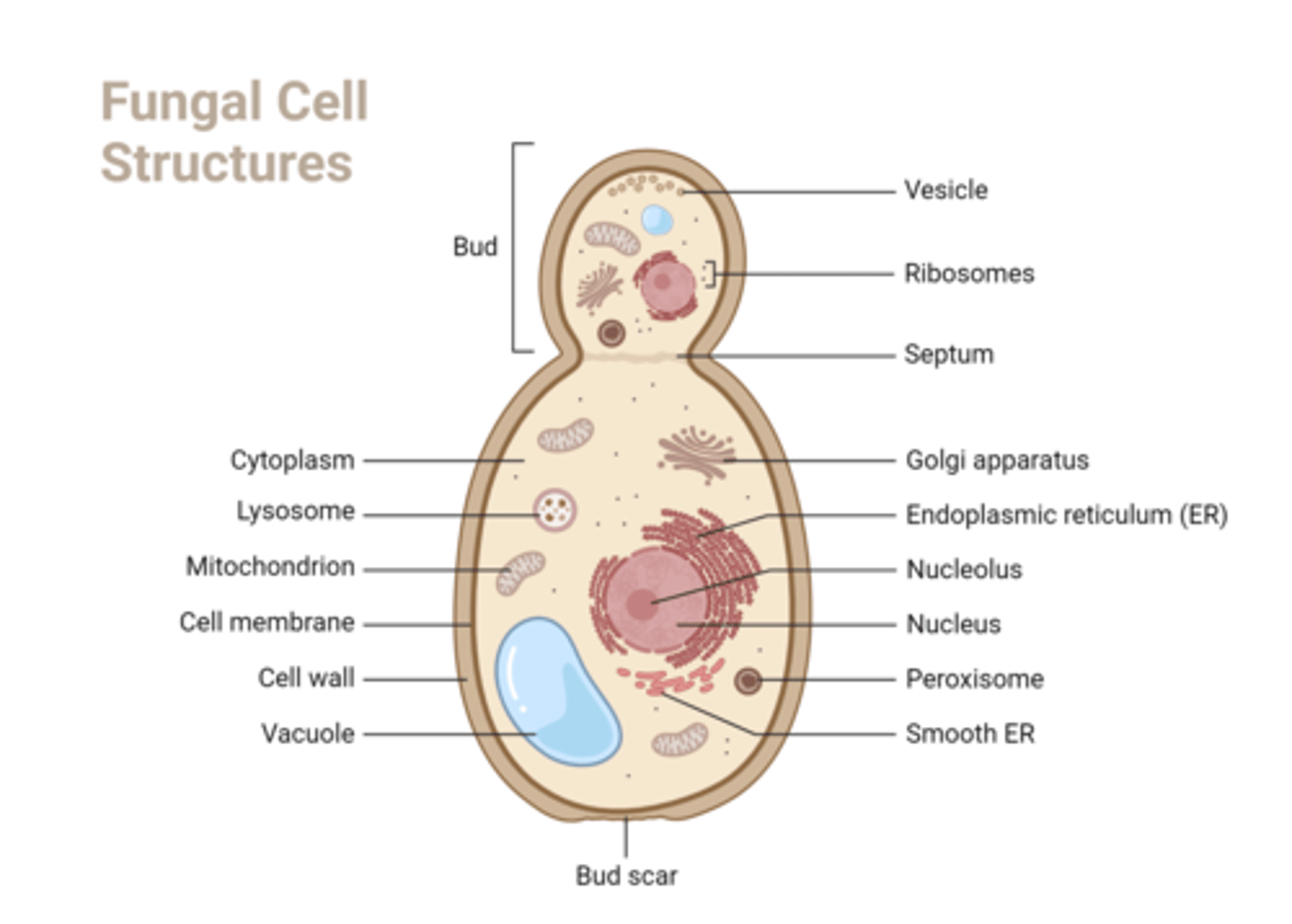
Fungal cell walls are made of ______.
chitin
Fungal cell membranes contain ______.
ergosterol
What are the specimen types that are acceptable for fungal culture?
Sputum, urine, skin, nails, hair/scalp, CSF/sterile BF, tissue, blood/BM, stool, genital
Media without ______ are used to isolate and identify fungi.
supplements
These media are 4-5% ______.
sugar
These media are kept at a pH of ______.
3.8-5.5
Most fungi grow at ______C, but some can grow at ______C.
22-28C, 30-37C
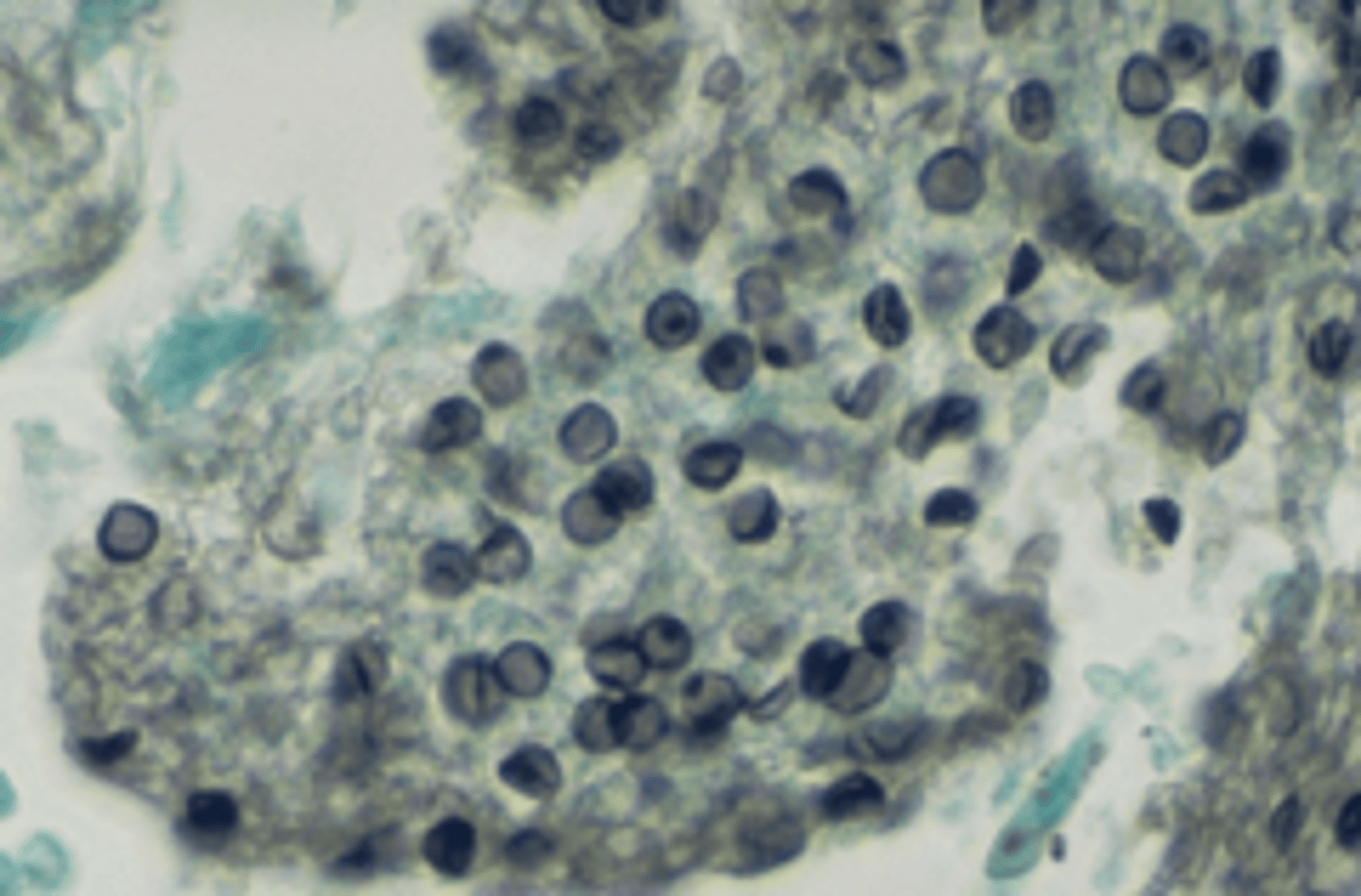
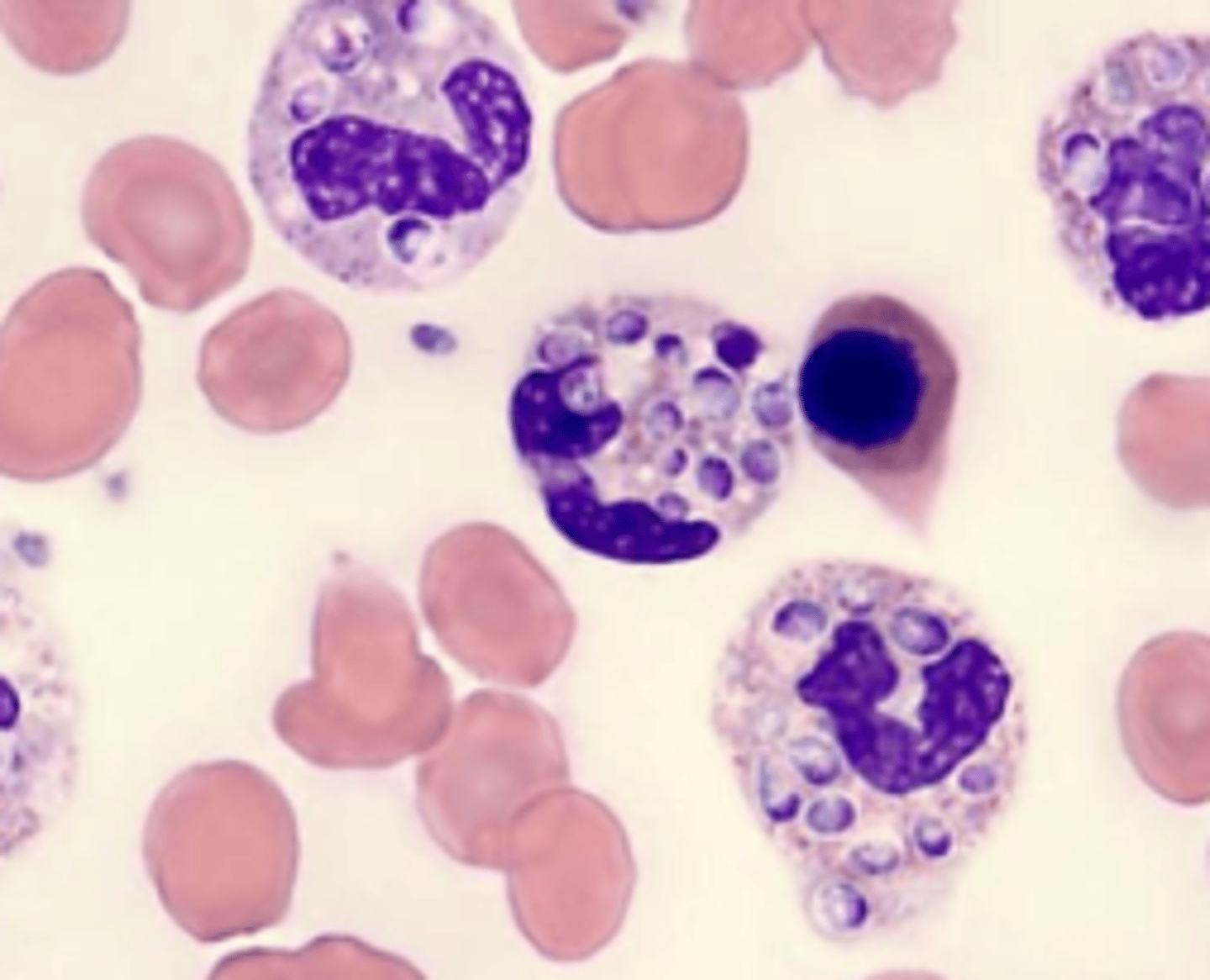
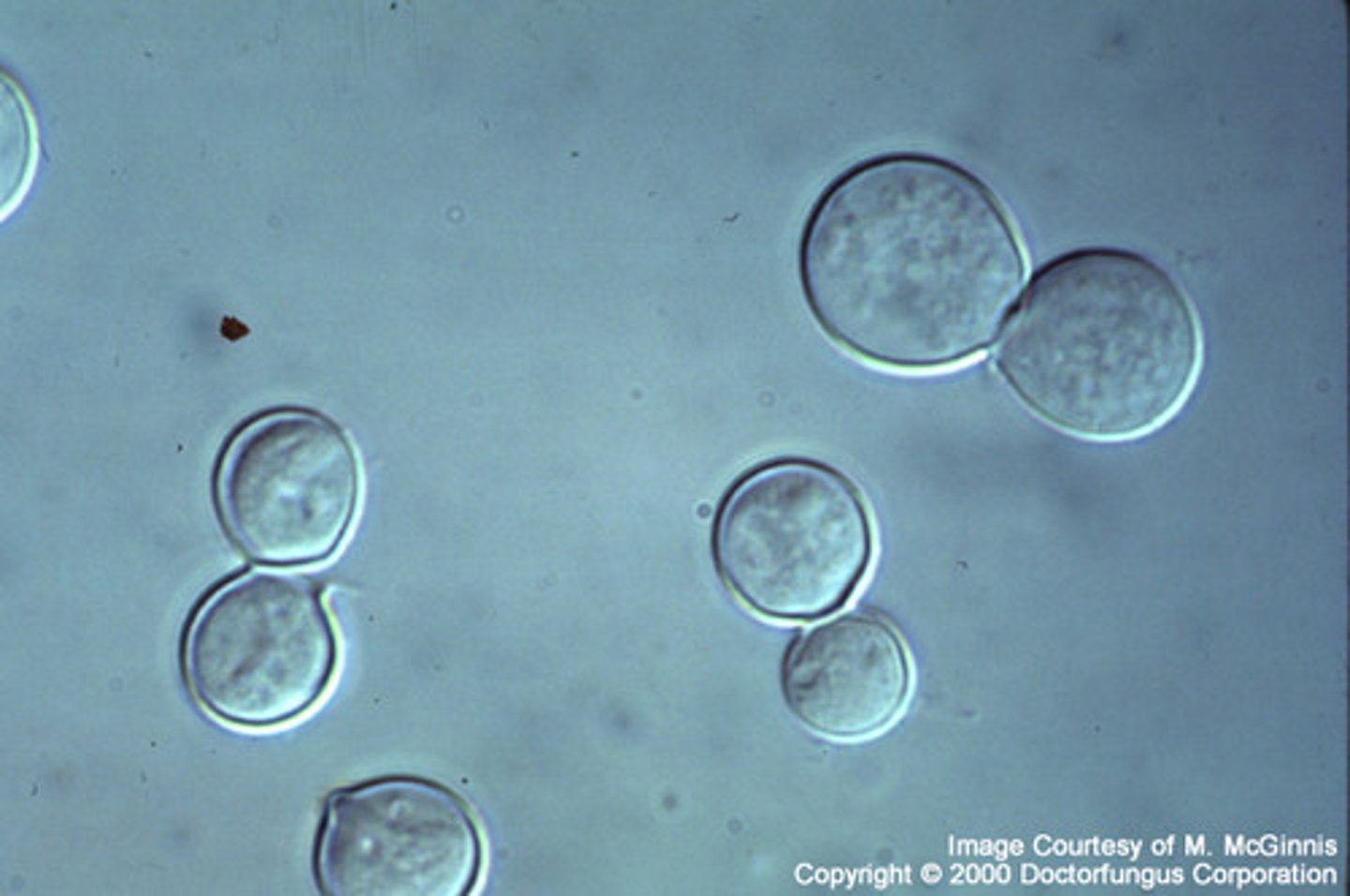
India Ink stain
Dark background
Shows capsule of cryptococcus neoformans
Gomori Methenamine Silver (GMS) stain is specific for ______.
pneumocystis (brown)
GMS will stain ______ and ______ yeast.
living, dead
Gram stain of yeast is ______.
accidental
Periodic Acid Schiff (PAS) will stain fungal cell walls ______.
dark magenta
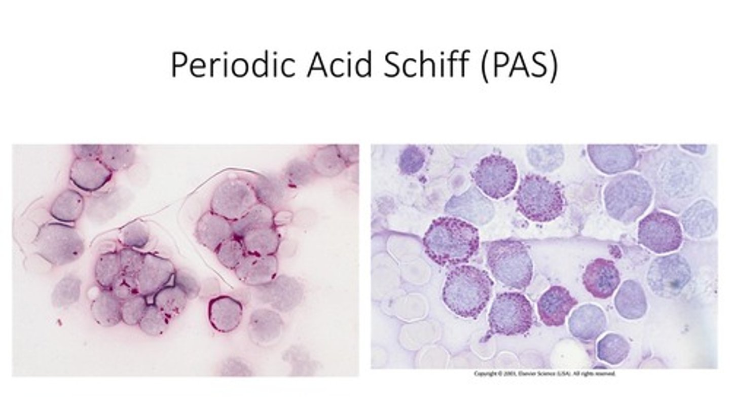
PAS stain only works on ______ fungi.
living
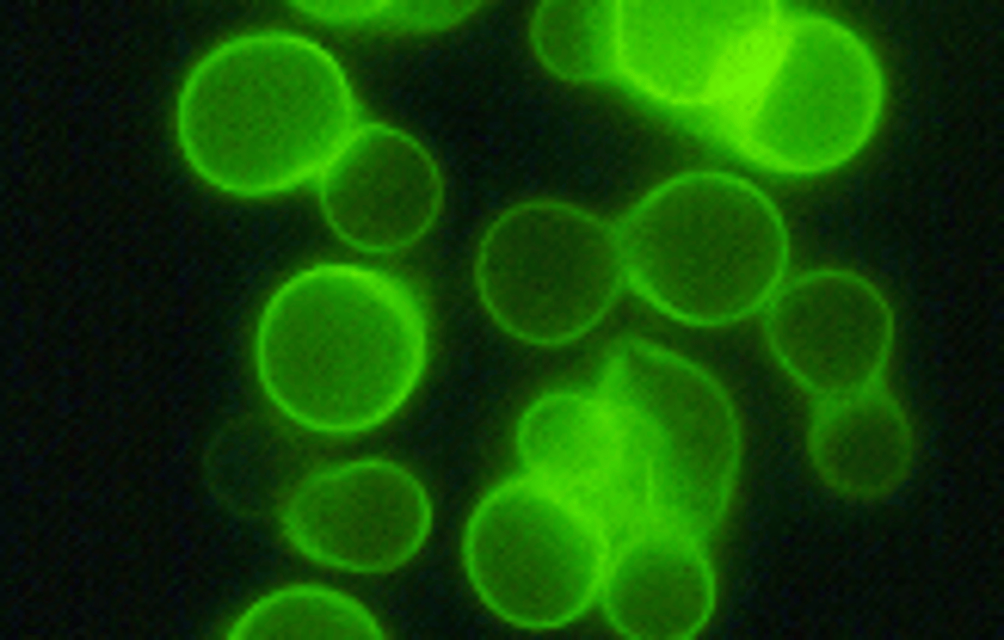
Calcofluor White stain is ______.
fluorescent
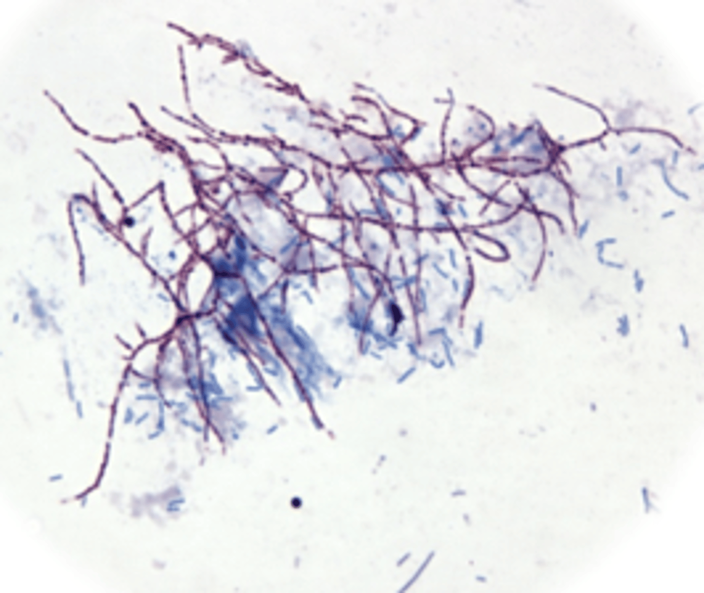
Modified Kinyoun will stain fungi ______.
purple
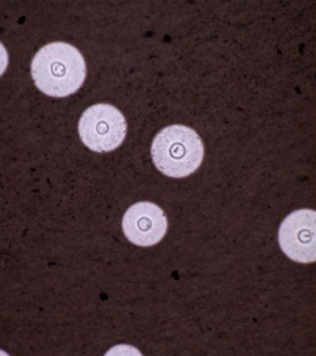
KOH preparation
10% potassium hydroxide
Giemsa/Wright stain
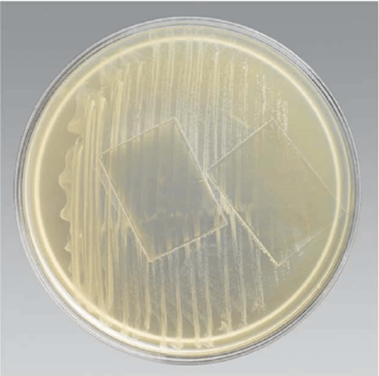
What are the nonselective media for fungi?
- Sabouraud's Dextrose
- Emmon's Modified Sabouraud's Dextrose
What are the selective media for fungi?
- Mycosel
- Inhibitory Mold Agar
What are the enriched media for fungi?
- Brain Heart Infusion (BHI) with 5% sheep RBCs
- SABHI
What are the special/differential media for fungi?
- Niger/Birdseed/Caffeic acid agar
- Trichophyton agar
- Dermatophyte test medium
- Morphology media
Niger/Birdseed/Caffeic acid agar is differential for ______.
Cryptococcus neoformans
Trichophyton agar is used for what types of organisms?
Those that frow on the skin, hair, and nails
Dermatophyte test medium differentiates ______.
Tinea (ringworm)
Dermatophyte test medium will change from ______ to ______ if there is a dermatophyte present.
yellow, red
What are the morphology media and what organisms are they for?
- Cornmeal agar (molds)
- Rice extract (molds)
- Potato dextrose (yeast)
- RIOT agar (yeast)
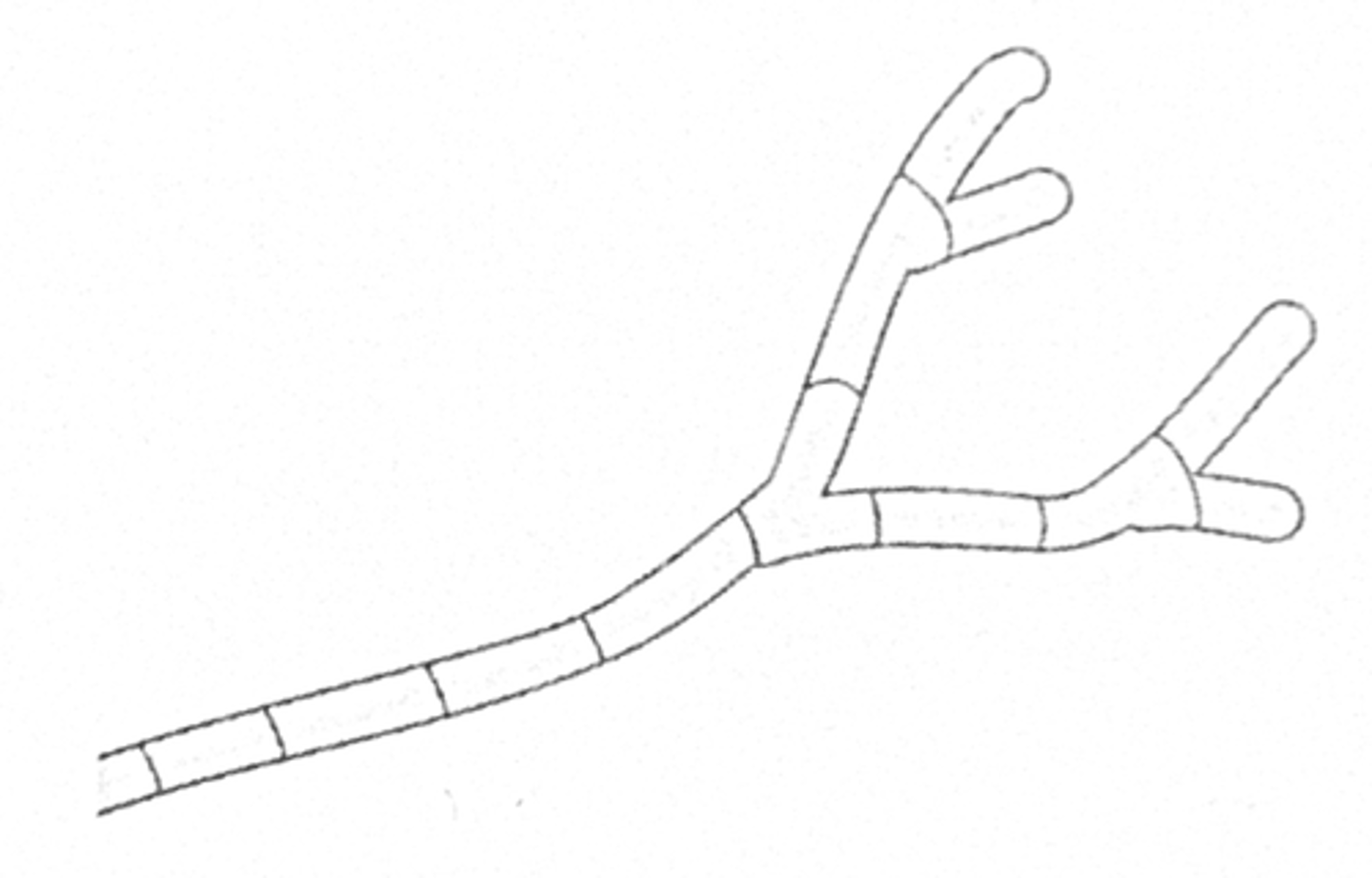
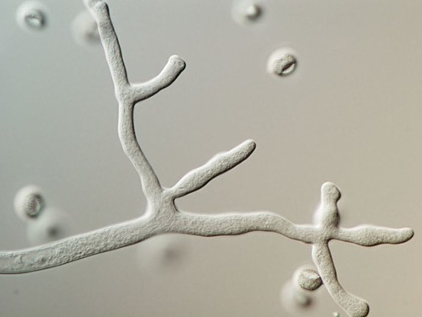
Septate hyphae
Cross walls within hyphae
Aseptate hyphae
No cross walls within hyphae
Pseudohyphae
Chains of cells formed by budding
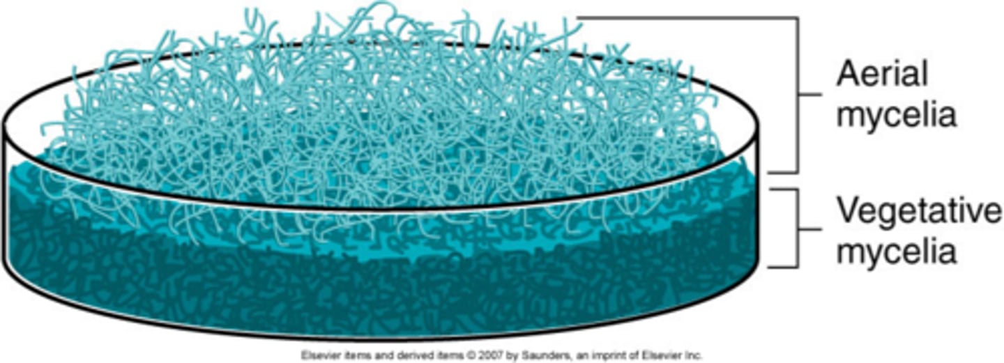
Aerial mycelium
Grows off surface
Gives rise to spores
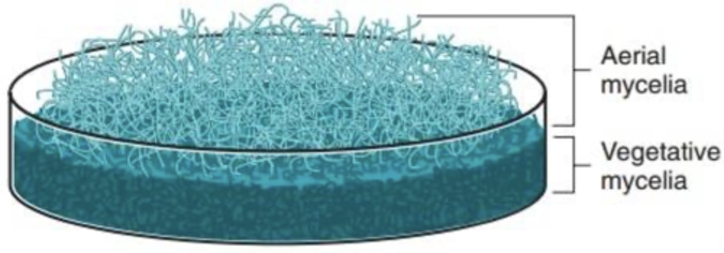
Vegetative mycelium
Grows into medium
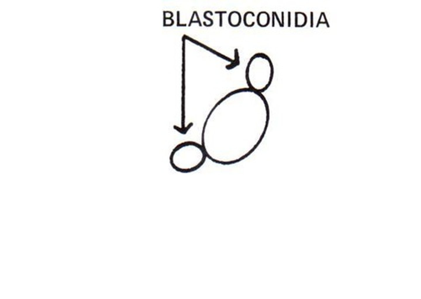
Blastoconidia
Formed by budding along hyphae, pseudohyphae or single cell
Arthroconidia
Breaking or fragmentation of hyphae at separation point
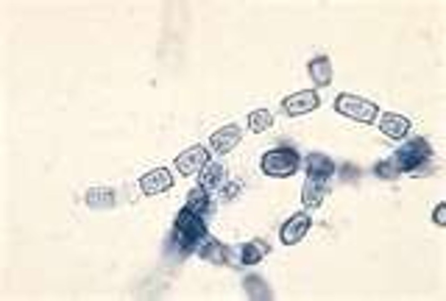
Arthroconidia are ______ or ______.
rectangular, barrel shaped
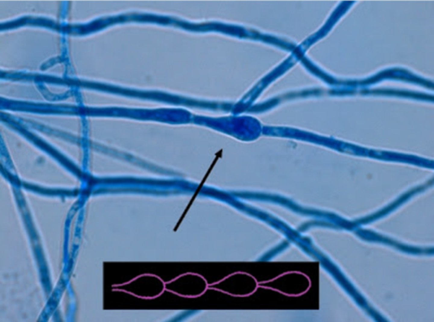
Chlamydoconidia
Enlarged cell, thick wall, contains stored food
Greater in diameter than hyphae
Sexual reproduction includes the joining of two ______ from compatible mating strains.
nuclei
Asexual reproduction is the formation of ______ following mitosis.
conidia
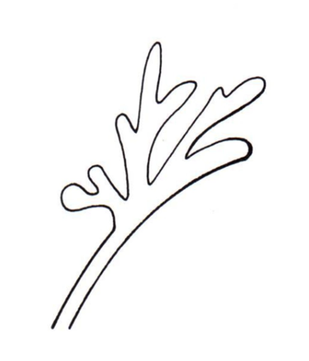
Favic chandeliers hyphae
Deer antlers/branching
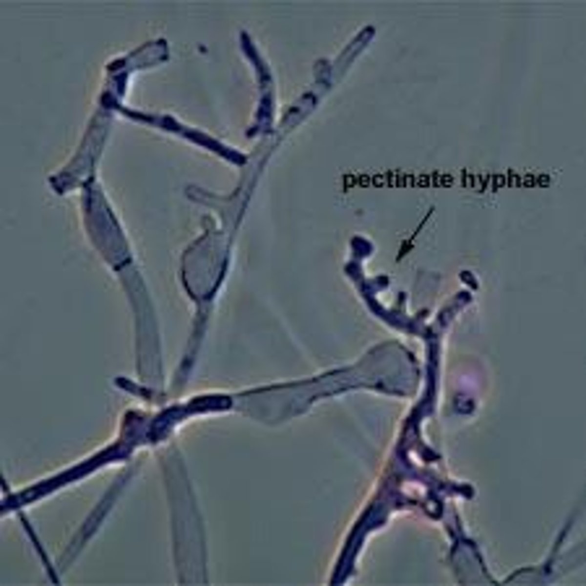
Pectinate hyphae
Wide-toothed comb
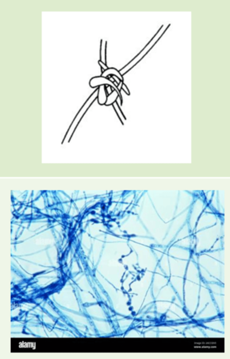
Nodular hyphae
Twisted, knotted
Racquet hyphae
Tennis racket shaped
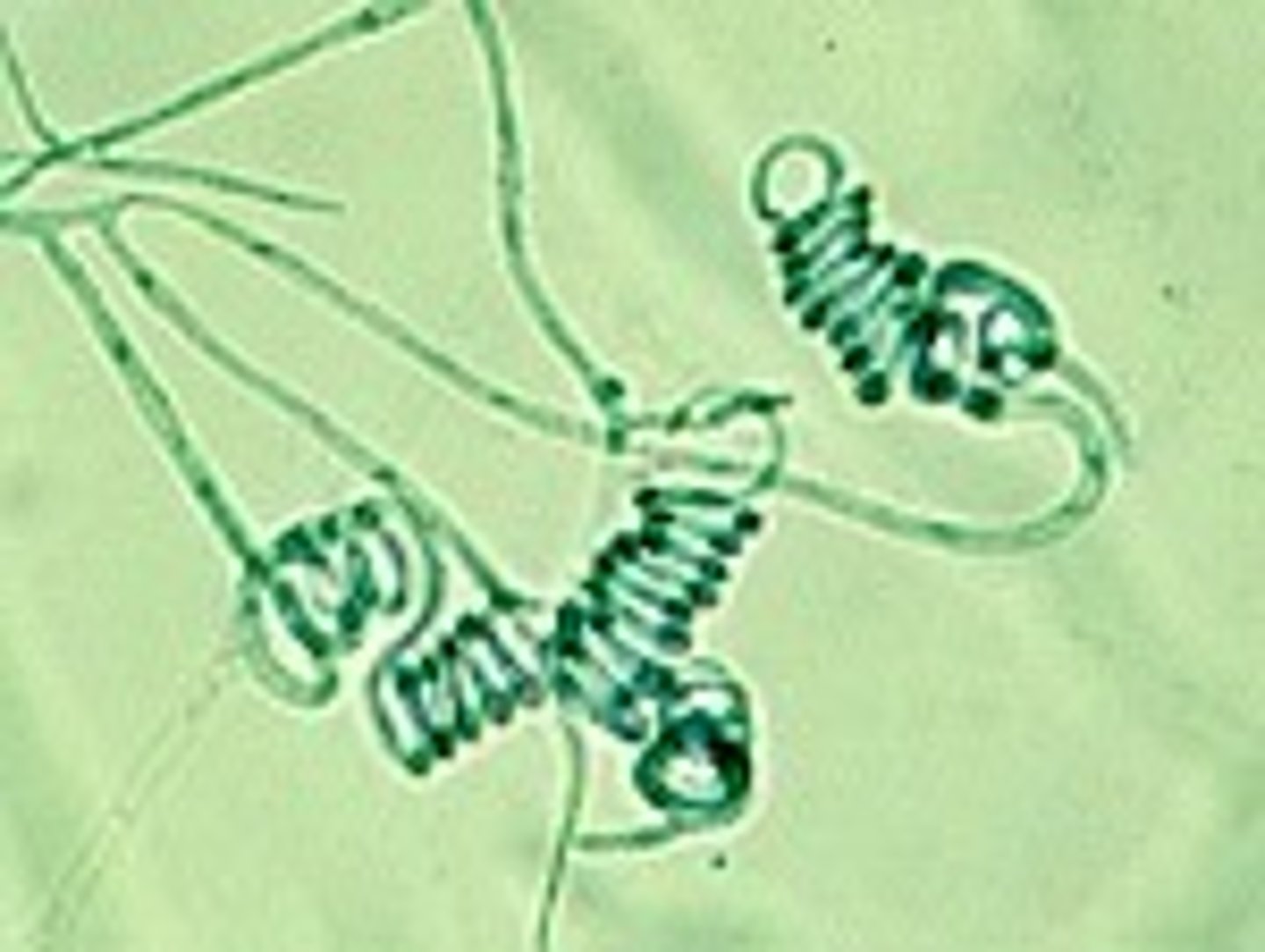
Spiral hyphae
Coiled
Dematiaceous
Dark colored
Hyaline
Light colored
Chromomycosis/Chromoblastomycosis
Color-related mycological infection
Mycetoma
Entrance point/inoculation into the skin
Phaeohyphomycosis
Broad term for infections caused by pigmented fungi that show dark-walled sepatate hyphae intissue
Sporotrichosis
Subcutaneous fungal infection caused by Sporothrix schenckii complex
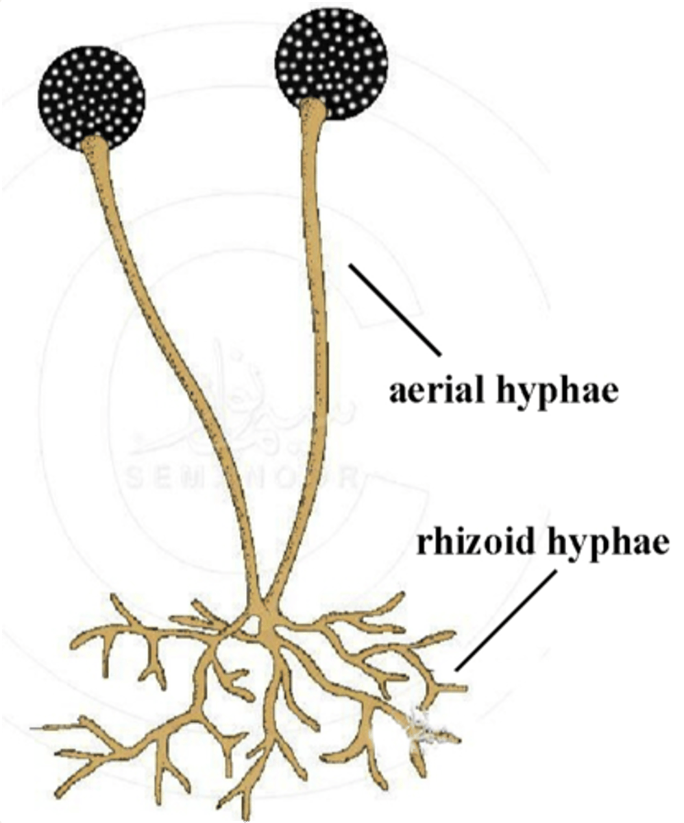
Rhizoid
Root-like structure
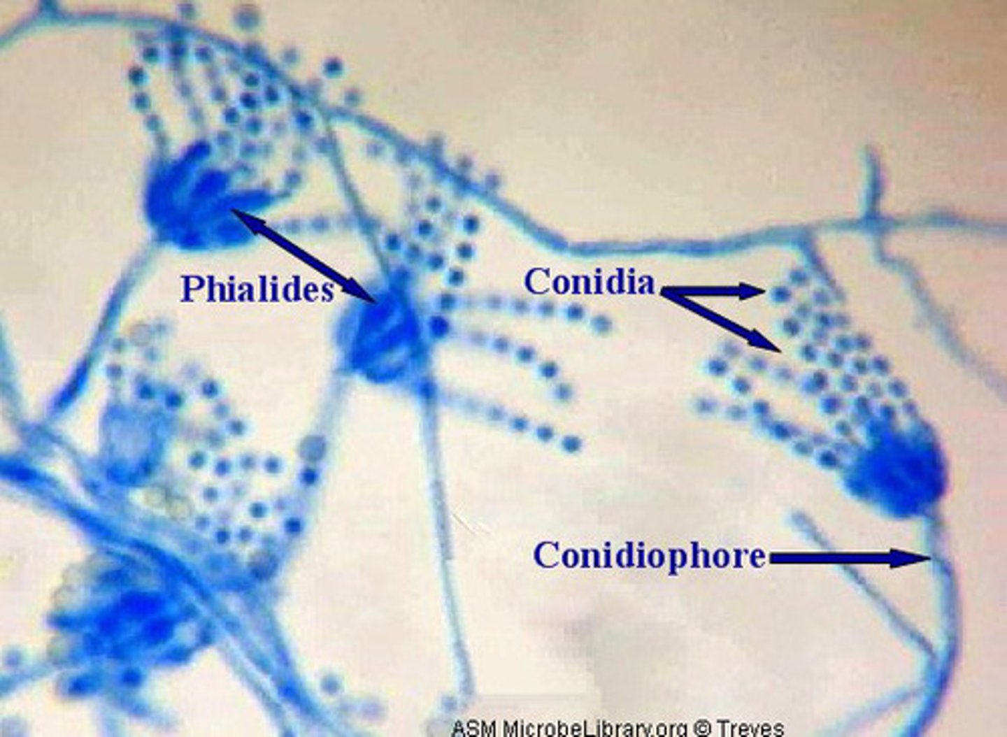
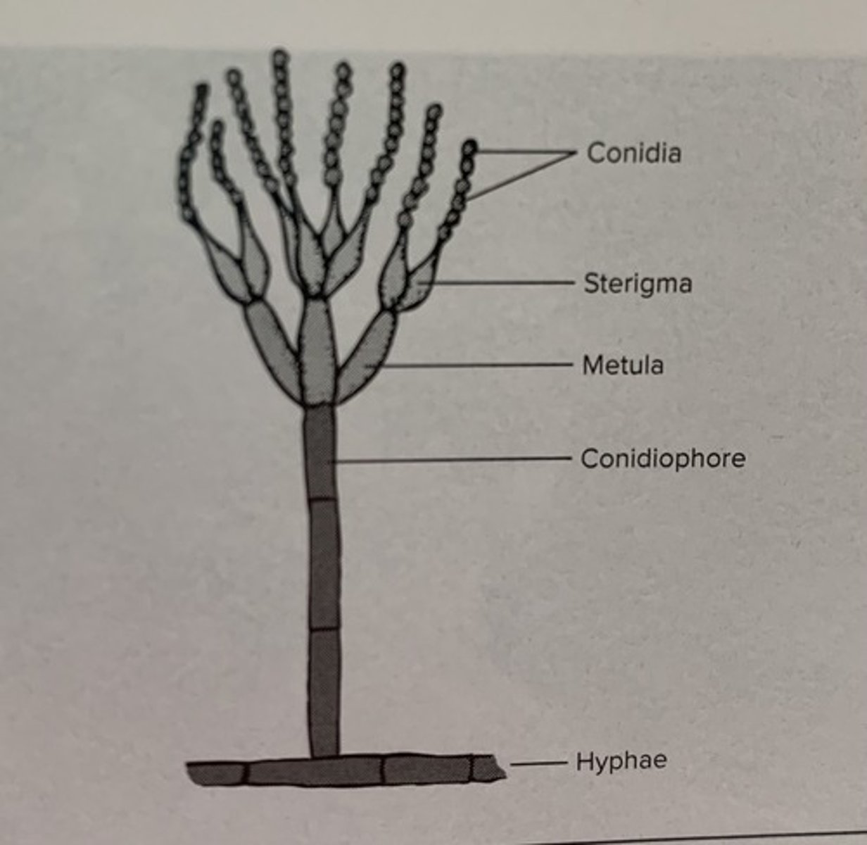
Phialid
Small, flask shaped
At the end of conidiophore
Bear conidia

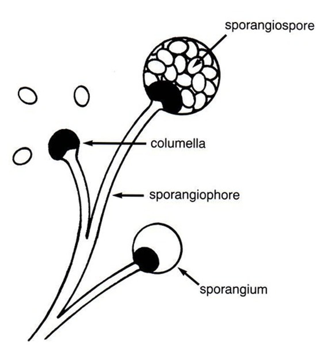
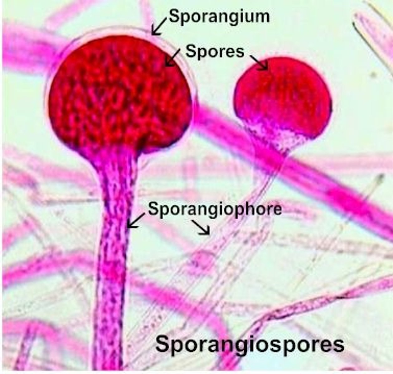
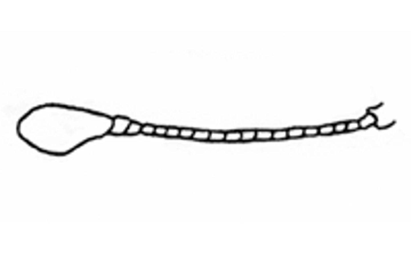
Sporangiospore
Asexual spores produced inside the sporangium
Vesicle
Enlarged area at the end of conidiophore
Sporangium
Sac-like structure with sporangiospores
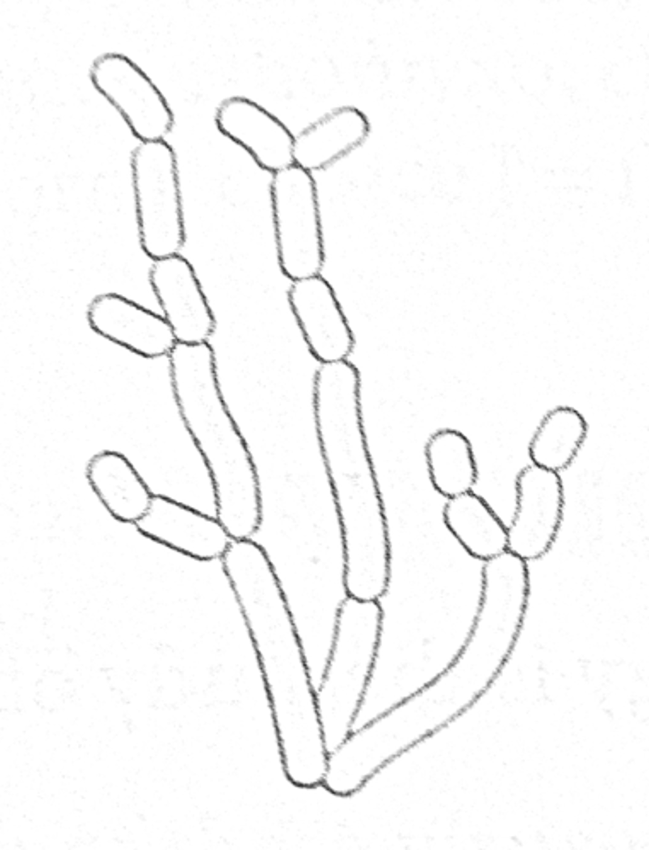
Conidia
Asexual spores produced by mitosis
Formed at the tips or sides of conidiophores
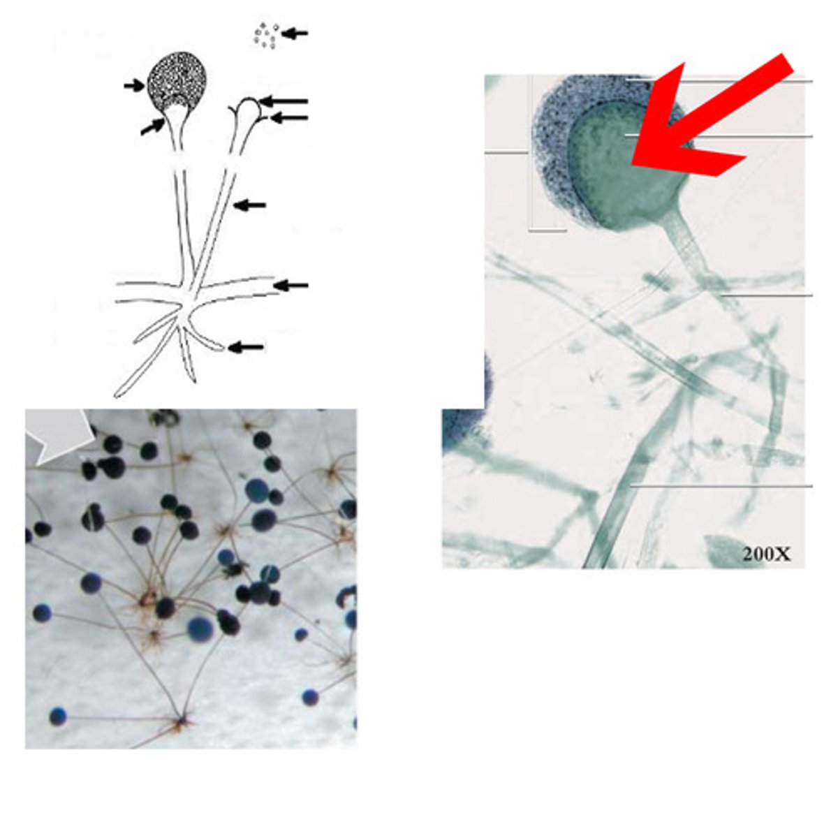
Columella
Enlarged, dome-shaped
Tip of sporangiophore
Conidiophore
Specialized hyphal structure
Stalk on which conidia form

Sporangiophore
Specialized hyphae
Bears sporangium

Metula
Stolon
Horizontal hyphae
Grows along surface
Bears rhizoids and sporangiophores
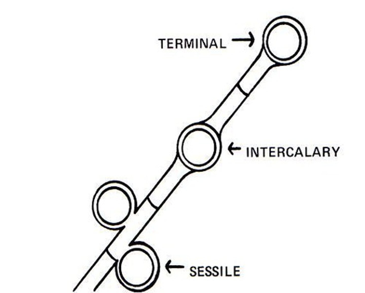
Macroconidia
Thick/thin walled, spiny/smooth, club/oval shaped
On conidiophore
Multi-celled
"Pod"
Cells are the "peas"
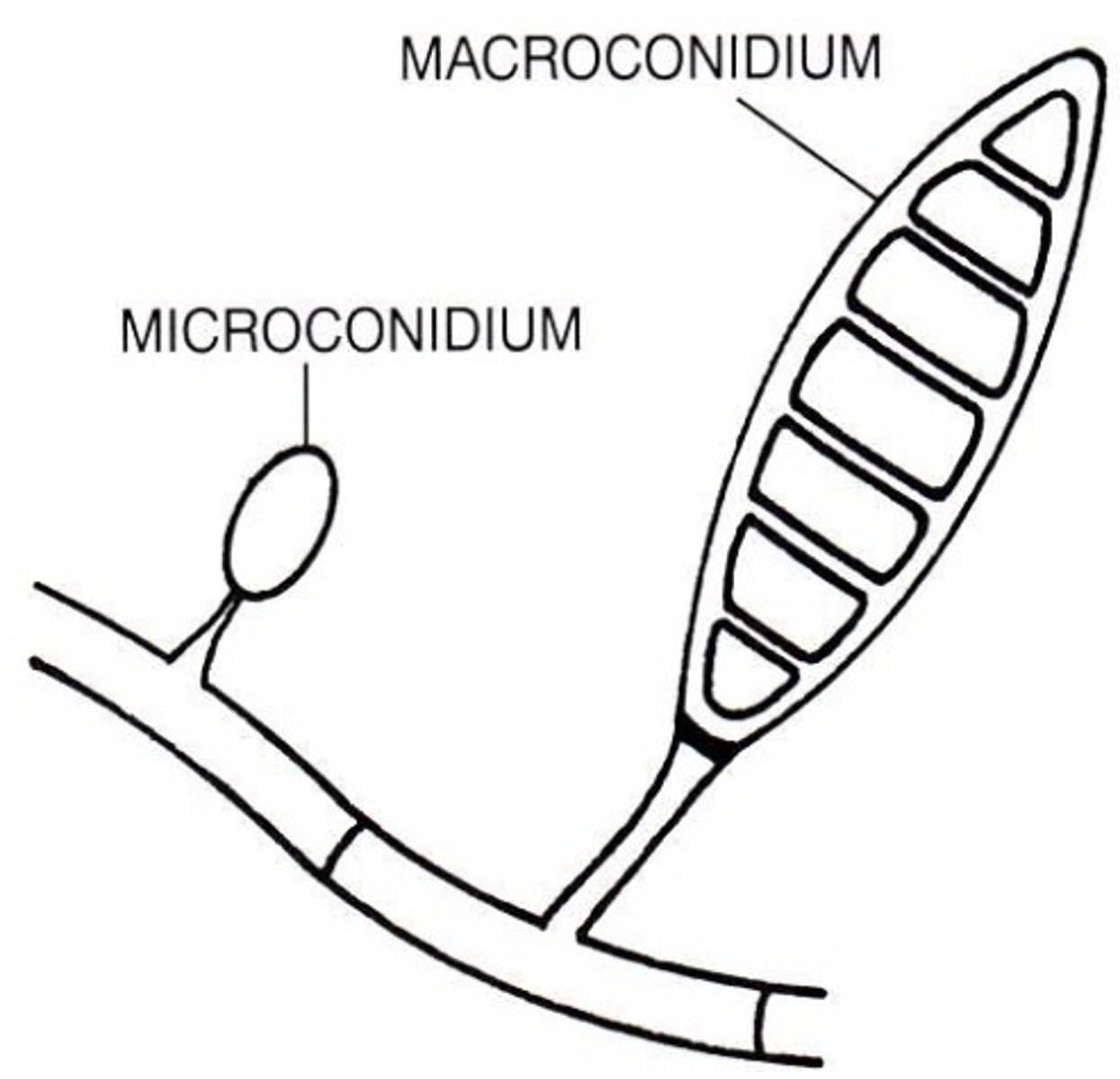
Microconidia
One-celled
On the outside of the macroconidia, like leaves

Verrucose
Wrinkled, bumpy surface
Clavate
Club-shaped
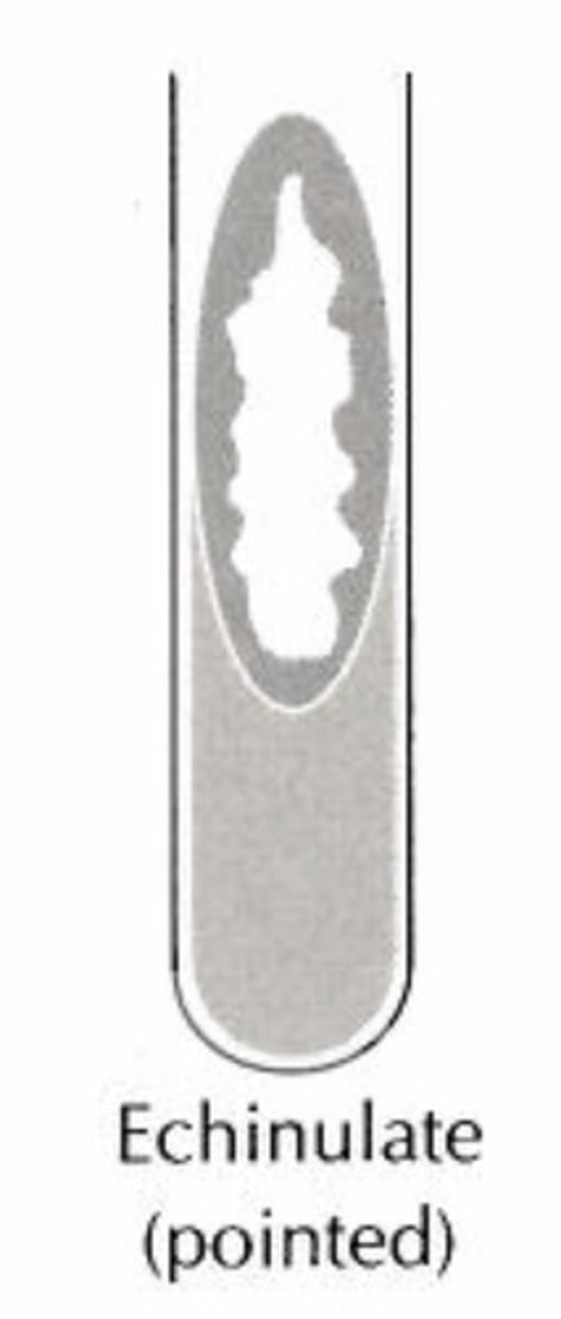
Echinulate
Globose
Spherical
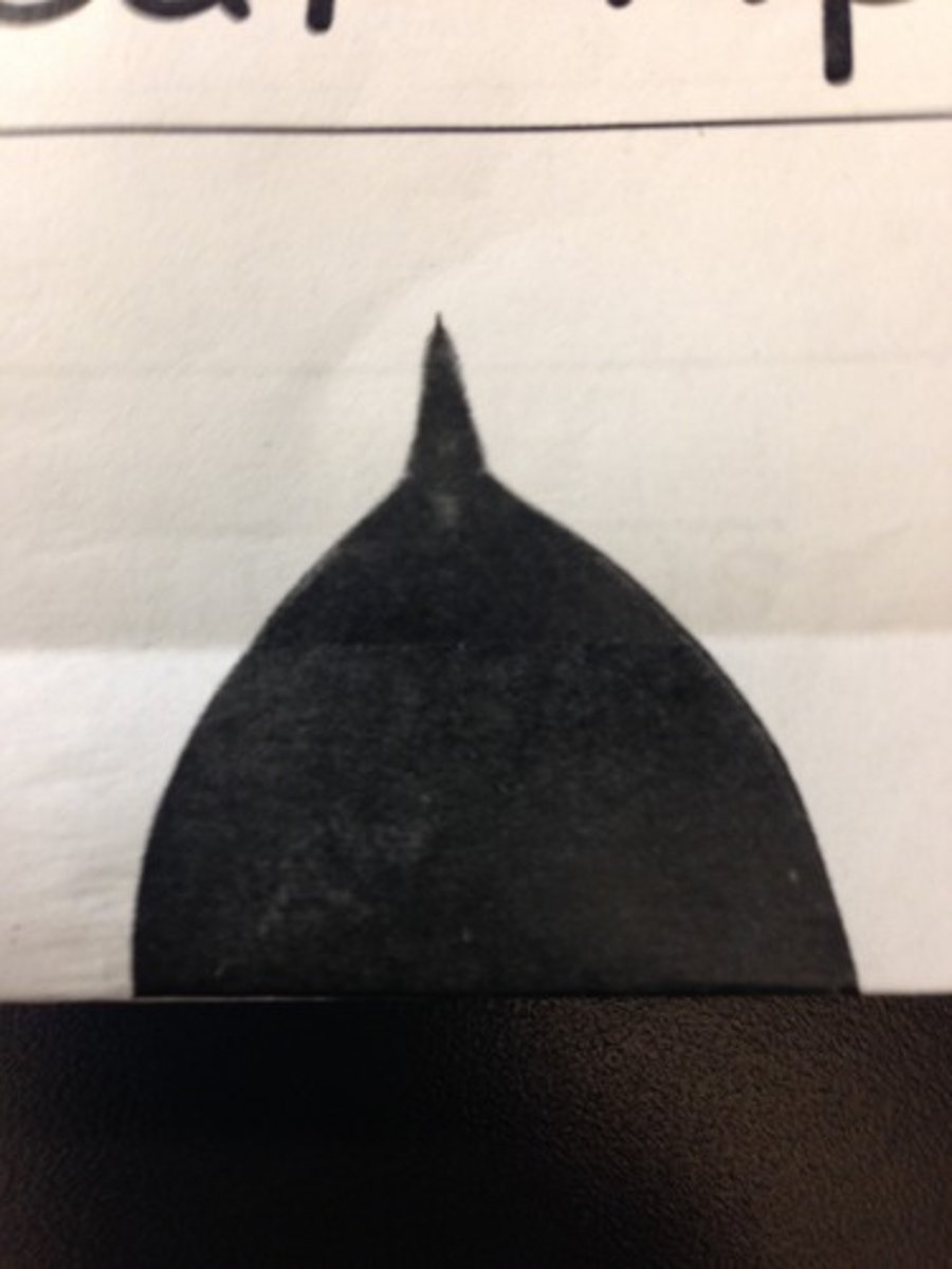
Apiculate
Ace of spades
Pyriform
Pear-shaped
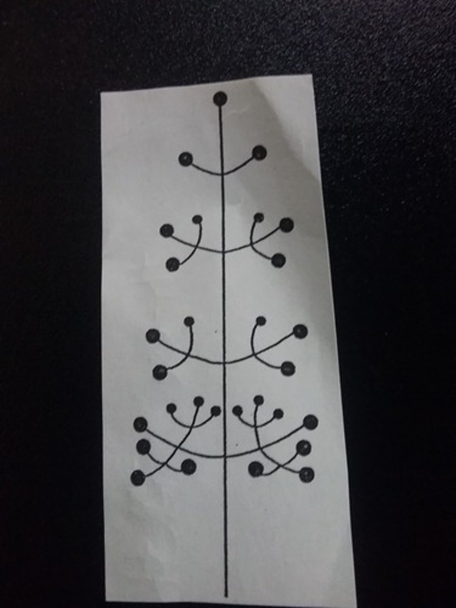
En thryse
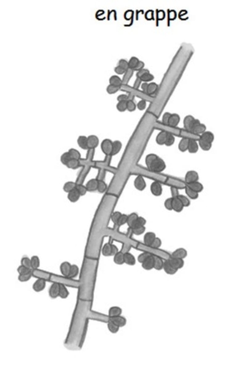
En grappe
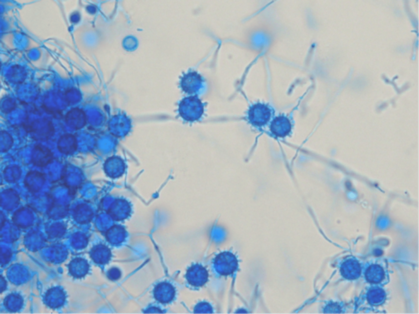
Lactophenol Cotton Blue
Stains fungal cell walls blue
Tease Prep
- Tease colony apart
- Add lactophenol cotton blue
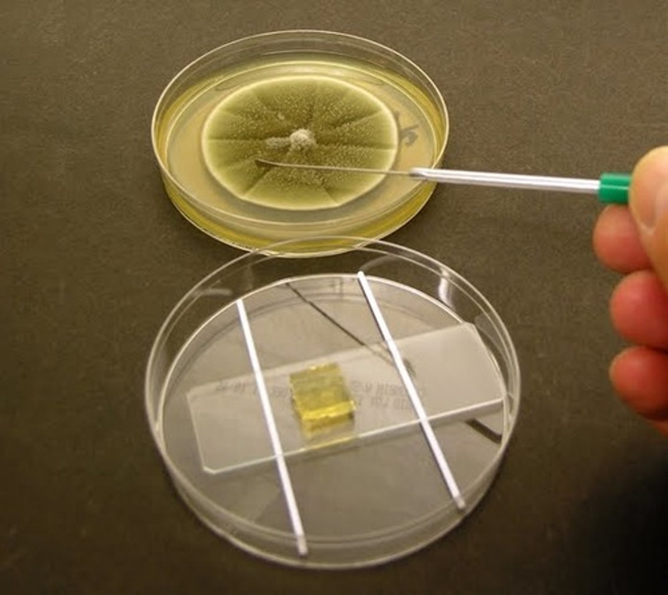
Cellophane Tape Prep
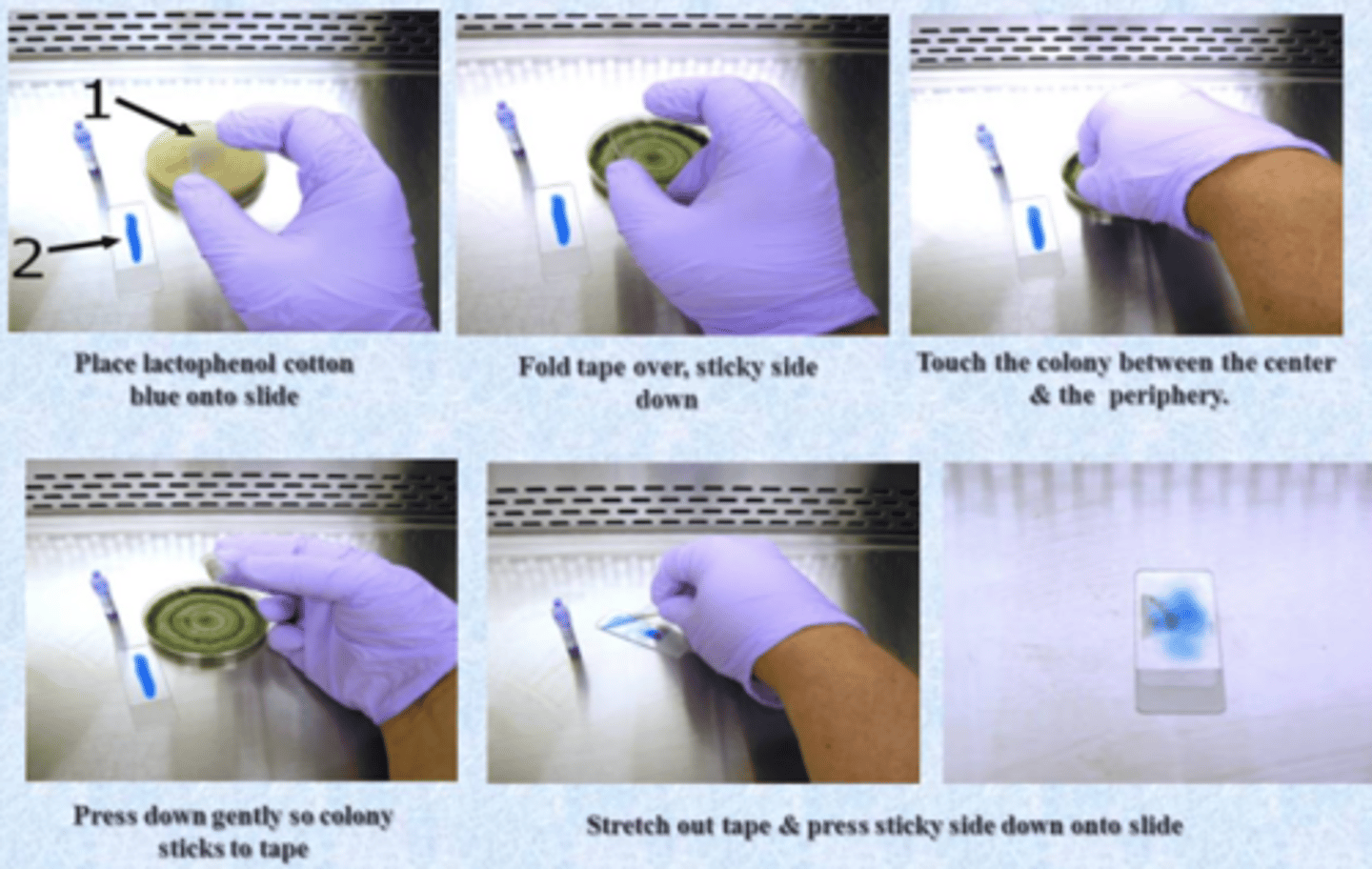
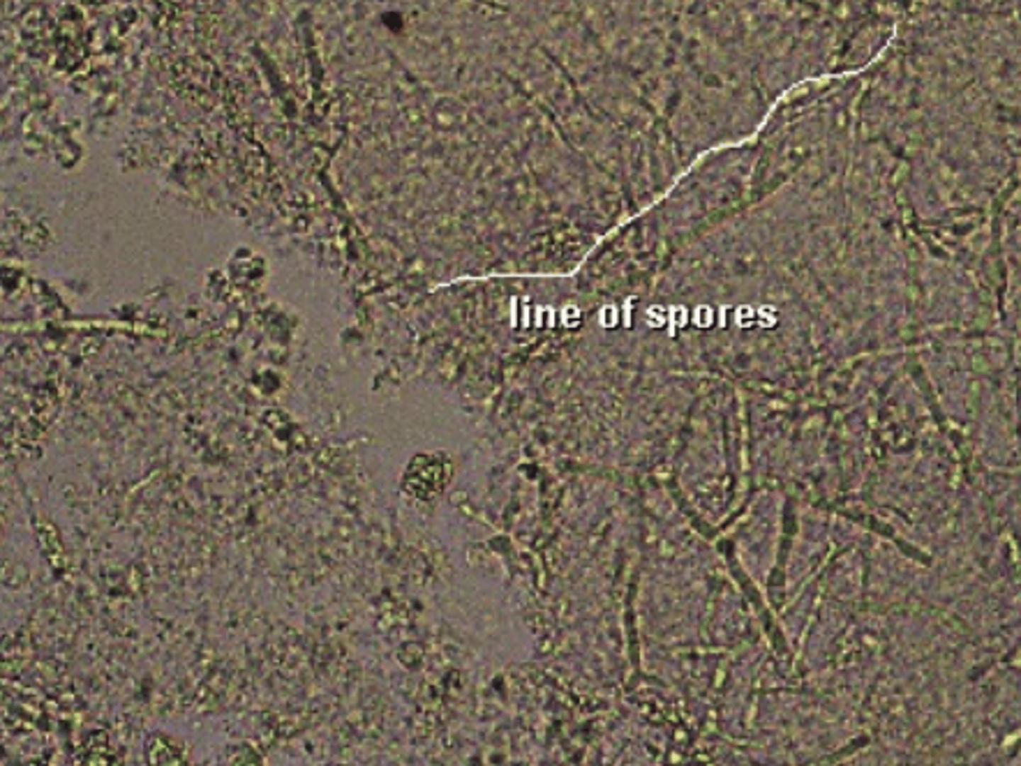
Slide culture
Best method for preserving and observing actual structures of fungi
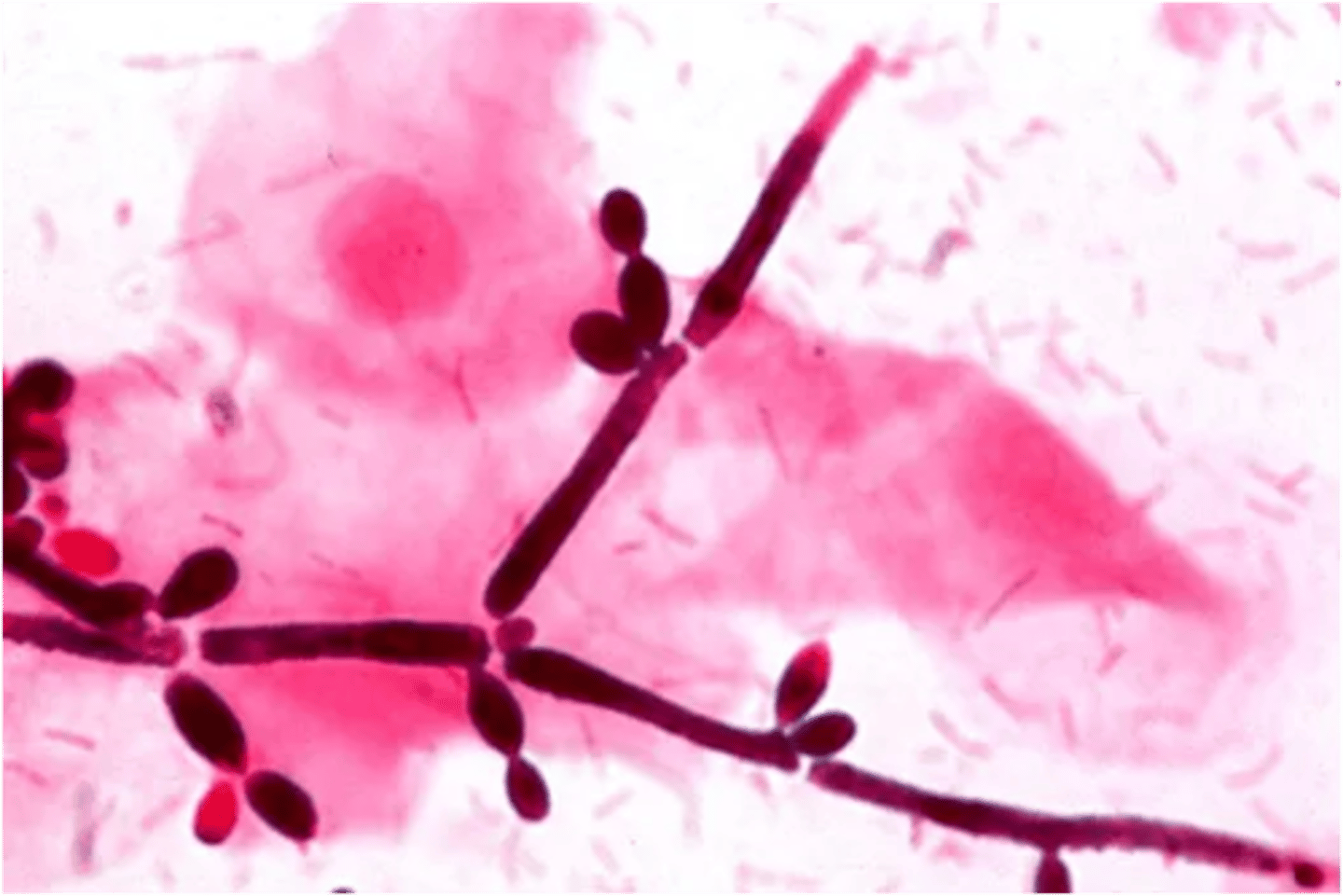
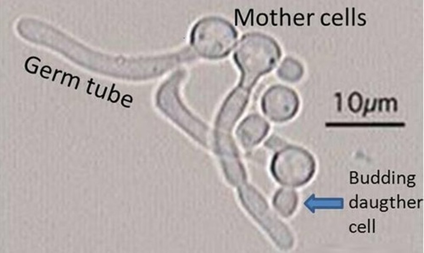
Germ tube test is used to identify ______.
yeast
RIOT agar
Rice Infusion, Oxgall, Polysorbate 80
Demonstrates sequential development of germ tubes and chlamydospores by Candida albicans